 Movie
Movie Controller
 Structure viewers
 About Yorodumi Papers


+Search query
-Structure paper
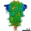
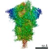
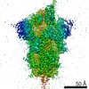
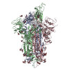
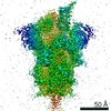
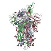
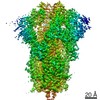
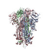
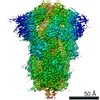
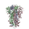
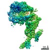
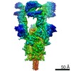
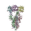
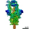
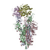
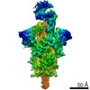
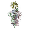
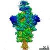
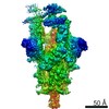
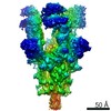
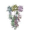
| Title | Cryo-EM Structures of SARS-CoV-2 Spike without and with ACE2 Reveal a pH-Dependent Switch to Mediate Endosomal Positioning of Receptor-Binding Domains. |
|---|---|
| Journal, issue, pages | Cell Host Microbe, Vol. 28, Issue 6, Page 867-879.e5, Year 2020 |
| Publish date | Dec 9, 2020 |
 Authors Authors | Tongqing Zhou / Yaroslav Tsybovsky / Jason Gorman / Micah Rapp / Gabriele Cerutti / Gwo-Yu Chuang / Phinikoula S Katsamba / Jared M Sampson / Arne Schön / Jude Bimela / Jeffrey C Boyington / Alexandra Nazzari / Adam S Olia / Wei Shi / Mallika Sastry / Tyler Stephens / Jonathan Stuckey / I-Ting Teng / Pengfei Wang / Shuishu Wang / Baoshan Zhang / Richard A Friesner / David D Ho / John R Mascola / Lawrence Shapiro / Peter D Kwong /  |
| PubMed Abstract | The SARS-CoV-2 spike employs mobile receptor-binding domains (RBDs) to engage the human ACE2 receptor and to facilitate virus entry, which can occur through low-pH-endosomal pathways. To understand ...The SARS-CoV-2 spike employs mobile receptor-binding domains (RBDs) to engage the human ACE2 receptor and to facilitate virus entry, which can occur through low-pH-endosomal pathways. To understand how ACE2 binding and low pH affect spike conformation, we determined cryo-electron microscopy structures-at serological and endosomal pH-delineating spike recognition of up to three ACE2 molecules. RBDs freely adopted "up" conformations required for ACE2 interaction, primarily through RBD movement combined with smaller alterations in neighboring domains. In the absence of ACE2, single-RBD-up conformations dominated at pH 5.5, resolving into a solitary all-down conformation at lower pH. Notably, a pH-dependent refolding region (residues 824-858) at the spike-interdomain interface displayed dramatic structural rearrangements and mediated RBD positioning through coordinated movements of the entire trimer apex. These structures provide a foundation for understanding prefusion-spike mechanics governing endosomal entry; we suggest that the low pH all-down conformation potentially facilitates immune evasion from RBD-up binding antibody. |
 External links External links | Cell Host Microbe / PubMed:33271067 / PubMed Central |
| Methods | EM (single particle) |
| Resolution | 2.4 - 3.93 Å |
| Structure data | EMDB-22251, PDB-6xlu: EMDB-22253, PDB-6xm0: EMDB-22254, PDB-6xm3: EMDB-22255, PDB-6xm4: EMDB-22256, PDB-6xm5: EMDB-22515, PDB-7jwy: EMDB-22922, PDB-7kmb: EMDB-22927, PDB-7kms: EMDB-22932, PDB-7kmz: EMDB-22941, PDB-7knb: EMDB-22943, PDB-7kne: EMDB-22949, PDB-7knh: EMDB-22950, PDB-7kni: |
| Chemicals |  ChemComp-NAG:  ChemComp-HOH: |
| Source |
|
 Keywords Keywords | VIRAL PROTEIN / SARS-CoV-2 spike COVID19 / SARS-CoV-2 spike / COVID19 / SARS-CoV-2 COVID19 / Hydrolase/Viral Protein / COVID / SARS-CoV2 / ACE2 / prefusion / Hydrolase-Viral Protein complex / VIRAL PROTEIN/Hydrolase / VIRAL PROTEIN-Hydrolase complex |

 severe acute respiratory syndrome coronavirus 2
 homo sapiens (human)