| タイトル | Coenzyme recognition and gene regulation by a flavin mononucleotide riboswitch. |
|---|
| ジャーナル・号・ページ | Nature, Vol. 458, Page 233-237, Year 2009 |
|---|
| 掲載日 | 2008年10月30日 (構造データの登録日) |
|---|
 著者 著者 | Serganov, A. / Huang, L. / Patel, D.J. |
|---|
 リンク リンク |  Nature / Nature /  PubMed:19169240 PubMed:19169240 |
|---|
| 手法 | X線回折 |
|---|
| 解像度 | 2.95 - 3.45 Å |
|---|
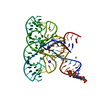
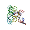
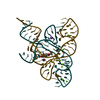
| 構造データ | PDB-3f2q:
Crystal structure of the FMN riboswitch bound to FMN
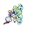
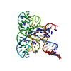
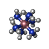
手法: X-RAY DIFFRACTION / 解像度: 2.95 Å PDB-3f2t:
Crystal structure of the FMN riboswitch bound to FMN, iridium hexamine soak.
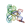
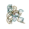
手法: X-RAY DIFFRACTION / 解像度: 3 Å PDB-3f2w:
Crystal structure of the FMn riboswitch bound to FMN, Ba2+ soak.
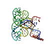
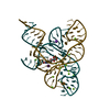
手法: X-RAY DIFFRACTION / 解像度: 3.45 Å PDB-3f2x:
Crystal structure of the FMN riboswitch bound to FMN, Cs+ soak.
手法: X-RAY DIFFRACTION / 解像度: 3.11 Å PDB-3f2y:
Crystal structure of the FMN riboswitch bound to FMN, Mn2+ soak.
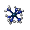
手法: X-RAY DIFFRACTION / 解像度: 3.2 Å PDB-3f30:
Crystal structure of the FMN riboswitch bound to FMN, cobalt hexammine soak.
手法: X-RAY DIFFRACTION / 解像度: 3.15 Å PDB-3f4e:
Crystal structure of the FMN riboswitch bound to FMN, split RNA.
手法: X-RAY DIFFRACTION / 解像度: 3.05 Å PDB-3f4g:
Crystal structure of the FMN riboswitch bound to riboflavin.
手法: X-RAY DIFFRACTION / 解像度: 3.01 Å PDB-3f4h:
Crystal structure of the FMN riboswitch bound to roseoflavin
手法: X-RAY DIFFRACTION / 解像度: 3 Å |
|---|
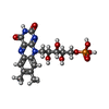
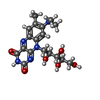
| 化合物 | ChemComp-RS3:
1-deoxy-1-[8-(dimethylamino)-7-methyl-2,4-dioxo-3,4-dihydrobenzo[g]pteridin-10(2H)-yl]-D-ribitol / ロセオフラビン
|
|---|
 キーワード キーワード | RNA / FMN riboswitch / transcription / FMN / riboswitch / barium / cesium / manganese / cobalt hexamine / riboflavin / Roseoflavin |
|---|
 著者
著者 リンク
リンク Nature /
  PubMed:19169240





 キーワード
キーワード ムービー
ムービー コントローラー
 構造ビューア
 万見文献について


