 ムービー
ムービー コントローラー
 構造ビューア
 EMN検索について


-検索条件
-検索結果
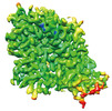
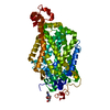
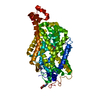
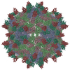
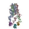
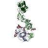
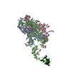
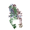
検索 (著者・登録者: zhu & k)の結果2,456件中、1から50件目までを表示しています
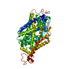
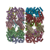
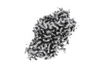
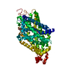
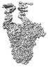
EMDB-37842: 
Cryo-EM structure of noradrenaline transporter in apo state
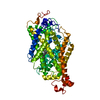
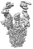
EMDB-37843:
Cryo-EM structure of noradrenaline transporter in complex with noradrenaline
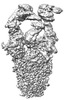
EMDB-37844:
Cryo-EM structure of noradrenaline transporter in complex with a x-MrlA analogue
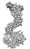
EMDB-37845:
Cryo-EM structure of noradrenaline transporter in complex with bupropion
EMDB-37846:
Cryo-EM structure of noradrenaline transporter in complex with ziprasidone
PDB-8wtu:
Cryo-EM structure of noradrenaline transporter in apo state
PDB-8wtv:
Cryo-EM structure of noradrenaline transporter in complex with noradrenaline
PDB-8wtw:
Cryo-EM structure of noradrenaline transporter in complex with a x-MrlA analogue
PDB-8wtx:
Cryo-EM structure of noradrenaline transporter in complex with bupropion
PDB-8wty:
Cryo-EM structure of noradrenaline transporter in complex with ziprasidone
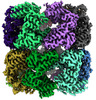
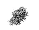
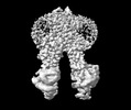
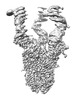
EMDB-45078:
E.coli GroEL + PBZ1587 inhibitor
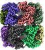
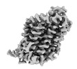
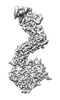
EMDB-45079:
E.coli GroEL apoenzyme
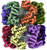
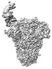
EMDB-45080:
E.Faecium GroEL

EMDB-45098:
E.coli GroEL + PBZ1587 inhibitor C1 reconstruction
PDB-9c0b:
E.coli GroEL + PBZ1587 inhibitor
PDB-9c0c:
E.coli GroEL apoenzyme
PDB-9c0d:
E.Faecium GroEL
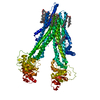
EMDB-37515:
Cryo-EM structure of inward-open state human norepinephrine transporter NET bound with antidepressant desipramine in KCl condition.
EMDB-37520:
Cryo-EM structure of inward-open state human norepinephrine transporter NET bound with norepinephrine in nanodisc.
EMDB-39729:
Cryo-EM structure of human norepinephrine transporter NET in the presence of the antidepressant atomoxetine in an outward-open state at resolution of 3.4 angstrom.
PDB-8wgr:
Cryo-EM structure of inward-open state human norepinephrine transporter NET bound with antidepressant desipramine in KCl condition.
PDB-8wgx:
Cryo-EM structure of inward-open state human norepinephrine transporter NET bound with norepinephrine in nanodisc.

PDB-8z1l:
Cryo-EM structure of human norepinephrine transporter NET in the presence of the antidepressant atomoxetine in an outward-open state at resolution of 3.4 angstrom.
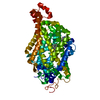
EMDB-38532:
Cryo-EM structure of human ABCC4
PDB-8xok:
Cryo-EM structure of human ABCC4
EMDB-32979:
Cryo-EM structure of Coxsackievirus B1 A-particle in complex with nAb 8A10 (CVB1-A:8A10)
PDB-7x35:
Cryo-EM structure of Coxsackievirus B1 A-particle in complex with nAb 8A10 (CVB1-A:8A10)
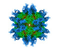
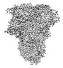
EMDB-39025:
Structure of HCoV-HKU1A spike in the functionally anchored-3up conformation with 3TMPRSS2
EMDB-39026:
Local structure of HCoV-HKU1A spike in complex with TMPRSS2 and glycan
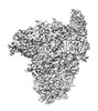
EMDB-39036:
Structure of HCoV-HKU1C spike in the functionally anchored-1up conformation with 1TMPRSS2
EMDB-39037:
Structure of HCoV-HKU1C spike in the functionally anchored-2up conformation with 2TMPRSS2
EMDB-39038:
Structure of HCoV-HKU1C spike in the functionally anchored-3up conformation with 2TMPRSS2
EMDB-39039:
Structure of HCoV-HKU1C spike in the functionally anchored-3up conformation with 3TMPRSS2
EMDB-39040:
Local structure of HCoV-HKU1C spike in complex with TMPRSS2 and glycan
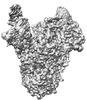
EMDB-39041:
Structure of HCoV-HKU1C spike in the inactive-closed conformation
EMDB-39042:
Structure of HCoV-HKU1C spike in the inactive-1up conformation
EMDB-39043:
Structure of HCoV-HKU1C spike in the inactive-2up conformation
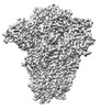
EMDB-39044:
Structure of HCoV-HKU1C spike in the glycan-activated-closed conformation
EMDB-39045:
Structure of HCoV-HKU1C spike in the glycan-activated-1up conformation
EMDB-39046:
Structure of HCoV-HKU1C spike in the glycan-activated-2up conformation
EMDB-39047:
Structure of HCoV-HKU1C spike in the glycan-activated-3up conformation
EMDB-39048:
Local structure of HCoV-HKU1C spike in complex with glycan
PDB-8y7x:
Structure of HCoV-HKU1A spike in the functionally anchored-3up conformation with 3TMPRSS2
PDB-8y7y:
Local structure of HCoV-HKU1A spike in complex with TMPRSS2 and glycan
PDB-8y87:
Structure of HCoV-HKU1C spike in the functionally anchored-1up conformation with 1TMPRSS2
PDB-8y88:
Structure of HCoV-HKU1C spike in the functionally anchored-2up conformation with 2TMPRSS2
PDB-8y89:
Structure of HCoV-HKU1C spike in the functionally anchored-3up conformation with 2TMPRSS2
PDB-8y8a:
Structure of HCoV-HKU1C spike in the functionally anchored-3up conformation with 3TMPRSS2
PDB-8y8b:
Local structure of HCoV-HKU1C spike in complex with TMPRSS2 and glycan
PDB-8y8c:
Structure of HCoV-HKU1C spike in the inactive-closed conformation
ページ:
 wwPDBはEMDBデータモデルのバージョン3へ移行します
