 Movie
Movie Controller
 Structure viewers
 About EMN search


-Search query
-Search result
Showing 1 - 50 of 5,412 items for (author: yang & t)
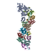
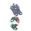
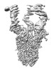
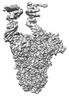
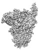
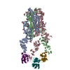
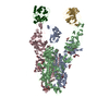
EMDB-39645: 
The structure of HKU1-B S protein with bsAb1
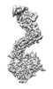
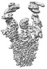
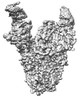
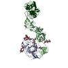
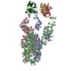
EMDB-39646:
the complex structure of the H4B6 Fab with the RBD of Omicron BA.5 S protein
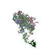
PDB-8yww:
The structure of HKU1-B S protein with bsAb1
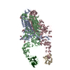
PDB-8ywx:
the complex structure of the H4B6 Fab with the RBD of Omicron BA.5 S protein

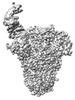
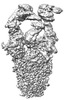
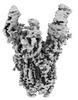
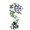
EMDB-37499:
Cryo-EM structure of CRISPR-Csm effector complex from Mycobacterium canettii
PDB-8wfx:
Cryo-EM structure of CRISPR-Csm effector complex from Mycobacterium canettii
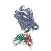
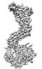
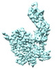
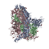
EMDB-19978:
Outward-open structure of Drosophila dopamine transporter bound to an atypical non-competitive inhibitor
EMDB-19979:
Inhibitor-free outward-open structure of Drosophila dopamine transporter
PDB-9euo:
Outward-open structure of Drosophila dopamine transporter bound to an atypical non-competitive inhibitor
PDB-9eup:
Inhibitor-free outward-open structure of Drosophila dopamine transporter
EMDB-44965:
Sub-tomogram average of the RSV M lattice from native virions released from RSV-infected BEAS-2B cells cultured on EM grids

EMDB-44966:
Sub-tomogram average of a pair of RSV F trimers from native virions released from RSV-infected BEAS-2B cells cultured on EM grids

EMDB-44968:
Sub-tomogram average of two pairs of RSV F trimers from the surface of native virions released from RSV-infected BEAS-2B cells cultured on EM grids

EMDB-44969:
Sub-tomogram average of two pairs of RSV F trimers from the surface of native virions released from RSV-infected BEAS-2B cells cultured on EM grids

EMDB-44971:
Sub-tomogram average of two pairs of RSV F trimers from the surface of native virions released from RSV-infected BEAS-2B cells cultured on EM grids
EMDB-60607:
A local Cryo-EM structure of Bitter taste receptor TAS2R14
EMDB-60608:
A Cryo-EM structure of Bitter taste receptor TAS2R14 with Ggust

EMDB-60626:
A Cryo-EM structure of Bitter taste receptor TAS2R14 with Gi complex
EMDB-60627:
A local Cryo-EM structure of Bitter taste receptor TAS2R14 with Gi complex
PDB-9iiw:
A local Cryo-EM structure of Bitter taste receptor TAS2R14
PDB-9iix:
A Cryo-EM structure of Bitter taste receptor TAS2R14 with Ggust
PDB-9ij9:
A Cryo-EM structure of Bitter taste receptor TAS2R14 with Gi complex
PDB-9ija:
A local Cryo-EM structure of Bitter taste receptor TAS2R14 with Gi complex
EMDB-38532:
Cryo-EM structure of human ABCC4
PDB-8xok:
Cryo-EM structure of human ABCC4
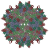
EMDB-32979:
Cryo-EM structure of Coxsackievirus B1 A-particle in complex with nAb 8A10 (CVB1-A:8A10)
PDB-7x35:
Cryo-EM structure of Coxsackievirus B1 A-particle in complex with nAb 8A10 (CVB1-A:8A10)
EMDB-39025:
Structure of HCoV-HKU1A spike in the functionally anchored-3up conformation with 3TMPRSS2
EMDB-39026:
Local structure of HCoV-HKU1A spike in complex with TMPRSS2 and glycan
EMDB-39036:
Structure of HCoV-HKU1C spike in the functionally anchored-1up conformation with 1TMPRSS2
EMDB-39037:
Structure of HCoV-HKU1C spike in the functionally anchored-2up conformation with 2TMPRSS2
EMDB-39038:
Structure of HCoV-HKU1C spike in the functionally anchored-3up conformation with 2TMPRSS2
EMDB-39039:
Structure of HCoV-HKU1C spike in the functionally anchored-3up conformation with 3TMPRSS2
EMDB-39040:
Local structure of HCoV-HKU1C spike in complex with TMPRSS2 and glycan
EMDB-39041:
Structure of HCoV-HKU1C spike in the inactive-closed conformation
EMDB-39042:
Structure of HCoV-HKU1C spike in the inactive-1up conformation
EMDB-39043:
Structure of HCoV-HKU1C spike in the inactive-2up conformation
EMDB-39044:
Structure of HCoV-HKU1C spike in the glycan-activated-closed conformation
EMDB-39045:
Structure of HCoV-HKU1C spike in the glycan-activated-1up conformation
EMDB-39046:
Structure of HCoV-HKU1C spike in the glycan-activated-2up conformation
EMDB-39047:
Structure of HCoV-HKU1C spike in the glycan-activated-3up conformation
EMDB-39048:
Local structure of HCoV-HKU1C spike in complex with glycan
PDB-8y7x:
Structure of HCoV-HKU1A spike in the functionally anchored-3up conformation with 3TMPRSS2
PDB-8y7y:
Local structure of HCoV-HKU1A spike in complex with TMPRSS2 and glycan
PDB-8y87:
Structure of HCoV-HKU1C spike in the functionally anchored-1up conformation with 1TMPRSS2
PDB-8y88:
Structure of HCoV-HKU1C spike in the functionally anchored-2up conformation with 2TMPRSS2
PDB-8y89:
Structure of HCoV-HKU1C spike in the functionally anchored-3up conformation with 2TMPRSS2
PDB-8y8a:
Structure of HCoV-HKU1C spike in the functionally anchored-3up conformation with 3TMPRSS2
PDB-8y8b:
Local structure of HCoV-HKU1C spike in complex with TMPRSS2 and glycan
PDB-8y8c:
Structure of HCoV-HKU1C spike in the inactive-closed conformation
Pages:
 wwPDB to switch to version 3 of the EMDB data model
