 Movie
Movie Controller
 Structure viewers
 About EMN search


-Search query
-Search result
Showing all 18 items for (author: ventura & santos & c)

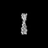
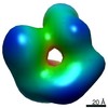
EMDB-16466: 
The structural architecture of alpha-synuclein oligomer
Method: single particle / : Cuellar J, Santos J, Pallares I, Ventura S, Valpuesta JM

EMDB-16528:
3D reconstruction of alpha-synuclein oligomer-PSMa3 complex
Method: single particle / : Cuellar J, Santos J, Pallares I, Ventura S, Valpuesta JM
EMDB-18475:
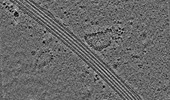
Cryo-electron tomogram of an induced S2 cell protrusion. The cell was treated with 2uM thapsigargin (5h) and with 2.5uM Cytochalasin D (2h).
Method: electron tomography / : Ventura Santos C, Carter AP
EMDB-16877:
Subtomogram averaging structure of cofilactin filament inside microtubule lumen of Drosophila S2 cell protrusion.
Method: subtomogram averaging / : Ventura Santos C, Carter AP

PDB-8oh4:
Subtomogram averaging structure of cofilactin filament inside microtubule lumen of Drosophila S2 cell protrusion.
Method: subtomogram averaging / : Ventura Santos C, Carter AP
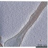
EMDB-16685:
Tomogram of an induced protrusion of a Drosophila S2 cell
Method: electron tomography / : Ventura Santos C, Carter AP
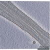
EMDB-16693:
Tomogram of an induced protrusion of a Drosophila S2 cell
Method: electron tomography / : Ventura Santos C, Carter AP
EMDB-16695:
Tomogram of an induced protrusion of a Drosophila S2 alpha-tubulin acetyltransferase knock-out (dTAT KO) cell with a filament inside the microtubule lumen
Method: electron tomography / : Ventura Santos C, Carter AP
EMDB-16720:
Tomogram of an induced protrusion of a Drosophila S2 cell with filaments inside the microtubule lumen.
Method: electron tomography / : Ventura Santos C, Carter AP
EMDB-16800:
Tomogram of an induced protrusion of a cofilin knock-down Drosophila S2 cell with filaments inside the microtubule lumen
Method: electron tomography / : Ventura Santos C, Carter AP
EMDB-16811:
Tomogram of an induced protrusion of a cofilin knock-down Drosophila S2 cell with a filament inside the microtubule lumen.
Method: electron tomography / : Ventura Santos C, Carter AP

EMDB-12639:
In situ subtomogram average of 13 protofilament microtubule from Mus musculus DRG axons
Method: subtomogram averaging / : Foster HE, Ventura Santos C, Carter AP
EMDB-12640:
In situ subtomogram average of microtubule inner protein from Mus musculus DRG axons
Method: subtomogram averaging / : Foster HE, Ventura Santos C, Carter AP
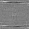
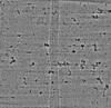
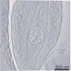
EMDB-13598:
Tomogram of a mouse dorsal root ganglion axon (dataset 2, TS_43).
Method: electron tomography / : Foster HE, Ventura Santos C, Carter AP
EMDB-13599:
Tomogram of mouse dorsal root ganglion axon (dataset 2, TS_41).
Method: electron tomography / : Foster HE, Ventura Santos C, Carter AP
EMDB-13600:
Tomogram of mouse dorsal root ganglion axon (dataset 1, TS_14).
Method: electron tomography / : Foster HE, Ventura Santos C, Carter AP
EMDB-13601:
Tomogram of mouse dorsal root ganglion axon (dataset 1, TS_20).
Method: electron tomography / : Foster HE, Ventura Santos C, Carter AP
EMDB-13602:
Tomogram of mouse dorsal root ganglion axon (dataset 1, TS_29).
Method: electron tomography / : Foster HE, Ventura Santos C, Carter AP
 wwPDB to switch to version 3 of the EMDB data model
