Movie
Movie Controller
 Structure viewers
 About EMN search



-Search query
-Search result
Showing all 37 items for (author: peterl & s)

EMDB-56664: 
Cryo-ET of IAV (WSN-M1-Udorn) supernatant
Method: electron tomography / : Peterl S, Chlanda P

EMDB-56667:
Cryo-ET of IAV (WSN-M1-Udorn) pellet
Method: electron tomography / : Peterl S, Chlanda P

EMDB-56668:
Cryo-ET of IAV (WSN-M1-Udorn) unfractionated
Method: electron tomography / : Peterl S, Chlanda P

EMDB-53448:
Cryo-electron tomography of an A549 cell infected with influenza A/WSN/33 virus
Method: electron tomography / : Peterl S, Chlanda P

EMDB-53449:
Cryo-electron tomography of an A549 cell infected with influenza A/WSN/33 virus
Method: electron tomography / : Peterl S, Chlanda P

EMDB-54375:
Tomogram showing an NA membrane in an A549wt cell infected with WSNdeltaHA at 16 hpi.
Method: electron tomography / : Wachsmuth-Melm M, Chlanda P

EMDB-51740:
Subtomogram average of nuclear helical M1 assemblies
Method: subtomogram averaging / : Wachsmuth-Melm M, Chlanda P

EMDB-51741:
Subtomogram average of zippered influenza A virus neuraminidase
Method: subtomogram averaging / : Wachsmuth-Melm M, Chlanda P

EMDB-51742:
Subtomogram average of influenza vRNPs in-situ.
Method: subtomogram averaging / : Wachsmuth-Melm M, Chlanda P

EMDB-51790:
Subtomogram average of intracellular influenza A virus hemagglutinin (subtype H1)
Method: subtomogram averaging / : Wachsmuth-Melm M, Chlanda P

EMDB-51811:
Tomogram showing M1 cylinders in a VeroE6 cell transfected with M1 (HK68) 24 hpt [figure 3].
Method: electron tomography / : Wachsmuth-Melm M, Chlanda P

EMDB-50067:
Tomogram showing an HA remodelled membrane in an A549wt cell infected with PR8 at 16 hpi [figure 1].
Method: electron tomography / : Wachsmuth-Melm M, Chlanda P

EMDB-50070:
Tomogram showing an HA remodelled membrane in an A549wt cell infected with HK68 at 16 hpi [figure 1].
Method: electron tomography / : Wachsmuth-Melm M, Chlanda P

EMDB-50078:
Tomogram showing an HA remodelled membrane in an A549wt cell infected with HK68 at 16 hpi [figure 1].
Method: electron tomography / : Wachsmuth-Melm M, Chlanda P

EMDB-50079:
Tomogram showing an HA remodelled membrane in an A549wt cell infected with HK68 at 16 hpi [figure 1].
Method: electron tomography / : Wachsmuth-Melm M, Chlanda P

EMDB-50080:
Tomogram showing an HA remodelled membrane in an A549wt cell infected with PR8 at 16 hpi [figure 1].
Method: electron tomography / : Wachsmuth-Melm M, Chlanda P

EMDB-50081:
Tomogram showing an HA remodelled membrane in an A549-Rab11wt cell infected with PR8 at 16 hpi [figure 2].
Method: electron tomography / : Wachsmuth-Melm M, Chlanda P

EMDB-50082:
A549-Rab11dn cell infected with PR8 virus at 16 hpi.
Method: electron tomography / : Wachsmuth-Melm M, Chlanda P

EMDB-50083:
A549wt cell infected with PR8 virus at 16 hpi.
Method: electron tomography / : Wachsmuth-Melm M, Chlanda P

EMDB-50084:
A549wt cell infected with PR8 virus at 8 hpi.
Method: electron tomography / : Wachsmuth-Melm M, Chlanda P

EMDB-50085:
A549wt cell infected with PR8 virus at 16 hpi.
Method: electron tomography / : Wachsmuth-Melm M, Chlanda P

EMDB-50086:
A549wt cell infected with PR8 virus at 16 hpi.
Method: electron tomography / : Wachsmuth-Melm M, Chlanda P

EMDB-50087:
A549wt cell infected with PR8 virus at 16 hpi.
Method: electron tomography / : Wachsmuth-Melm M, Chlanda P

EMDB-50088:
A549wt cell infected with HK68 virus at 16 hpi.
Method: electron tomography / : Wachsmuth-Melm M, Chlanda P

EMDB-16128:
Tomogram of a late endosome of A549 cell infected with influenza A virus.
Method: electron tomography / : Klein S, Chlanda P

EMDB-16129:
Tomogram of a late endosome of A549 cell infected with influenza A virus (Figure 6C).
Method: electron tomography / : Klein S, Chlanda P

EMDB-16130:
Tomogram of a late endosome of A549 cell infected with influenza A virus (Figure 6E)
Method: electron tomography / : Klein S, Chlanda P

EMDB-16131:
Tomogram of a late endosome of A549 cell infected with influenza A virus (Figure 6G)
Method: electron tomography / : Klein S, Chlanda P

EMDB-16132:
Tomogram of a late endosome of A549 cell infected with influenza A virus (Figure 6I,K,M)
Method: electron tomography / : Klein S, Chlanda P

EMDB-16133:
Tomogram of a late endosome of A549 cell infected with influenza A virus (Figure 6O)
Method: electron tomography / : Klein S, Chlanda P
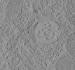
EMDB-15705:
Tomogram of a late endosome of A549 cell (Figure 1R)
Method: electron tomography / : Klein S, Chlanda P
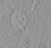
EMDB-15707:
Tomogram of a late endosome of A549 cell treated with IFN-beta (Figure 1T)
Method: electron tomography / : Klein S, Chlanda P
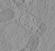
EMDB-15708:
Tomogram of a late endosome of A549-IFITM3 cell (Figure 1V)
Method: electron tomography / : Klein S, Chlanda P
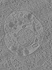
EMDB-15131:
Tomogram of a late endosome of A549-IFITM3 cells infected with influenza A virus (Figure S7)
Method: electron tomography / : Klein S, Chlanda P
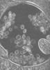
EMDB-15130:
Tomogram of a late endosome of A549-IFITM3 cells infected with influenza A virus (Figure 4E)
Method: electron tomography / : Klein S, Chlanda P
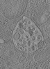
EMDB-15132:
Tomogram of a late endosome of A549-IFITM3 cells infected with influenza A virus (Figure S8)
Method: electron tomography / : Klein S, Chlanda P

EMDB-15133:
Tomogram of a late endosome of A549-IFITM3 cells infected with influenza A virus (Figure S9)
Method: electron tomography / : Klein S, Chlanda P
 wwPDB to switch to version 3 of the EMDB data model