 ムービー
ムービー コントローラー
 構造ビューア
 EMN検索について


-検索条件
-検索結果
検索 (著者・登録者: ma & x)の結果12,807件中、1から50件目までを表示しています
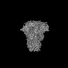
EMDB-19978: 
Outward-open structure of Drosophila dopamine transporter bound to an atypical non-competitive inhibitor
EMDB-19979:
Inhibitor-free outward-open structure of Drosophila dopamine transporter
PDB-9euo:
Outward-open structure of Drosophila dopamine transporter bound to an atypical non-competitive inhibitor
PDB-9eup:
Inhibitor-free outward-open structure of Drosophila dopamine transporter

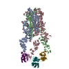
EMDB-19758:
Cryo-EM Structure of the R388 plasmid conjugative pilus reveals a helical polymer characterised by an unusual pilin/phospholipid binary complex

PDB-8s6h:
Cryo-EM Structure of the R388 plasmid conjugative pilus reveals a helical polymer characterised by an unusual pilin/phospholipid binary complex

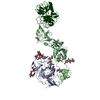
EMDB-38873:
cryo-EM structure of Staphylococcus aureus(ATCC 29213) 50S ribosome in complex with MCX-190.

EMDB-38874:
Cryo-EM structure of Staphylococcus aureus (15B196) 50S ribosome in complex with MCX-190.

EMDB-38875:
Cryo-EM structure of Staphylococcus aureus 70S ribosome (strain 15B196) in complex with MCX-190.

EMDB-38876:
cryo-EM structure of Staphylococcus aureus(ATCC 29213) 70S ribosome in complex with MCX-190.

PDB-8y36:
cryo-EM structure of Staphylococcus aureus(ATCC 29213) 50S ribosome in complex with MCX-190.

PDB-8y37:
Cryo-EM structure of Staphylococcus aureus (15B196) 50S ribosome in complex with MCX-190.

PDB-8y38:
Cryo-EM structure of Staphylococcus aureus 70S ribosome (strain 15B196) in complex with MCX-190.

PDB-8y39:
cryo-EM structure of Staphylococcus aureus(ATCC 29213) 70S ribosome in complex with MCX-190.

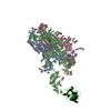
EMDB-43762:
Aca2 from Pectobacterium phage ZF40 bound to RNA

PDB-8w35:
Aca2 from Pectobacterium phage ZF40 bound to RNA
EMDB-50621:
Structure of heteromeric amyloid filament of TDP-43 and AXNA11 from FTLD-TDP Type C (variant 2)
EMDB-50628:
Structure of heteromeric amyloid filament of TDP-43 and AXNA11 from FTLD-TDP Type C (variant 1)

PDB-9fof:
Structure of heteromeric amyloid filament of TDP-43 and AXNA11 from FTLD-TDP Type C (variant 2)
PDB-9for:
Structure of heteromeric amyloid filament of TDP-43 and AXNA11 from FTLD-TDP Type C (variant 1)

EMDB-18950:
70S Escherichia coli ribosome with Paenilamicin B2 bound with A- and P-site tRNA.
EMDB-19004:
70S Escherichia coli ribosome with Paenilamicin B2 bound with hybrid A/P- and hybrid P/E-tRNA.

PDB-8r6c:
70S Escherichia coli ribosome with Paenilamicin B2 bound with A- and P-site tRNA.

PDB-8r8m:
70S Escherichia coli ribosome with Paenilamicin B2 bound with hybrid A/P- and hybrid P/E-tRNA.
EMDB-32979:
Cryo-EM structure of Coxsackievirus B1 A-particle in complex with nAb 8A10 (CVB1-A:8A10)
PDB-7x35:
Cryo-EM structure of Coxsackievirus B1 A-particle in complex with nAb 8A10 (CVB1-A:8A10)
EMDB-18307:
Native eisosome lattice bound to plasma membrane microdomain
EMDB-18308:
Helical reconstruction of yeast eisosome protein Pil1 bound to membrane composed of lipid mixture -PIP2/+sterol (DOPC, DOPE, DOPS, cholesterol 30:20:20:30)
EMDB-18309:
Helical reconstruction of yeast eisosome protein Pil1 bound to membrane composed of lipid mixture +PIP2/-sterol (DOPC, DOPE, DOPS, PI(4,5)P2 50:20:20:10)
EMDB-18310:
Helical reconstruction of yeast eisosome protein Pil1 bound to membrane composed of lipid mixture +PIP2/+sterol (DOPC, DOPE, DOPS, cholesterol, PI(4,5)P2 35:20:20:15:10)
EMDB-18311:
Compact state - Native eisosome lattice bound to plasma membrane microdomain
EMDB-18312:
Stretched state - Native eisosome lattice bound to plasma membrane microdomain

EMDB-39025:
Structure of HCoV-HKU1A spike in the functionally anchored-3up conformation with 3TMPRSS2

EMDB-39026:
Local structure of HCoV-HKU1A spike in complex with TMPRSS2 and glycan

EMDB-39036:
Structure of HCoV-HKU1C spike in the functionally anchored-1up conformation with 1TMPRSS2
EMDB-39037:
Structure of HCoV-HKU1C spike in the functionally anchored-2up conformation with 2TMPRSS2
EMDB-39038:
Structure of HCoV-HKU1C spike in the functionally anchored-3up conformation with 2TMPRSS2

EMDB-39039:
Structure of HCoV-HKU1C spike in the functionally anchored-3up conformation with 3TMPRSS2

EMDB-39040:
Local structure of HCoV-HKU1C spike in complex with TMPRSS2 and glycan
EMDB-39041:
Structure of HCoV-HKU1C spike in the inactive-closed conformation
EMDB-39042:
Structure of HCoV-HKU1C spike in the inactive-1up conformation

EMDB-39043:
Structure of HCoV-HKU1C spike in the inactive-2up conformation
EMDB-39044:
Structure of HCoV-HKU1C spike in the glycan-activated-closed conformation

EMDB-39045:
Structure of HCoV-HKU1C spike in the glycan-activated-1up conformation

EMDB-39046:
Structure of HCoV-HKU1C spike in the glycan-activated-2up conformation

EMDB-39047:
Structure of HCoV-HKU1C spike in the glycan-activated-3up conformation

EMDB-39048:
Local structure of HCoV-HKU1C spike in complex with glycan
PDB-8y7x:
Structure of HCoV-HKU1A spike in the functionally anchored-3up conformation with 3TMPRSS2
PDB-8y7y:
Local structure of HCoV-HKU1A spike in complex with TMPRSS2 and glycan
PDB-8y87:
Structure of HCoV-HKU1C spike in the functionally anchored-1up conformation with 1TMPRSS2
ページ:
 wwPDBはEMDBデータモデルのバージョン3へ移行します
