 Movie
Movie Controller


+ Open data

- Basic information
| Entry | Database: PDB / ID: 1gru | ||||||
|---|---|---|---|---|---|---|---|
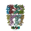
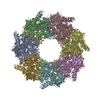
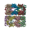
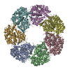
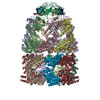
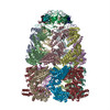
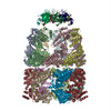
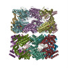
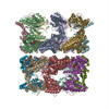
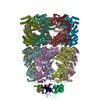
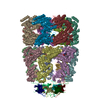
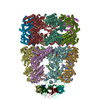
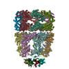
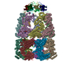
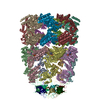
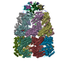
| Title | SOLUTION STRUCTURE OF GROES-ADP7-GROEL-ATP7 COMPLEX BY CRYO-EM | ||||||
 Components Components |
| ||||||
 Keywords Keywords | CHAPERONE / CHAPERONIN / HSP60 / GROEL-GROES / MOLECULAR CHAPERONE / ADP | ||||||
| Function / homology |  Function and homology information Function and homology informationGroEL-GroES complex / chaperonin ATPase / virion assembly / chaperone cofactor-dependent protein refolding / protein folding chaperone / isomerase activity / ATP-dependent protein folding chaperone / response to radiation / unfolded protein binding / protein folding ...GroEL-GroES complex / chaperonin ATPase / virion assembly / chaperone cofactor-dependent protein refolding / protein folding chaperone / isomerase activity / ATP-dependent protein folding chaperone / response to radiation / unfolded protein binding / protein folding / protein-folding chaperone binding / protein refolding / response to heat / magnesium ion binding / ATP hydrolysis activity / ATP binding / identical protein binding / membrane / metal ion binding / cytoplasm / cytosol Similarity search - Function | ||||||
| Biological species |  | ||||||
| Method | ELECTRON MICROSCOPY / single particle reconstruction / cryo EM / Resolution: 12.5 Å | ||||||
 Authors Authors | Ranson, N.A. / Farr, G.W. / Roseman, A.M. / Gowen, B. / Fenton, W.A. / Horwich, A.L. / Saibil, H.R. | ||||||
 Citation Citation | Journal: Cell / Year: 2001 Title: ATP-bound states of GroEL captured by cryo-electron microscopy. Authors: N A Ranson / G W Farr / A M Roseman / B Gowen / W A Fenton / A L Horwich / H R Saibil /  Abstract: The chaperonin GroEL drives its protein-folding cycle by cooperatively binding ATP to one of its two rings, priming that ring to become folding-active upon GroES binding, while simultaneously ...The chaperonin GroEL drives its protein-folding cycle by cooperatively binding ATP to one of its two rings, priming that ring to become folding-active upon GroES binding, while simultaneously discharging the previous folding chamber from the opposite ring. The GroEL-ATP structure, determined by cryo-EM and atomic structure fitting, shows that the intermediate domains rotate downward, switching their intersubunit salt bridge contacts from substrate binding to ATP binding domains. These observations, together with the effects of ATP binding to a GroEL-GroES-ADP complex, suggest structural models for the ATP-induced reduction in affinity for polypeptide and for cooperativity. The model for cooperativity, based on switching of intersubunit salt bridge interactions around the GroEL ring, may provide general insight into cooperativity in other ring complexes and molecular machines. | ||||||
| History |
| ||||||
| Remark 700 | SHEET THE SHEET STRUCTURE OF THIS MOLECULE IS BIFURCATED. IN ORDER TO REPRESENT THIS FEATURE IN ... SHEET THE SHEET STRUCTURE OF THIS MOLECULE IS BIFURCATED. IN ORDER TO REPRESENT THIS FEATURE IN THE SHEET RECORDS BELOW, TWO SHEETS ARE DEFINED. |
- Structure visualization
| Movie |
Movie viewer |
|---|---|
| Structure viewer | Molecule: MolmilJmol/JSmol |
- Downloads & links
-Download
| PDBx/mmCIF format | 1gru.cif.gz | 1.3 MB | Display | PDBx/mmCIF format |
|---|---|---|---|---|
| PDB format | pdb1gru.ent.gz | 1.1 MB | Display | PDB format |
| PDBx/mmJSON format | 1gru.json.gz | Tree view | PDBx/mmJSON format | |
| Others |  Other downloads Other downloads |
-Validation report
| Summary document | 1gru_validation.pdf.gz | 933.9 KB | Display | wwPDB validaton report |
|---|---|---|---|---|
| Full document | 1gru_full_validation.pdf.gz | 1.6 MB | Display | |
| Data in XML | 1gru_validation.xml.gz | 278.3 KB | Display | |
| Data in CIF | 1gru_validation.cif.gz | 414.9 KB | Display | |
| Arichive directory | https://data.pdbj.org/pub/pdb/validation_reports/gr/1gruftp://data.pdbj.org/pub/pdb/validation_reports/gr/1gru | HTTPS FTP |
-Related structure data
| Related structure data |  1046MC 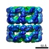 1042C 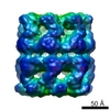 1047C 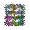 1gr5C 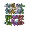 2c7eC C: citing same article ( M: map data used to model this data |
|---|---|
| Similar structure data |
-Links
 PDBj
PDBj

- Assembly
| Deposited unit | 
|
|---|---|
| 1 |
|
-Components
| #1: Protein | Mass: 57260.504 Da / Num. of mol.: 14 Source method: isolated from a genetically manipulated source Source: (gene. exp.) #2: Protein | Mass: 10400.938 Da / Num. of mol.: 7 Source method: isolated from a genetically manipulated source Source: (gene. exp.) Compound details | CRYO-EM MAP OF GROEL-GROES COMPLEX WITH ADP IN THE (CIS)RING WITH GROES BOUND AND ATP IN THE OPEN ...CRYO-EM MAP OF GROEL-GROES COMPLEX WITH ADP IN THE (CIS)RING WITH GROES BOUND AND ATP IN THE OPEN (TRANS) RING. | |
|---|
-Experimental details
-Experiment
| Experiment | Method: ELECTRON MICROSCOPY |
|---|---|
| EM experiment | Aggregation state: PARTICLE / 3D reconstruction method: single particle reconstruction |
- Sample preparation
| Component | Name: GROEL (C7) / Type: COMPLEX |
|---|---|
| Buffer solution | Name: HEPES / pH: 7.5 / Details: HEPES |
| Specimen | Conc.: 1 mg/ml / Embedding applied: NO / Shadowing applied: NO / Staining applied: NO / Vitrification applied: YES |
| Specimen support | Details: HOLEY CARBON |
| Vitrification | Instrument: HOMEMADE PLUNGER / Cryogen name: ETHANE / Details: LIQUID ETHANE |
| Crystal grow | *PLUS Method: cryo-electron microscopy |
- Electron microscopy imaging
| Experimental equipment |  Model: Tecnai F20 / Image courtesy: FEI Company |
|---|---|
| Microscopy | Model: FEI TECNAI F20 / Date: Jul 1, 2001 |
| Electron gun | Electron source:  FIELD EMISSION GUN / Accelerating voltage: 200 kV / Illumination mode: FLOOD BEAM FIELD EMISSION GUN / Accelerating voltage: 200 kV / Illumination mode: FLOOD BEAM |
| Electron lens | Mode: BRIGHT FIELD / Nominal magnification: 50000 X / Nominal defocus max: 3000 nm / Nominal defocus min: 900 nm |
| Specimen holder | Temperature: 95 K / Tilt angle min: 0 ° |
| Image recording | Electron dose: 20 e/Å2 / Film or detector model: KODAK SO-163 FILM |
| Radiation wavelength | Relative weight: 1 |
- Processing
| EM software |
| ||||||||||||
|---|---|---|---|---|---|---|---|---|---|---|---|---|---|
| CTF correction | Details: CLASS AVERAGES | ||||||||||||
| Symmetry | Point symmetry: C7 (7 fold cyclic) | ||||||||||||
| 3D reconstruction | Method: MODEL BASED ANGULAR REFINEMENT / Resolution: 12.5 Å / Num. of particles: 1448 / Nominal pixel size: 2.8 Å / Actual pixel size: 2.8 Å Details: ATOMIC COORDINATES FROM 1AON FITTED BY HAND. CIS RING FITTED WHOLE. TRANS EQUATORIAL AND INTERMEDIATE DOMAINS ALSO FITTED AS 7MER. A SINGLE TRANS APICAL WAS FITTED BY HAND AND THEN THE OTHER ...Details: ATOMIC COORDINATES FROM 1AON FITTED BY HAND. CIS RING FITTED WHOLE. TRANS EQUATORIAL AND INTERMEDIATE DOMAINS ALSO FITTED AS 7MER. A SINGLE TRANS APICAL WAS FITTED BY HAND AND THEN THE OTHER 6 POSITIONS CREATED FROM SYMMETRY. Symmetry type: POINT | ||||||||||||
| Atomic model building | Protocol: OTHER / Space: REAL / Details: METHOD--BY HAND REFINEMENT PROTOCOL--X-RAY | ||||||||||||
| Atomic model building | PDB-ID: 1GRU Accession code: 1GRU / Source name: PDB / Type: experimental model | ||||||||||||
| Refinement | Highest resolution: 12.5 Å | ||||||||||||
| Refinement step | Cycle: LAST / Highest resolution: 12.5 Å
|
