 Movie
Movie Controller

[English] 日本語
 Yorodumi
- EMDB-8251: Subtomogram-averaged map of in vitro assembled rubella virus caps... -
+ Open data

- Basic information
| Entry | Database: EMDB / ID: EMD-8251 | |||||||||
|---|---|---|---|---|---|---|---|---|---|---|
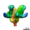
| Title | Subtomogram-averaged map of in vitro assembled rubella virus capsid protein tetramer complex | |||||||||
 Map data Map data | Subtomogram-averaged map of in vitro assembled rubella virus capsid protein tetramer complex, unfiltered and unmasked | |||||||||
 Sample Sample |
| |||||||||
| Function / homology |  Function and homology information Function and homology informationT=4 icosahedral viral capsid / host cell Golgi membrane / host cell mitochondrion / viral nucleocapsid / clathrin-dependent endocytosis of virus by host cell / fusion of virus membrane with host endosome membrane / viral envelope / virion attachment to host cell / virion membrane / RNA binding ...T=4 icosahedral viral capsid / host cell Golgi membrane / host cell mitochondrion / viral nucleocapsid / clathrin-dependent endocytosis of virus by host cell / fusion of virus membrane with host endosome membrane / viral envelope / virion attachment to host cell / virion membrane / RNA binding / membrane / metal ion binding Similarity search - Function | |||||||||
| Biological species |  Rubella virus strain M33 Rubella virus strain M33 | |||||||||
| Method | subtomogram averaging / cryo EM / Resolution: 35.0 Å | |||||||||
 Authors Authors | Mangala Prasad V / Klose T / Rossmann MG | |||||||||
 Citation Citation | Journal: PLoS Pathog / Year: 2017 Title: Assembly, maturation and three-dimensional helical structure of the teratogenic rubella virus. Authors: Vidya Mangala Prasad / Thomas Klose / Michael G Rossmann /  Abstract: Viral infections during pregnancy are a significant cause of infant morbidity and mortality. Of these, rubella virus infection is a well-substantiated example that leads to miscarriages or severe ...Viral infections during pregnancy are a significant cause of infant morbidity and mortality. Of these, rubella virus infection is a well-substantiated example that leads to miscarriages or severe fetal defects. However, structural information about the rubella virus has been lacking due to the pleomorphic nature of the virions. Here we report a helical structure of rubella virions using cryo-electron tomography. Sub-tomogram averaging of the surface spikes established the relative positions of the viral glycoproteins, which differed from the earlier icosahedral models of the virus. Tomographic analyses of in vitro assembled nucleocapsids and virions provide a template for viral assembly. Comparisons of immature and mature virions show large rearrangements in the glycoproteins that may be essential for forming the infectious virions. These results present the first known example of a helical membrane-enveloped virus, while also providing a structural basis for its assembly and maturation pathway. | |||||||||
| History |
|
- Structure visualization
| Movie |
Movie viewer |
|---|---|
| Structure viewer | EM map: SurfViewMolmilJmol/JSmol |
| Supplemental images |

- Downloads & links
-EMDB archive
| Map data | emd_8251.map.gz | 454 KB | EMDB map data format | |
|---|---|---|---|---|
| Header (meta data) | emd-8251-v30.xmlemd-8251.xml | 11.4 KB 11.4 KB | Display Display | EMDB header |
| Images | 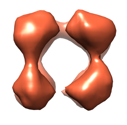 emd_8251.png emd_8251.png | 88.3 KB | ||
| Archive directory |  http://ftp.pdbj.org/pub/emdb/structures/EMD-8251ftp://ftp.pdbj.org/pub/emdb/structures/EMD-8251 http://ftp.pdbj.org/pub/emdb/structures/EMD-8251ftp://ftp.pdbj.org/pub/emdb/structures/EMD-8251 | HTTPS FTP |
-Validation report
| Summary document | emd_8251_validation.pdf.gz | 408.3 KB | Display | EMDB validaton report |
|---|---|---|---|---|
| Full document | emd_8251_full_validation.pdf.gz | 407.9 KB | Display | |
| Data in XML | emd_8251_validation.xml.gz | 5.1 KB | Display | |
| Data in CIF | emd_8251_validation.cif.gz | 5.6 KB | Display | |
| Arichive directory | https://ftp.pdbj.org/pub/emdb/validation_reports/EMD-8251ftp://ftp.pdbj.org/pub/emdb/validation_reports/EMD-8251 | HTTPS FTP |
-Related structure data
| Related structure data |  8248C  8249C 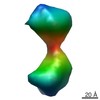 8250C 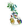 5khcC 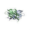 5kheC  5khfC C: citing same article ( |
|---|---|
| Similar structure data |

-Links
| EMDB pages | EMDB (EBI/PDBe) / EMDataResource |
|---|---|
| Related items in Molecule of the Month |

-Map
| File | Download / File: emd_8251.map.gz / Format: CCP4 / Size: 489.3 KB / Type: IMAGE STORED AS FLOATING POINT NUMBER (4 BYTES) | ||||||||||||||||||||||||||||||||||||||||||||||||||||||||||||
|---|---|---|---|---|---|---|---|---|---|---|---|---|---|---|---|---|---|---|---|---|---|---|---|---|---|---|---|---|---|---|---|---|---|---|---|---|---|---|---|---|---|---|---|---|---|---|---|---|---|---|---|---|---|---|---|---|---|---|---|---|---|
| Annotation | Subtomogram-averaged map of in vitro assembled rubella virus capsid protein tetramer complex, unfiltered and unmasked | ||||||||||||||||||||||||||||||||||||||||||||||||||||||||||||
| Voxel size | X=Y=Z: 5.28 Å | ||||||||||||||||||||||||||||||||||||||||||||||||||||||||||||
| Density |
| ||||||||||||||||||||||||||||||||||||||||||||||||||||||||||||
| Symmetry | Space group: 1 | ||||||||||||||||||||||||||||||||||||||||||||||||||||||||||||
| Details | EMDB XML:
CCP4 map header:
| ||||||||||||||||||||||||||||||||||||||||||||||||||||||||||||
-Supplemental data
- Sample components
-Entire : Rubella virus capsid protein
| Entire | Name: Rubella virus capsid protein |
|---|---|
| Components |
|
-Supramolecule #1: Rubella virus capsid protein
| Supramolecule | Name: Rubella virus capsid protein / type: complex / ID: 1 / Parent: 0 / Macromolecule list: all |
|---|---|
| Source (natural) | Organism: Rubella virus strain M33 |
| Recombinant expression | Organism:  |
| Molecular weight | Experimental: 256 KDa |
-Macromolecule #1: Rubella virus capsid protein
| Macromolecule | Name: Rubella virus capsid protein / type: protein_or_peptide / ID: 1 / Enantiomer: LEVO |
|---|---|
| Source (natural) | Organism: Rubella virus strain M33 |
| Recombinant expression | Organism: |
| Sequence | String: MEDLQKALEA QSRALRAELA AGASQSRRPR PPRQRDSSTS GDDSGRDSGG PRRRRGNRGR GQRRDWSRAP PPPEERQETR SQTPAPKPSR APPQQPQPPR MQTGRGGSAP RPELGPPTNP FQAAVARGLR PPLHDPDTEA PTEACVTSWL WSEGEGAVFY RVDLHFTNLG ...String: MEDLQKALEA QSRALRAELA AGASQSRRPR PPRQRDSSTS GDDSGRDSGG PRRRRGNRGR GQRRDWSRAP PPPEERQETR SQTPAPKPSR APPQQPQPPR MQTGRGGSAP RPELGPPTNP FQAAVARGLR PPLHDPDTEA PTEACVTSWL WSEGEGAVFY RVDLHFTNLG TPPLDEDGRW DPALMYNPCG PEPPAHVVRA YNQPAGDVRG VWGKGERTYA EQDFRVGGTR WHRLLRMPVR GLDGDSAPLP PHTTERIETR SARHPWRIR |
-Experimental details
-Structure determination
| Method | cryo EM |
|---|---|
 Processing Processing | subtomogram averaging |
| Aggregation state | particle |
-Sample preparation
| Concentration | 1 mg/mL | |||||||||
|---|---|---|---|---|---|---|---|---|---|---|
| Buffer | pH: 7.2 Component:
| |||||||||
| Grid | Model: Quantifoil R1.2/1.3 / Material: COPPER / Mesh: 200 / Support film - Material: CARBON | |||||||||
| Vitrification | Cryogen name: ETHANE |
- Electron microscopy
| Microscope | FEI TITAN KRIOS |
|---|---|
| Image recording | Film or detector model: GATAN K2 SUMMIT (4k x 4k) / Detector mode: SUPER-RESOLUTION / Average electron dose: 90.0 e/Å2 |
| Electron beam | Acceleration voltage: 300 kV / Electron source:  FIELD EMISSION GUN FIELD EMISSION GUN |
| Electron optics | Illumination mode: SPOT SCAN / Imaging mode: BRIGHT FIELD / Nominal defocus max: 0.005 µm / Nominal magnification: 11000 |
| Experimental equipment |  Model: Titan Krios / Image courtesy: FEI Company |
-Image processing
| Final reconstruction | Applied symmetry - Point group: C1 (asymmetric) / Resolution.type: BY AUTHOR / Resolution: 35.0 Å / Resolution method: OTHER / Software - Name: PEET (ver. 1.11.0) / Number subtomograms used: 18 |
|---|---|
| Extraction | Number tomograms: 15 / Number images used: 20 / Method: manual picking / Software - Name: IMOD/PEEt (ver. 4.8.40/1.11.0) |
| CTF correction | Software - Name: IMOD (ver. 4.8.40) |
| Final angle assignment | Type: OTHER |
