 Movie
Movie Controller

[English] 日本語
 Yorodumi
- PDB-1thd: COMPLEX ORGANIZATION OF DENGUE VIRUS E PROTEIN AS REVEALED BY 9.5... -
+ Open data

- Basic information
| Entry | Database: PDB / ID: 1thd | ||||||
|---|---|---|---|---|---|---|---|
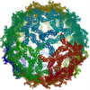
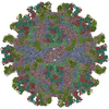
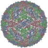
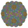
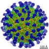
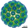
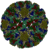
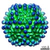
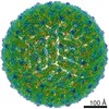
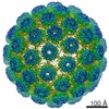
| Title | COMPLEX ORGANIZATION OF DENGUE VIRUS E PROTEIN AS REVEALED BY 9.5 ANGSTROM CRYO-EM RECONSTRUCTION | ||||||
 Components Components | Major envelope protein E | ||||||
 Keywords Keywords |  VIRUS / FLAVIVIRUS / FLAVIVIRIDAE / DENGUE VIRUS / GLYCOPROTEIN E / CRYO-EM / Icosahedral virus VIRUS / FLAVIVIRUS / FLAVIVIRIDAE / DENGUE VIRUS / GLYCOPROTEIN E / CRYO-EM / Icosahedral virus | ||||||
| Function / homology |  Function and homology information Function and homology informationsymbiont-mediated suppression of host JAK-STAT cascade via inhibition of host TYK2 activity / flavivirin / host cell mitochondrion / symbiont-mediated suppression of host JAK-STAT cascade via inhibition of STAT2 activity / symbiont-mediated suppression of host cytoplasmic pattern recognition receptor signaling pathway via inhibition of MAVS activity / viral capsid / : / nucleoside-triphosphate phosphatase / double-stranded RNA binding / protein complex oligomerization ...symbiont-mediated suppression of host JAK-STAT cascade via inhibition of host TYK2 activity / flavivirin / host cell mitochondrion / symbiont-mediated suppression of host JAK-STAT cascade via inhibition of STAT2 activity / symbiont-mediated suppression of host cytoplasmic pattern recognition receptor signaling pathway via inhibition of MAVS activity / viral capsid / : / nucleoside-triphosphate phosphatase / double-stranded RNA binding / protein complex oligomerization / monoatomic ion channel activity / mRNA (guanine-N7)-methyltransferase / methyltransferase cap1 / clathrin-dependent endocytosis of virus by host cell / mRNA (nucleoside-2'-O-)-methyltransferase activity / mRNA 5'-cap (guanine-N7-)-methyltransferase activity / RNA helicase activity / host cell endoplasmic reticulum membrane / host cell perinuclear region of cytoplasm / protein dimerization activity / RNA helicase / induction by virus of host autophagy / RNA-directed RNA polymerase / viral RNA genome replication / RNA-dependent RNA polymerase activity / serine-type endopeptidase activity / fusion of virus membrane with host endosome membrane / viral envelope / symbiont-mediated suppression of host type I interferon-mediated signaling pathway / host cell nucleus / virion attachment to host cell / virion membrane / structural molecule activity / ATP hydrolysis activity / proteolysis / extracellular region / ATP binding / membrane / metal ion bindingSimilarity search - Function | ||||||
| Biological species |  Dengue virus 2 Puerto Rico/PR159-S1/1969 Dengue virus 2 Puerto Rico/PR159-S1/1969 | ||||||
| Method | ELECTRON MICROSCOPY / single particle reconstruction / cryo EM / Resolution: 9.5 Å | ||||||
 Authors Authors | Zhang, Y. / Zhang, W. / Ogata, S. / Clements, D. / Strauss, J.H. / Baker, T.S. / Kuhn, R.J. / Rossmann, M.G. | ||||||
 Citation Citation | Journal: Structure / Year: 2004 Title: Conformational changes of the flavivirus E glycoprotein. Authors: Ying Zhang / Wei Zhang / Steven Ogata / David Clements / James H Strauss / Timothy S Baker / Richard J Kuhn / Michael G Rossmann /  Abstract: Dengue virus, a member of the Flaviviridae family, has a surface composed of 180 copies each of the envelope (E) glycoprotein and the membrane (M) protein. The crystal structure of an N-terminal ...Dengue virus, a member of the Flaviviridae family, has a surface composed of 180 copies each of the envelope (E) glycoprotein and the membrane (M) protein. The crystal structure of an N-terminal fragment of E has been determined and compared with a previously described structure. The primary difference between these structures is a 10 degrees rotation about a hinge relating the fusion domain DII to domains DI and DIII. These two rigid body components were used for independent fitting of E into the cryo-electron microscopy maps of both immature and mature dengue viruses. The fitted E structures in these two particles showed a difference of 27 degrees between the two components. Comparison of the E structure in its postfusion state with that in the immature and mature virions shows a rotation approximately around the same hinge. Flexibility of E is apparently a functional requirement for assembly and infection of flaviviruses. #1: Journal: Nat Struct Biol / Year: 2003Title: Visualization of membrane protein domains by cryo-electron microscopy of dengue virus. Authors: Wei Zhang / Paul R Chipman / Jeroen Corver / Peter R Johnson / Ying Zhang / Suchetana Mukhopadhyay / Timothy S Baker / James H Strauss / Michael G Rossmann / Richard J Kuhn / Abstract: Improved technology for reconstructing cryo-electron microscopy (cryo-EM) images has now made it possible to determine secondary structural features of membrane proteins in enveloped viruses. The ...Improved technology for reconstructing cryo-electron microscopy (cryo-EM) images has now made it possible to determine secondary structural features of membrane proteins in enveloped viruses. The structure of mature dengue virus particles was determined to a resolution of 9.5 A by cryo-EM and image reconstruction techniques, establishing the secondary structural disposition of the 180 envelope (E) and 180 membrane (M) proteins in the lipid envelope. The alpha-helical 'stem' regions of the E molecules, as well as part of the N-terminal section of the M proteins, are buried in the outer leaflet of the viral membrane. The 'anchor' regions of E and the M proteins each form antiparallel E-E and M-M transmembrane alpha-helices, leaving their C termini on the exterior of the viral membrane, consistent with the predicted topology of the unprocessed polyprotein. This is one of only a few determinations of the disposition of transmembrane proteins in situ and shows that the nucleocapsid core and envelope proteins do not have a direct interaction in the mature virus. | ||||||
| History |
| ||||||
| Remark 999 | SEQUENCE AUTHORS SUBMITTED COORDINATES FOR CA ATOMS ONLY. |
- Structure visualization
| Movie |
Movie viewer |
|---|---|
| Structure viewer | Molecule: MolmilJmol/JSmol |
- Downloads & links
-Download
| PDBx/mmCIF format | 1thd.cif.gz | 49.1 KB | Display | PDBx/mmCIF format |
|---|---|---|---|---|
| PDB format | pdb1thd.ent.gz | 29.9 KB | Display | PDB format |
| PDBx/mmJSON format | 1thd.json.gz | Tree view | PDBx/mmJSON format | |
| Others |  Other downloads Other downloads |
-Validation report
| Arichive directory | https://data.pdbj.org/pub/pdb/validation_reports/th/1thdftp://data.pdbj.org/pub/pdb/validation_reports/th/1thd | HTTPS FTP |
|---|
-Related structure data


-Links
 PDBj
PDBj


- Assembly
| Deposited unit | 
|
|---|---|
| 1 | x 60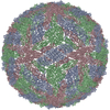
|
| 2 |
|
| 3 | x 5
|
| 4 | x 6
|
| 5 | 
|
| Symmetry | Point symmetry: (Hermann–Mauguin notation: 532 / Schoenflies symbol: I (icosahedral)) |
-Components
| #1: Protein | Mass: 43863.398 Da / Num. of mol.: 3 / Source method: isolated from a natural source / Source: (natural) Dengue virus 2 Puerto Rico/PR159-S1/1969 / Genus: Flavivirus / Species: Dengue virus / Strain: PR159-S1 / References: UniProt: P12823 |
|---|
-Experimental details
-Experiment
| Experiment | Method: ELECTRON MICROSCOPY |
|---|---|
| EM experiment | Aggregation state: PARTICLE / 3D reconstruction method: single particle reconstruction |
- Sample preparation
| Component | Name: DENGUE VIRUS / Type: VIRUS |
|---|---|
| Buffer solution | Name: 50 mM TRIS, 75 mM NACL, 1 mM EDTA / pH: 7.6 / Details: 50 mM TRIS, 75 mM NACL, 1 mM EDTA |
| Specimen | Embedding applied: NO / Shadowing applied: NO / Staining applied: NO / Vitrification applied: YES |
| Vitrification | Details: SAMPLES WERE PREPARED AS THIN LAYERS OF VITREOUS ICE AND MAINTAINED AT LIQUID NITROGEN TEMPERATURE IN THE ELECTRON MICROSCOPE |
- Electron microscopy imaging
| Microscopy | Model: FEI/PHILIPS CM200T / Date: Jun 27, 2000 |
|---|---|
| Electron gun | Electron source: FIELD EMISSION GUN / Accelerating voltage: 200 kV / Illumination mode: FLOOD BEAM |
| Electron lens | Mode: BRIGHT FIELDBright-field microscopy / Nominal magnification: 50000 X / Nominal defocus max: 4800 nm / Nominal defocus min: 800 nm / Cs: 2 mm |
| Specimen holder | Temperature: 87 K / Tilt angle max: 0 ° / Tilt angle min: 0 ° |
| Image recording | Electron dose: 27 e/Å2 / Film or detector model: KODAK SO-163 FILM |
- Processing
| EM software |
| ||||||||||||
|---|---|---|---|---|---|---|---|---|---|---|---|---|---|
| CTF correction | Details: EACH VIRAL IMAGE WAS CTF CORRECTED BEFORE RECONSTRUCTION, BASED ON THE FOLLOWING EQUATION: F(CORR)=F(OBS)/[|CTF|+WIENER*(1-|CTF|)] | ||||||||||||
| Symmetry | Point symmetry: I (icosahedral) | ||||||||||||
| 3D reconstruction | Method: FOURIER-BESSEL METHOD / Resolution: 9.5 Å / Nominal pixel size: 2.8 Å Details: THE RECONSTRUCTION WAS COMPUTED FROM 1691 DENGUE VIRUS IMAGES THAT WERE SELECTED FROM 78 MICROGRAPHS. ORIENTATIONS WERE DETERMINED BY THE MODEL-BASED POLAR-FOURIER TRANSFORM METHOD (BAKER ...Details: THE RECONSTRUCTION WAS COMPUTED FROM 1691 DENGUE VIRUS IMAGES THAT WERE SELECTED FROM 78 MICROGRAPHS. ORIENTATIONS WERE DETERMINED BY THE MODEL-BASED POLAR-FOURIER TRANSFORM METHOD (BAKER AND CHENG, 1996, J.STRUCT.BIOL. 116, 120-130) AND REFINED BY THE MODEL-BASED FOURIER TRANSFORM REFINEMENT PROCEDURE (HTTP://BOND.CS.UCF.EDU/COMPUTATIONALBIOLOGY/PROJECTS/POR/HOME.HTML). Symmetry type: POINT | ||||||||||||
| Atomic model building | Protocol: OTHER / Details: METHOD--please see citation | ||||||||||||
| Atomic model building | PDB-ID: 1TG8 Accession code: 1TG8 / Source name: PDB / Type: experimental model | ||||||||||||
| Refinement step | Cycle: LAST
|
