 Movie
Movie Controller


+ Open data

- Basic information
| Entry | Database: EMDB / ID: EMD-9232 | |||||||||
|---|---|---|---|---|---|---|---|---|---|---|
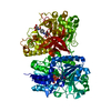
| Title | Cryo-EM structure of human AA amyloid fibril | |||||||||
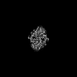
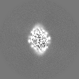
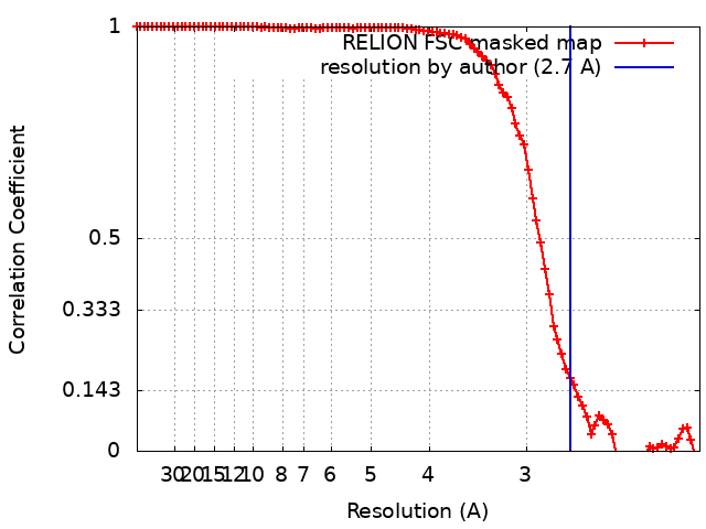
 Map data Map data | Cryo-EM reconstruction of human Serum Amyloid A fibrils, extracted from diseased human kidney. The fibril shows a helical rise of 2.40 A, a helical twist of 180.79 degree and a resolution of 2.7 A. | |||||||||
 Sample Sample |
| |||||||||
 Keywords Keywords | AA-amyloidosis / Serum Amyloid A / cross-beta / helical / protein fibril | |||||||||
| Function / homology |  Function and homology information Function and homology informationlymphocyte chemotaxis / Scavenging by Class B Receptors / positive regulation of interleukin-1 production / high-density lipoprotein particle / Formyl peptide receptors bind formyl peptides and many other ligands / regulation of protein secretion / TRAF6 mediated NF-kB activation / macrophage chemotaxis / Advanced glycosylation endproduct receptor signaling / cytoplasmic microtubule ...lymphocyte chemotaxis / Scavenging by Class B Receptors / positive regulation of interleukin-1 production / high-density lipoprotein particle / Formyl peptide receptors bind formyl peptides and many other ligands / regulation of protein secretion / TRAF6 mediated NF-kB activation / macrophage chemotaxis / Advanced glycosylation endproduct receptor signaling / cytoplasmic microtubule / neutrophil chemotaxis / endocytic vesicle lumen / positive regulation of cell adhesion / positive regulation of cytokine production / acute-phase response / TAK1-dependent IKK and NF-kappa-B activation / platelet activation / G protein-coupled receptor binding / negative regulation of inflammatory response / heparin binding / positive regulation of cytosolic calcium ion concentration / Interleukin-4 and Interleukin-13 signaling / G alpha (i) signalling events / G alpha (q) signalling events / Amyloid fiber formation / extracellular exosome / extracellular region Similarity search - Function | |||||||||
| Biological species |  Homo sapiens (human) Homo sapiens (human) | |||||||||
| Method | helical reconstruction / cryo EM / Resolution: 2.7 Å | |||||||||
 Authors Authors | Rennegarbe M / Liberta F / Fandrich M / Schmidt M | |||||||||
| Funding support |  Germany, 2 items Germany, 2 items
| |||||||||
 Citation Citation | Journal: Nat Commun / Year: 2019 Title: Cryo-EM fibril structures from systemic AA amyloidosis reveal the species complementarity of pathological amyloids. Authors: Falk Liberta / Sarah Loerch / Matthies Rennegarbe / Angelika Schierhorn / Per Westermark / Gunilla T Westermark / Bouke P C Hazenberg / Nikolaus Grigorieff / Marcus Fändrich / Matthias Schmidt /    Abstract: Systemic AA amyloidosis is a worldwide occurring protein misfolding disease of humans and animals. It arises from the formation of amyloid fibrils from the acute phase protein serum amyloid A. Here, ...Systemic AA amyloidosis is a worldwide occurring protein misfolding disease of humans and animals. It arises from the formation of amyloid fibrils from the acute phase protein serum amyloid A. Here, we report the purification and electron cryo-microscopy analysis of amyloid fibrils from a mouse and a human patient with systemic AA amyloidosis. The obtained resolutions are 3.0 Å and 2.7 Å for the murine and human fibril, respectively. The two fibrils differ in fundamental properties, such as presence of right-hand or left-hand twisted cross-β sheets and overall fold of the fibril proteins. Yet, both proteins adopt highly similar β-arch conformations within the N-terminal ~21 residues. Our data demonstrate the importance of the fibril protein N-terminus for the stability of the analyzed amyloid fibril morphologies and suggest strategies of combating this disease by interfering with specific fibril polymorphs. | |||||||||
| History |
|
- Structure visualization
| Movie |
Movie viewer |
|---|---|
| Structure viewer | EM map: SurfViewMolmilJmol/JSmol |
| Supplemental images |

- Downloads & links
-EMDB archive
| Map data | emd_9232.map.gz | 4.8 MB | EMDB map data format | |
|---|---|---|---|---|
| Header (meta data) | emd-9232-v30.xmlemd-9232.xml | 16.4 KB 16.4 KB | Display Display | EMDB header |
| FSC (resolution estimation) | emd_9232_fsc.xml | 9.5 KB | Display | FSC data file |
| Images | 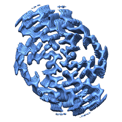 emd_9232.png emd_9232.png | 185.9 KB | ||
| Masks | emd_9232_msk_1.map | 75.1 MB | Mask map | |
| Filedesc metadata | emd-9232.cif.gz | 5.8 KB | ||
| Others | emd_9232_half_map_1.map.gzemd_9232_half_map_2.map.gz | 58.3 MB 58.4 MB | ||
| Archive directory |  http://ftp.pdbj.org/pub/emdb/structures/EMD-9232ftp://ftp.pdbj.org/pub/emdb/structures/EMD-9232 http://ftp.pdbj.org/pub/emdb/structures/EMD-9232ftp://ftp.pdbj.org/pub/emdb/structures/EMD-9232 | HTTPS FTP |
-Related structure data
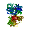
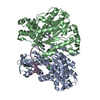
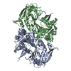
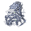
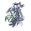
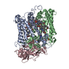
| Related structure data | 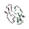 6mstMC 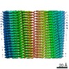 8910C 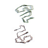 6dsoC M: atomic model generated by this map C: citing same article ( |
|---|---|
| Similar structure data | |
| EM raw data | EMPIAR-10734 (Title: Cryo electron microscopy of ex-vivo human SAA amyloid fibrils Data size: 1.1 TB Data #1: Unaligned multiframe micrographs of ex-vivo human SAA1 [micrographs - multiframe]) |

-Links
| EMDB pages | EMDB (EBI/PDBe) / EMDataResource |
|---|---|
| Related items in Molecule of the Month |


-Map
| File | Download / File: emd_9232.map.gz / Format: CCP4 / Size: 75.1 MB / Type: IMAGE STORED AS FLOATING POINT NUMBER (4 BYTES) | ||||||||||||||||||||||||||||||||||||||||||||||||||||||||||||
|---|---|---|---|---|---|---|---|---|---|---|---|---|---|---|---|---|---|---|---|---|---|---|---|---|---|---|---|---|---|---|---|---|---|---|---|---|---|---|---|---|---|---|---|---|---|---|---|---|---|---|---|---|---|---|---|---|---|---|---|---|---|
| Annotation | Cryo-EM reconstruction of human Serum Amyloid A fibrils, extracted from diseased human kidney. The fibril shows a helical rise of 2.40 A, a helical twist of 180.79 degree and a resolution of 2.7 A. | ||||||||||||||||||||||||||||||||||||||||||||||||||||||||||||
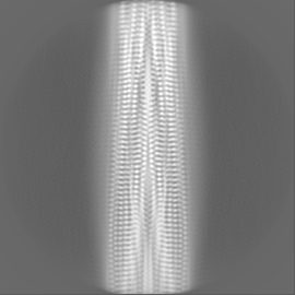
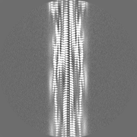
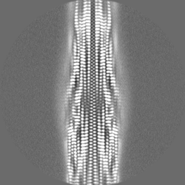
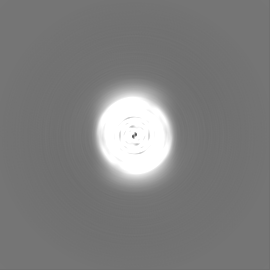
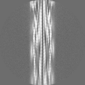
| Projections & slices | Image control
Images are generated by Spider. | ||||||||||||||||||||||||||||||||||||||||||||||||||||||||||||
| Voxel size | X=Y=Z: 1.04 Å | ||||||||||||||||||||||||||||||||||||||||||||||||||||||||||||
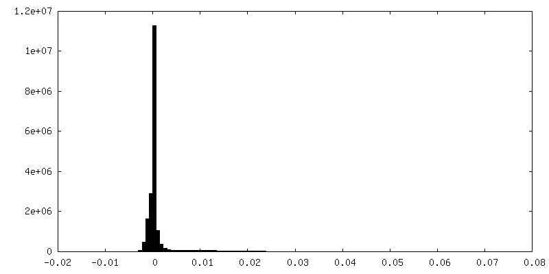
| Density |
| ||||||||||||||||||||||||||||||||||||||||||||||||||||||||||||
| Symmetry | Space group: 1 | ||||||||||||||||||||||||||||||||||||||||||||||||||||||||||||
| Details | EMDB XML:
CCP4 map header:
| ||||||||||||||||||||||||||||||||||||||||||||||||||||||||||||
 Z (Sec.)
 Y (Row.)
 X (Col.)


-Supplemental data
-Mask #1
| File | emd_9232_msk_1.map | ||||||||||||
|---|---|---|---|---|---|---|---|---|---|---|---|---|---|
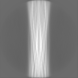
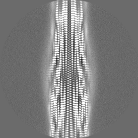
| Projections & Slices |
| ||||||||||||
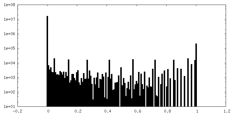
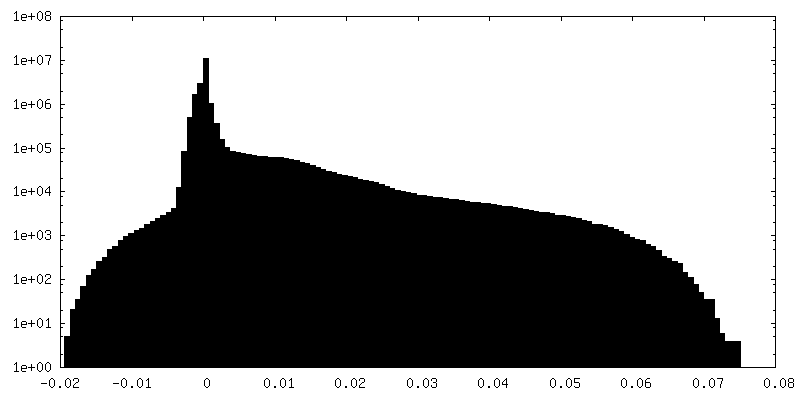
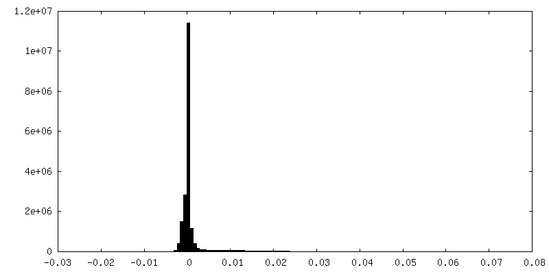
| Density Histograms |


-Half map: One of the two independently refined half maps.
| File | emd_9232_half_map_1.map | ||||||||||||
|---|---|---|---|---|---|---|---|---|---|---|---|---|---|
| Annotation | One of the two independently refined half maps. | ||||||||||||
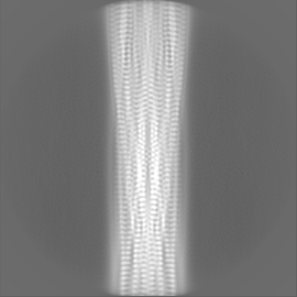
| Projections & Slices |
| ||||||||||||
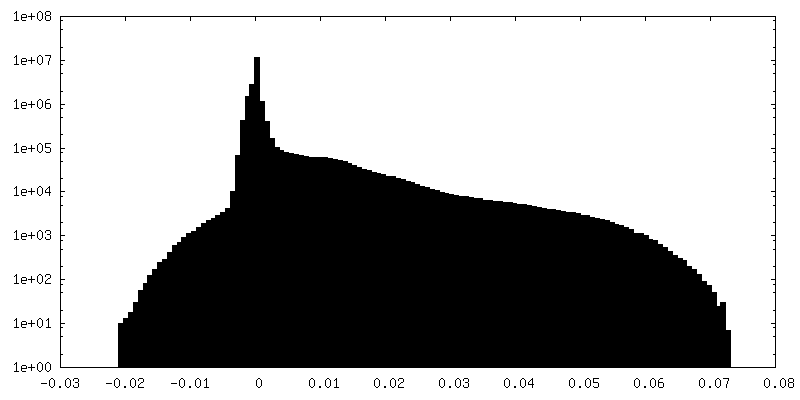
| Density Histograms |

-Half map: One of the two independently refined half maps.
| File | emd_9232_half_map_2.map | ||||||||||||
|---|---|---|---|---|---|---|---|---|---|---|---|---|---|
| Annotation | One of the two independently refined half maps. | ||||||||||||
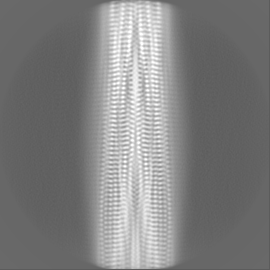
| Projections & Slices |
| ||||||||||||
| Density Histograms |

- Sample components
-Entire : human AA amyloid fibril
| Entire | Name: human AA amyloid fibril |
|---|---|
| Components |
|
-Supramolecule #1: human AA amyloid fibril
| Supramolecule | Name: human AA amyloid fibril / type: complex / ID: 1 / Parent: 0 / Macromolecule list: all |
|---|---|
| Source (natural) | Organism: Homo sapiens (human) / Organ: kidney |
-Macromolecule #1: Serum amyloid A-1 protein
| Macromolecule | Name: Serum amyloid A-1 protein / type: protein_or_peptide / ID: 1 / Number of copies: 12 / Enantiomer: LEVO |
|---|---|
| Source (natural) | Organism: Homo sapiens (human) |
| Molecular weight | Theoretical: 7.481154 KDa |
| Sequence | String: SFFSFLGEAF DGARDMWRAY SDMREANYIG SDKYFHARGN YDAAKRGPGG VWAAEAISDA RENIQR UniProtKB: Serum amyloid A-1 protein |
-Experimental details
-Structure determination
| Method | cryo EM |
|---|---|
 Processing Processing | helical reconstruction |
| Aggregation state | helical array |
-Sample preparation
| Concentration | 0.2 mg/mL |
|---|---|
| Buffer | pH: 7 / Component - Formula: ddH2O |
| Grid | Model: C-flat-1.2/1.3 4C / Material: COPPER / Pretreatment - Type: GLOW DISCHARGE / Pretreatment - Time: 40 sec. / Pretreatment - Atmosphere: OTHER / Pretreatment - Pressure: 0.025 kPa / Details: 15 mA |
| Vitrification | Cryogen name: ETHANE / Chamber humidity: 95 % / Chamber temperature: 293 K / Instrument: FEI VITROBOT MARK III |
- Electron microscopy
| Microscope | FEI TITAN KRIOS |
|---|---|
| Image recording | Film or detector model: GATAN K2 SUMMIT (4k x 4k) / Detector mode: COUNTING / Average exposure time: 12.0 sec. / Average electron dose: 40.0 e/Å2 |
| Electron beam | Acceleration voltage: 300 kV / Electron source:  FIELD EMISSION GUN FIELD EMISSION GUN |
| Electron optics | C2 aperture diameter: 50.0 µm / Illumination mode: FLOOD BEAM / Imaging mode: BRIGHT FIELD / Cs: 2.7 mm / Nominal defocus max: -2.5 µm / Nominal defocus min: -0.5 µm / Nominal magnification: 130000 |
| Sample stage | Cooling holder cryogen: NITROGEN |
| Experimental equipment |  Model: Titan Krios / Image courtesy: FEI Company |
+Image processing
-Atomic model buiding 1
| Refinement | Protocol: AB INITIO MODEL |
|---|---|
| Output model | PDB-6mst: |
