 Movie
Movie Controller

[English] 日本語
 Yorodumi
- EMDB-7878: Noodle-like structure in infectious microvesicles (Class 3) from ... -
+ Open data

- Basic information
| Entry | Database: EMDB / ID: EMD-7878 | |||||||||
|---|---|---|---|---|---|---|---|---|---|---|
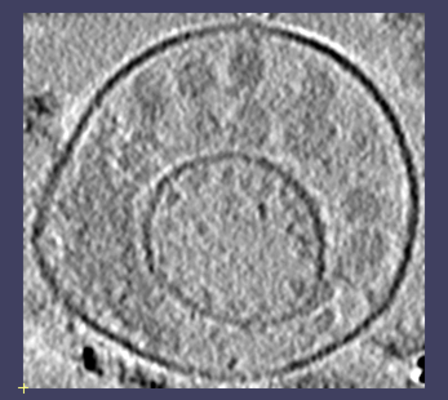
| Title | Noodle-like structure in infectious microvesicles (Class 3) from poliovirus-infected HeLa cells | |||||||||
 Map data Map data | Noodle-like structures in infectious microvesicles (Class 3) from poliovirus-infected cells | |||||||||
 Sample Sample |
| |||||||||
| Biological species |   Human poliovirus 1 Mahoney Human poliovirus 1 Mahoney | |||||||||
| Method | electron tomography / cryo EM | |||||||||
 Authors Authors | Yang JE | |||||||||
| Funding support |  United States, 2 items United States, 2 items
| |||||||||
 Citation Citation | Journal: Sci Rep / Year: 2020 Title: Complexity and ultrastructure of infectious extracellular vesicles from cells infected by non-enveloped virus. Authors: Jie E Yang / Evan D Rossignol / Deborah Chang / Joseph Zaia / Isaac Forrester / Kiran Raja / Holly Winbigler / Daniela Nicastro / William T Jackson / Esther Bullitt / Abstract: Enteroviruses support cell-to-cell viral transmission prior to their canonical lytic spread of virus. Poliovirus (PV), a prototype for human pathogenic positive-sense RNA enteroviruses, and ...Enteroviruses support cell-to-cell viral transmission prior to their canonical lytic spread of virus. Poliovirus (PV), a prototype for human pathogenic positive-sense RNA enteroviruses, and picornaviruses in general, transport multiple virions en bloc via infectious extracellular vesicles, 100~1000 nm in diameter, secreted from host cells. Using biochemical and biophysical methods we identify multiple components in secreted microvesicles, including mature PV virions; positive-sense genomic and negative-sense replicative, template viral RNA; essential viral replication proteins; and cellular proteins. Using cryo-electron tomography, we visualize the near-native three-dimensional architecture of secreted infectious microvesicles containing both virions and a unique morphological component that we describe as a mat-like structure. While the composition of these mat-like structures is not yet known, based on our biochemical data they are expected to be comprised of unencapsidated RNA and proteins. In addition to infectious microvesicles, CD9-positive exosomes released from PV-infected cells are also infectious and transport virions. Thus, our data show that, prior to cell lysis, non-enveloped viruses are secreted within infectious vesicles that also transport viral unencapsidated RNAs, viral and host proteins. Understanding the structure and function of these infectious particles helps elucidate the mechanism by which extracellular vesicles contribute to the spread of non-enveloped virus infection. | |||||||||
| History |
|
- Structure visualization
| Movie |
 Movie viewer Movie viewer |
|---|---|
| Supplemental images |
- Downloads & links
-EMDB archive
| Map data | emd_7878.map.gz | 14.9 MB | EMDB map data format | |
|---|---|---|---|---|
| Header (meta data) | emd-7878-v30.xmlemd-7878.xml | 10.4 KB 10.4 KB | Display Display | EMDB header |
| Images |  emd_7878.png emd_7878.png | 135.4 KB | ||
| Archive directory |  http://ftp.pdbj.org/pub/emdb/structures/EMD-7878ftp://ftp.pdbj.org/pub/emdb/structures/EMD-7878 http://ftp.pdbj.org/pub/emdb/structures/EMD-7878ftp://ftp.pdbj.org/pub/emdb/structures/EMD-7878 | HTTPS FTP |
-Validation report
| Summary document | emd_7878_validation.pdf.gz | 76.9 KB | Display | EMDB validaton report |
|---|---|---|---|---|
| Full document | emd_7878_full_validation.pdf.gz | 76 KB | Display | |
| Data in XML | emd_7878_validation.xml.gz | 499 B | Display | |
| Arichive directory | https://ftp.pdbj.org/pub/emdb/validation_reports/EMD-7878ftp://ftp.pdbj.org/pub/emdb/validation_reports/EMD-7878 | HTTPS FTP |
-Related structure data


-Links
| EMDB pages | EMDB (EBI/PDBe) / EMDataResource |
|---|---|
| Related items in Molecule of the Month |

-Map
| File | Download / File: emd_7878.map.gz / Format: CCP4 / Size: 29.8 MB / Type: IMAGE STORED AS SIGNED BYTE | ||||||||||||||||||||||||||||||||||||||||||||||||||||||||||||||||||||
|---|---|---|---|---|---|---|---|---|---|---|---|---|---|---|---|---|---|---|---|---|---|---|---|---|---|---|---|---|---|---|---|---|---|---|---|---|---|---|---|---|---|---|---|---|---|---|---|---|---|---|---|---|---|---|---|---|---|---|---|---|---|---|---|---|---|---|---|---|---|
| Annotation | Noodle-like structures in infectious microvesicles (Class 3) from poliovirus-infected cells | ||||||||||||||||||||||||||||||||||||||||||||||||||||||||||||||||||||
| Voxel size | X=Y=Z: 8.2 Å | ||||||||||||||||||||||||||||||||||||||||||||||||||||||||||||||||||||
| Density |
| ||||||||||||||||||||||||||||||||||||||||||||||||||||||||||||||||||||
| Symmetry | Space group: 1 | ||||||||||||||||||||||||||||||||||||||||||||||||||||||||||||||||||||
| Details | EMDB XML:
CCP4 map header:
| ||||||||||||||||||||||||||||||||||||||||||||||||||||||||||||||||||||
-Supplemental data
- Sample components
-Entire : Human poliovirus 1 Mahoney
| Entire | Name: Human poliovirus 1 Mahoney |
|---|---|
| Components |
|
-Supramolecule #1: Human poliovirus 1 Mahoney
| Supramolecule | Name: Human poliovirus 1 Mahoney / type: virus / ID: 1 / Parent: 0 Details: virion-containing microvesicles collected from supernatant of poliovirus-infected cells at 8 hours post-infection NCBI-ID: 12081 / Sci species name: Human poliovirus 1 Mahoney / Virus type: VIRUS-LIKE PARTICLE / Virus isolate: STRAIN / Virus enveloped: No / Virus empty: No |
|---|---|
| Host (natural) | Organism:  Homo sapiens (human) Homo sapiens (human) |
-Experimental details
-Structure determination
| Method | cryo EM |
|---|---|
 Processing Processing | electron tomography |
| Aggregation state | particle |
-Sample preparation
| Concentration | 1.5 mg/mL |
|---|---|
| Buffer | pH: 7 / Details: 1% glutaraldehyde in 1x PBS |
| Grid | Material: COPPER / Mesh: 200 / Pretreatment - Type: GLOW DISCHARGE / Pretreatment - Atmosphere: AIR Details: The grid was coated with an extra layer of carbon (holes not covered) to provide extra support. |
| Vitrification | Cryogen name: ETHANE / Chamber humidity: 100 % / Chamber temperature: 283.15 K / Instrument: FEI VITROBOT MARK II |
| Details | The microvesicles from poliovirus-infected cells were collected through a series of centrifugations to enrich 100-1000 nm diameter membrane particles, followed by an annexin-V column purification to enrich phosphatidylserine-containing microvesicles. |
| Sectioning | Other: NO SECTIONING |
| Fiducial marker | Manufacturer: TED PELLA INC / Diameter: 10 nm |
- Electron microscopy
| Microscope | FEI TECNAI F20 |
|---|---|
| Image recording | Film or detector model: TVIPS TEMCAM-F416 (4k x 4k) / Average electron dose: 4.8 e/Å2 |
| Electron beam | Acceleration voltage: 160 kV / Electron source:  FIELD EMISSION GUN FIELD EMISSION GUN |
| Electron optics | Illumination mode: FLOOD BEAM / Imaging mode: BRIGHT FIELD |
| Experimental equipment |  Model: Tecnai F20 / Image courtesy: FEI Company |
-Image processing
| Final reconstruction | Algorithm: BACK PROJECTION / Software - Name: eTomo / Number images used: 47 |
|---|
