Movie
Movie Controller

[English] 日本語
 Yorodumi
- EMDB-6698: Structure of a eukaryotic voltage-gated sodium channel at near at... -
+ Open data

- Basic information
| Entry | Database: EMDB / ID: EMD-6698 | |||||||||
|---|---|---|---|---|---|---|---|---|---|---|
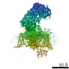
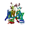
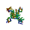
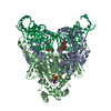
| Title | Structure of a eukaryotic voltage-gated sodium channel at near atomic resolution | |||||||||
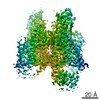
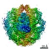
 Map data Map data | overall map | |||||||||
 Sample Sample |
| |||||||||
| Function / homology |  Function and homology information Function and homology informationvoltage-gated sodium channel complex / membrane depolarization during action potential / voltage-gated sodium channel activity / neuronal action potential / axon Similarity search - Function | |||||||||
| Biological species |  Periplaneta americana (American cockroach) Periplaneta americana (American cockroach) | |||||||||
| Method | single particle reconstruction / cryo EM / Resolution: 3.8 Å | |||||||||
 Authors Authors | Shen H / Zhou Q / Pan X / Li Z / Wu J / Yan N | |||||||||
| Funding support |  China, 2 items China, 2 items
| |||||||||
 Citation Citation | Journal: Science / Year: 2017 Title: Structure of a eukaryotic voltage-gated sodium channel at near-atomic resolution. Authors: Huaizong Shen / Qiang Zhou / Xiaojing Pan / Zhangqiang Li / Jianping Wu / Nieng Yan / Abstract: Voltage-gated sodium (Na) channels are responsible for the initiation and propagation of action potentials. They are associated with a variety of channelopathies and are targeted by multiple ...Voltage-gated sodium (Na) channels are responsible for the initiation and propagation of action potentials. They are associated with a variety of channelopathies and are targeted by multiple pharmaceutical drugs and natural toxins. Here, we report the cryogenic electron microscopy structure of a putative Na channel from American cockroach (designated NaPaS) at 3.8 angstrom resolution. The voltage-sensing domains (VSDs) of the four repeats exhibit distinct conformations. The entrance to the asymmetric selectivity filter vestibule is guarded by heavily glycosylated and disulfide bond-stabilized extracellular loops. On the cytoplasmic side, a conserved amino-terminal domain is placed below VSD, and a carboxy-terminal domain binds to the III-IV linker. The structure of NaPaS establishes an important foundation for understanding function and disease mechanism of Na and related voltage-gated calcium channels. | |||||||||
| History |
|
- Structure visualization
| Movie |
Movie viewer |
|---|---|
| Structure viewer | EM map: SurfViewMolmilJmol/JSmol |
| Supplemental images |

- Downloads & links
-EMDB archive
| Map data | emd_6698.map.gz | 28.5 MB | EMDB map data format | |
|---|---|---|---|---|
| Header (meta data) | emd-6698-v30.xmlemd-6698.xml | 16.1 KB 16.1 KB | Display Display | EMDB header |
| Images | 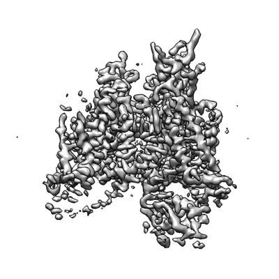 emd_6698.png emd_6698.png | 51.5 KB | ||
| Others | emd_6698_additional.map.gz | 28.6 MB | ||
| Archive directory |  http://ftp.pdbj.org/pub/emdb/structures/EMD-6698ftp://ftp.pdbj.org/pub/emdb/structures/EMD-6698 http://ftp.pdbj.org/pub/emdb/structures/EMD-6698ftp://ftp.pdbj.org/pub/emdb/structures/EMD-6698 | HTTPS FTP |
-Validation report
| Summary document | emd_6698_validation.pdf.gz | 487.6 KB | Display | EMDB validaton report |
|---|---|---|---|---|
| Full document | emd_6698_full_validation.pdf.gz | 487.2 KB | Display | |
| Data in XML | emd_6698_validation.xml.gz | 5.7 KB | Display | |
| Data in CIF | emd_6698_validation.cif.gz | 6.4 KB | Display | |
| Arichive directory | https://ftp.pdbj.org/pub/emdb/validation_reports/EMD-6698ftp://ftp.pdbj.org/pub/emdb/validation_reports/EMD-6698 | HTTPS FTP |
-Related structure data
| Related structure data |  5x0mMC M: atomic model generated by this map C: citing same article ( |
|---|---|
| Similar structure data |


-Links
| EMDB pages | EMDB (EBI/PDBe) / EMDataResource |
|---|---|
| Related items in Molecule of the Month |

-Map
| File | Download / File: emd_6698.map.gz / Format: CCP4 / Size: 30.5 MB / Type: IMAGE STORED AS FLOATING POINT NUMBER (4 BYTES) | ||||||||||||||||||||||||||||||||||||||||||||||||||||||||||||
|---|---|---|---|---|---|---|---|---|---|---|---|---|---|---|---|---|---|---|---|---|---|---|---|---|---|---|---|---|---|---|---|---|---|---|---|---|---|---|---|---|---|---|---|---|---|---|---|---|---|---|---|---|---|---|---|---|---|---|---|---|---|
| Annotation | overall map | ||||||||||||||||||||||||||||||||||||||||||||||||||||||||||||
| Projections & slices | Image control
Images are generated by Spider. | ||||||||||||||||||||||||||||||||||||||||||||||||||||||||||||
| Voxel size | X=Y=Z: 1.29 Å | ||||||||||||||||||||||||||||||||||||||||||||||||||||||||||||
| Density |
| ||||||||||||||||||||||||||||||||||||||||||||||||||||||||||||
| Symmetry | Space group: 1 | ||||||||||||||||||||||||||||||||||||||||||||||||||||||||||||
| Details | EMDB XML:
CCP4 map header:
| ||||||||||||||||||||||||||||||||||||||||||||||||||||||||||||
 Z (Sec.)
 Y (Row.)
 X (Col.)


-Supplemental data
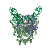
-Additional map: overall map with B factor of -300
| File | emd_6698_additional.map | ||||||||||||
|---|---|---|---|---|---|---|---|---|---|---|---|---|---|
| Annotation | overall map with B factor of -300 | ||||||||||||
| Projections & Slices |
| ||||||||||||
| Density Histograms |


- Sample components
-Entire : voltage-gated sodium channel
| Entire | Name: voltage-gated sodium channel |
|---|---|
| Components |
|
-Supramolecule #1: voltage-gated sodium channel
| Supramolecule | Name: voltage-gated sodium channel / type: organelle_or_cellular_component / ID: 1 / Parent: 0 / Macromolecule list: #1 |
|---|---|
| Source (natural) | Organism: Periplaneta americana (American cockroach) |
| Recombinant expression | Organism:  Homo sapiens (human) Homo sapiens (human) |
-Macromolecule #1: Sodium channel protein
| Macromolecule | Name: Sodium channel protein / type: protein_or_peptide / ID: 1 / Number of copies: 1 / Enantiomer: LEVO |
|---|---|
| Source (natural) | Organism: Periplaneta americana (American cockroach) |
| Molecular weight | Theoretical: 183.875422 KDa |
| Recombinant expression | Organism: Homo sapiens (human) |
| Sequence | String: MASWSHPQFE KGGGARGGSG GGSWSHPQFE KGFDYKDDDD KGTMADNSPL IREERQRLFR PYTRAMLTAP SAQPAKENGK TEENKDNSR DKGRGANKDR DGSAHPDQAL EQGSRLPARM RNIFPAELAS TPLEDFDPFY KNKKTFVVVT KAGDIFRFSG E KSLWMLDP ...String: MASWSHPQFE KGGGARGGSG GGSWSHPQFE KGFDYKDDDD KGTMADNSPL IREERQRLFR PYTRAMLTAP SAQPAKENGK TEENKDNSR DKGRGANKDR DGSAHPDQAL EQGSRLPARM RNIFPAELAS TPLEDFDPFY KNKKTFVVVT KAGDIFRFSG E KSLWMLDP FTPIRRVAIS TMVQPIFSYF IMITILIHCI FMIMPATQTT YILELVFLSI YTIEVVVKVL ARGFILHPFA YL RDPWNWL DFLVTLIGYI TLVVDLGHLY ALRAFRVLRS WRTVTIVPGW RTIVDALSLS ITSLKDLVLL LLFSLFVFAV LGL QIYMGV LTQKCVKHFP ADGSWGNFTD ERWFNYTSNS SHWYIPDDWI EYPLCGNSSG AGMCPPGYTC LQGYGGNPNY GYTS FDTFG WAFLSVFRLV TLDYWEDLYQ LALRSAGPWH ILFFIIVVFY GTFCFLNFIL AVVVMSYTHM VKRADEEKAA ERELK KEKK AASVANNTAN GQEQTTIEMN GDEAVVIDNN DQAARQQSDP ETPAPSVTQR LTDFLCVWDC CVPWQKLQGA IGAVVL SPF FELFIAVIIV LNITFMALDH HDMNIEFERI LRTGNYIFTS IYIVEAVLKI IALSPKFYFK DSWNVFDFII VVFAILE LG LEGVQGLSVF RSFRLLRVFR LAKFWPTLNN FMSVMTKSYG AFVNVMYVMF LLLFIFAIIG MQLFGMNYID NMERFPDG D LPRWNFTDFL HSFMIVFRAL CGEWIESMWD CMLVGDWSCI PFFVAVFFVG NLVILNLLIA LLLNNYGSFC TSPTSDEED SKDEDALAQI VRIFKRFKPN LNAVKLSPMK PDSEDIVESQ EIQGNNIADA EDVLAGEFPP DCCCNAFYKC FPSRPARDSS VQRMWSNIR RVCFLLAKNK YFQKFVTAVL VITSVLLALE DIYLPQRPVL VNITLYVDYV LTAFFVIEMI IMLFAVGFKK Y FTSKWYWL DFIVVVAYLL NFVLMCAGIE ALQTLRLLRV FRLFRPLSKV NGMQVVTSTL VEAVPHIFNV ILVGIFFWLV FA IMGVQLF AGKFYKCVDE NSTVLSHEIT MDRNDCLHEN YTWENSPMNF DHVGNAYLSL LQVATFKGWL QIMNDAIDSR EVH KQPIRE TNIYMYLYFI FFIVFGSFFI LKLFVCILID IFRQQRRKAE GLSATDSRTQ LIYRRAVMRT MSAKPVKRIP KPTC HPQSL MYDISVNRKF EYTMMILIIL NVAVMAIDHY GQSMEFSEVL DYLNLIFIII FFVECVIKVS GLRHHYFKDP WNIID FLYV VLAIAGLMLS DVIEKYFISP TLLRILRILR VGRLLRYFQS ARGMRLLLLA LRKALRTLFN VSFLLFVIMF VYAVFG MEF FMHIRDAGAI DDVYNFKTFG QSIILLFQLA TSAGWDGVYF AIANEEDCRA PDHELGYPGN CGSRALGIAY LVSYLII TC LVVINMYAAV ILDYVLEVYE DSKEGLTDDD YDMFFEVWQQ FDPEATQYIR YDQLSELLEA LQPPLQVQKP NKYKILSM N IPICKDDHIF YKDVLEALVK DVFSRRGSPV EAGDVQAPNV DEAEYKPVSS TLQRQREEYC VRLIQNAWRK HKQQN |
-Macromolecule #2: N-ACETYL-D-GLUCOSAMINE
| Macromolecule | Name: N-ACETYL-D-GLUCOSAMINE / type: ligand / ID: 2 / Number of copies: 13 / Formula: NAG |
|---|---|
| Molecular weight | Theoretical: 221.208 Da |
-Macromolecule #3: BETA-D-MANNOSE
| Macromolecule | Name: BETA-D-MANNOSE / type: ligand / ID: 3 / Number of copies: 7 / Formula: BMA |
|---|---|
| Molecular weight | Theoretical: 180.156 Da |
-Experimental details
-Structure determination
| Method | cryo EM |
|---|---|
 Processing Processing | single particle reconstruction |
| Aggregation state | particle |
-Sample preparation
| Concentration | 1 mg/mL |
|---|---|
| Buffer | pH: 7.4 Details: 25 mM Tris-HCl, pH 7.4, 50mM NaCl, 0.1% digitonin, 2.5 mM D-Desthiobiotin and protease inhibitor cocktail |
| Grid | Model: Quantifoil R1.2/1.3 / Material: COPPER / Mesh: 200 / Pretreatment - Type: GLOW DISCHARGE |
| Vitrification | Cryogen name: ETHANE / Chamber humidity: 100 % / Chamber temperature: 281 K Details: Grids were blotted for 3.5 s and flash-frozen in liquid ethane cooled by liquid nitrogen.. |
| Details | This sample was monodisperse |
- Electron microscopy
| Microscope | FEI TITAN KRIOS |
|---|---|
| Image recording | Film or detector model: GATAN K2 SUMMIT (4k x 4k) / Detector mode: SUPER-RESOLUTION / Digitization - Frames/image: 1-32 / Average exposure time: 0.25 sec. / Average electron dose: 1.5625 e/Å2 |
| Electron beam | Acceleration voltage: 300 kV / Electron source:  FIELD EMISSION GUN FIELD EMISSION GUN |
| Electron optics | Calibrated defocus max: 2.6 µm / Calibrated defocus min: 1.7 µm / Illumination mode: SPOT SCAN / Imaging mode: BRIGHT FIELD / Cs: 2.7 mm / Nominal magnification: 22500 |
| Experimental equipment |  Model: Titan Krios / Image courtesy: FEI Company |