 Movie
Movie Controller

[English] 日本語
 Yorodumi
- PDB-5x0m: Structure of a eukaryotic voltage-gated sodium channel at near at... -
+ Open data

- Basic information
| Entry | Database: PDB / ID: 5x0m | |||||||||
|---|---|---|---|---|---|---|---|---|---|---|
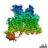
| Title | Structure of a eukaryotic voltage-gated sodium channel at near atomic resolution | |||||||||
 Components Components | Sodium channel protein | |||||||||
 Keywords Keywords | MEMBRANE PROTEIN / ion channel | |||||||||
| Function / homology |  Function and homology information Function and homology informationmembrane depolarization during action potential / voltage-gated sodium channel complex / voltage-gated sodium channel activity / voltage-gated monoatomic cation channel activity / neuronal action potential Similarity search - Function | |||||||||
| Biological species |  Periplaneta americana (American cockroach) Periplaneta americana (American cockroach) | |||||||||
| Method | ELECTRON MICROSCOPY / single particle reconstruction / cryo EM / Resolution: 3.8 Å | |||||||||
 Authors Authors | Shen, H. / Zhou, Q. / Pan, X. / Li, Z. / Wu, J. / Yan, N. | |||||||||
| Funding support |  China, 2items China, 2items
| |||||||||
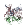
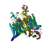
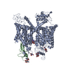
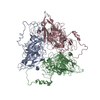
 Citation Citation | Journal: Science / Year: 2017 Title: Structure of a eukaryotic voltage-gated sodium channel at near-atomic resolution. Authors: Huaizong Shen / Qiang Zhou / Xiaojing Pan / Zhangqiang Li / Jianping Wu / Nieng Yan / Abstract: Voltage-gated sodium (Na) channels are responsible for the initiation and propagation of action potentials. They are associated with a variety of channelopathies and are targeted by multiple ...Voltage-gated sodium (Na) channels are responsible for the initiation and propagation of action potentials. They are associated with a variety of channelopathies and are targeted by multiple pharmaceutical drugs and natural toxins. Here, we report the cryogenic electron microscopy structure of a putative Na channel from American cockroach (designated NaPaS) at 3.8 angstrom resolution. The voltage-sensing domains (VSDs) of the four repeats exhibit distinct conformations. The entrance to the asymmetric selectivity filter vestibule is guarded by heavily glycosylated and disulfide bond-stabilized extracellular loops. On the cytoplasmic side, a conserved amino-terminal domain is placed below VSD, and a carboxy-terminal domain binds to the III-IV linker. The structure of NaPaS establishes an important foundation for understanding function and disease mechanism of Na and related voltage-gated calcium channels. | |||||||||
| History |
|
- Structure visualization
| Movie |
Movie viewer |
|---|---|
| Structure viewer | Molecule: MolmilJmol/JSmol |
- Downloads & links
-Download
| PDBx/mmCIF format | 5x0m.cif.gz | 294.5 KB | Display | PDBx/mmCIF format |
|---|---|---|---|---|
| PDB format | pdb5x0m.ent.gz | 224.3 KB | Display | PDB format |
| PDBx/mmJSON format | 5x0m.json.gz | Tree view | PDBx/mmJSON format | |
| Others |  Other downloads Other downloads |
-Validation report
| Arichive directory | https://data.pdbj.org/pub/pdb/validation_reports/x0/5x0mftp://data.pdbj.org/pub/pdb/validation_reports/x0/5x0m | HTTPS FTP |
|---|
-Related structure data
| Related structure data |  6698MC M: map data used to model this data C: citing same article ( |
|---|---|
| Similar structure data |


-Links
 PDBj
PDBj
- Assembly
| Deposited unit | 
|
|---|---|
| 1 |
|
-Components
| #1: Protein | Mass: 183875.422 Da / Num. of mol.: 1 Source method: isolated from a genetically manipulated source Source: (gene. exp.) Periplaneta americana (American cockroach)Production host:  Homo sapiens (human) / References: UniProt: D0E0C2 Homo sapiens (human) / References: UniProt: D0E0C2 | ||||||
|---|---|---|---|---|---|---|---|
| #2: Polysaccharide | beta-D-mannopyranose-(1-3)-[beta-D-mannopyranose-(1-6)]beta-D-mannopyranose-(1-4)-2-acetamido-2- ...beta-D-mannopyranose-(1-3)-[beta-D-mannopyranose-(1-6)]beta-D-mannopyranose-(1-4)-2-acetamido-2-deoxy-beta-D-glucopyranose-(1-4)-2-acetamido-2-deoxy-beta-D-glucopyranose Source method: isolated from a genetically manipulated source | ||||||
| #3: Polysaccharide | beta-D-mannopyranose-(1-4)-2-acetamido-2-deoxy-beta-D-glucopyranose-(1-4)-2-acetamido-2-deoxy-beta- ...beta-D-mannopyranose-(1-4)-2-acetamido-2-deoxy-beta-D-glucopyranose-(1-4)-2-acetamido-2-deoxy-beta-D-glucopyranose Source method: isolated from a genetically manipulated source #4: Polysaccharide | 2-acetamido-2-deoxy-beta-D-glucopyranose-(1-4)-2-acetamido-2-deoxy-beta-D-glucopyranose | Source method: isolated from a genetically manipulated source #5: Sugar | ChemComp-NAG / |   Type: D-saccharide, beta linking / Mass: 221.208 Da / Num. of mol.: 1 Type: D-saccharide, beta linking / Mass: 221.208 Da / Num. of mol.: 1Source method: isolated from a genetically manipulated source Formula: C8H15NO6 Has protein modification | Y | |
-Experimental details
-Experiment
| Experiment | Method: ELECTRON MICROSCOPY |
|---|---|
| EM experiment | Aggregation state: PARTICLE / 3D reconstruction method: single particle reconstruction |
- Sample preparation
| Component | Name: voltage-gated sodium channel / Type: ORGANELLE OR CELLULAR COMPONENT / Entity ID: #1 / Source: RECOMBINANT |
|---|---|
| Source (natural) | Organism: Periplaneta americana (American cockroach) |
| Source (recombinant) | Organism: Homo sapiens (human) |
| Buffer solution | pH: 7.4 Details: 25 mM Tris-HCl, pH 7.4, 50mM NaCl, 0.1% digitonin, 2.5 mM D-Desthiobiotin and protease inhibitor cocktail |
| Specimen | Conc.: 1 mg/ml / Embedding applied: NO / Shadowing applied: NO / Staining applied: NO / Vitrification applied: YES / Details: This sample was monodisperse |
| Specimen support | Grid material: COPPER / Grid mesh size: 200 divisions/in. / Grid type: Quantifoil R1.2/1.3 |
| Vitrification | Cryogen name: ETHANE / Humidity: 100 % / Chamber temperature: 281 K Details: Grids were blotted for 3.5 s and flash-frozen in liquid ethane cooled by liquid nitrogen. |
- Electron microscopy imaging
| Experimental equipment |  Model: Titan Krios / Image courtesy: FEI Company |
|---|---|
| Microscopy | Model: FEI TITAN KRIOS |
| Electron gun | Electron source:  FIELD EMISSION GUN / Accelerating voltage: 300 kV / Illumination mode: SPOT SCAN FIELD EMISSION GUN / Accelerating voltage: 300 kV / Illumination mode: SPOT SCAN |
| Electron lens | Mode: BRIGHT FIELD / Nominal magnification: 22500 X / Calibrated defocus min: 1700 nm / Calibrated defocus max: 2600 nm / Cs: 2.7 mm |
| Image recording | Average exposure time: 0.25 sec. / Electron dose: 1.5625 e/Å2 / Detector mode: SUPER-RESOLUTION / Film or detector model: GATAN K2 SUMMIT (4k x 4k) |
| Image scans | Movie frames/image: 32 / Used frames/image: 1-32 |
- Processing
| EM software |
| ||||||||||||||||||||||||
|---|---|---|---|---|---|---|---|---|---|---|---|---|---|---|---|---|---|---|---|---|---|---|---|---|---|
| CTF correction | Type: PHASE FLIPPING AND AMPLITUDE CORRECTION | ||||||||||||||||||||||||
| Particle selection | Num. of particles selected: 4739175 | ||||||||||||||||||||||||
| Symmetry | Point symmetry: C1 (asymmetric) | ||||||||||||||||||||||||
| 3D reconstruction | Resolution: 3.8 Å / Resolution method: FSC 0.143 CUT-OFF / Num. of particles: 1373581 / Symmetry type: POINT |
