 Movie
Movie Controller

[English] 日本語
 Yorodumi
- EMDB-6408: In situ structures of the segmented genome and RNA polymerase com... -
+ Open data

- Basic information
| Entry | Database: EMDB / ID: EMD-6408 | |||||||||
|---|---|---|---|---|---|---|---|---|---|---|
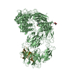
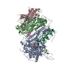
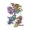
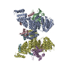
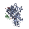
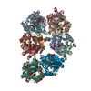
| Title | In situ structures of the segmented genome and RNA polymerase complex inside a dsRNA virus | |||||||||
 Map data Map data | Averaged qCPV TEC | |||||||||
 Sample Sample |
| |||||||||
 Keywords Keywords | cryo-EM / dsRNA genome organization / viral polymerase | |||||||||
| Function / homology |  Function and homology information Function and homology informationviral genome replication / RNA-directed RNA polymerase / RNA-directed RNA polymerase activity / RNA binding Similarity search - Function | |||||||||
| Biological species |   Bombyx mori cypovirus 1 Bombyx mori cypovirus 1 | |||||||||
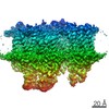
| Method | single particle reconstruction / cryo EM / Resolution: 3.3 Å | |||||||||
 Authors Authors | Zhang X / Ding K / Yu XK / Chang W / Sun JC / Zhou ZH | |||||||||
 Citation Citation | Journal: Nature / Year: 2015 Title: In situ structures of the segmented genome and RNA polymerase complex inside a dsRNA virus. Authors: Xing Zhang / Ke Ding / Xuekui Yu / Winston Chang / Jingchen Sun / Z Hong Zhou /   Abstract: Viruses in the Reoviridae, like the triple-shelled human rotavirus and the single-shelled insect cytoplasmic polyhedrosis virus (CPV), all package a genome of segmented double-stranded RNAs (dsRNAs) ...Viruses in the Reoviridae, like the triple-shelled human rotavirus and the single-shelled insect cytoplasmic polyhedrosis virus (CPV), all package a genome of segmented double-stranded RNAs (dsRNAs) inside the viral capsid and carry out endogenous messenger RNA synthesis through a transcriptional enzyme complex (TEC). By direct electron-counting cryoelectron microscopy and asymmetric reconstruction, we have determined the organization of the dsRNA genome inside quiescent CPV (q-CPV) and the in situ atomic structures of TEC within CPV in both quiescent and transcribing (t-CPV) states. We show that the ten segmented dsRNAs in CPV are organized with ten TECs in a specific, non-symmetric manner, with each dsRNA segment attached directly to a TEC. The TEC consists of two extensively interacting subunits: an RNA-dependent RNA polymerase (RdRP) and an NTPase VP4. We find that the bracelet domain of RdRP undergoes marked conformational change when q-CPV is converted to t-CPV, leading to formation of the RNA template entry channel and access to the polymerase active site. An amino-terminal helix from each of two subunits of the capsid shell protein (CSP) interacts with VP4 and RdRP. These findings establish the link between sensing of environmental cues by the external proteins and activation of endogenous RNA transcription by the TEC inside the virus. | |||||||||
| History |
|
- Structure visualization
| Movie |
Movie viewer |
|---|---|
| Structure viewer | EM map: SurfViewMolmilJmol/JSmol |
| Supplemental images |

- Downloads & links
-EMDB archive
| Map data | emd_6408.map.gz | 2.4 MB | EMDB map data format | |
|---|---|---|---|---|
| Header (meta data) | emd-6408-v30.xmlemd-6408.xml | 9.6 KB 9.6 KB | Display Display | EMDB header |
| Images | 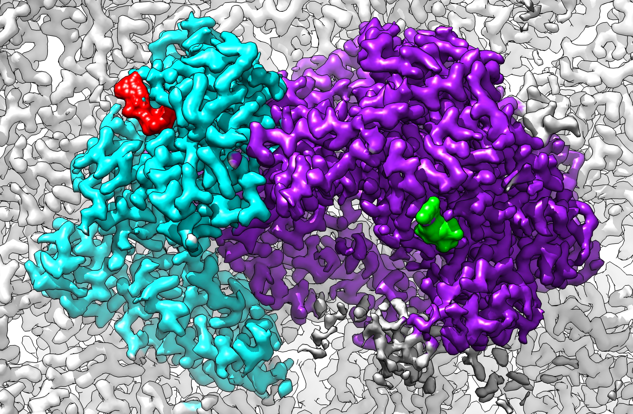 emd_6408.jpg emd_6408.jpg | 984.9 KB | ||
| Archive directory |  http://ftp.pdbj.org/pub/emdb/structures/EMD-6408ftp://ftp.pdbj.org/pub/emdb/structures/EMD-6408 http://ftp.pdbj.org/pub/emdb/structures/EMD-6408ftp://ftp.pdbj.org/pub/emdb/structures/EMD-6408 | HTTPS FTP |
-Related structure data
| Related structure data | 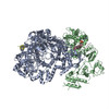 3jb6MC 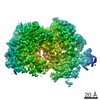 6404C 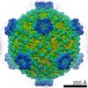 6405C 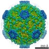 6406C 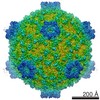 6407C 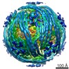 6409C  3jb7C M: atomic model generated by this map C: citing same article ( |
|---|---|
| Similar structure data |


-Links
| EMDB pages | EMDB (EBI/PDBe) / EMDataResource |
|---|
-Map
| File | Download / File: emd_6408.map.gz / Format: CCP4 / Size: 11.4 MB / Type: IMAGE STORED AS FLOATING POINT NUMBER (4 BYTES) | ||||||||||||||||||||||||||||||||||||||||||||||||||||||||||||||||||||
|---|---|---|---|---|---|---|---|---|---|---|---|---|---|---|---|---|---|---|---|---|---|---|---|---|---|---|---|---|---|---|---|---|---|---|---|---|---|---|---|---|---|---|---|---|---|---|---|---|---|---|---|---|---|---|---|---|---|---|---|---|---|---|---|---|---|---|---|---|---|
| Annotation | Averaged qCPV TEC | ||||||||||||||||||||||||||||||||||||||||||||||||||||||||||||||||||||
| Projections & slices | Image control
Images are generated by Spider. generated in cubic-lattice coordinate | ||||||||||||||||||||||||||||||||||||||||||||||||||||||||||||||||||||
| Voxel size | X=Y=Z: 1.01 Å | ||||||||||||||||||||||||||||||||||||||||||||||||||||||||||||||||||||
| Density |
| ||||||||||||||||||||||||||||||||||||||||||||||||||||||||||||||||||||
| Symmetry | Space group: 1 | ||||||||||||||||||||||||||||||||||||||||||||||||||||||||||||||||||||
| Details | EMDB XML:
CCP4 map header:
| ||||||||||||||||||||||||||||||||||||||||||||||||||||||||||||||||||||
 Z (Sec.)
 X (Row.)
 Y (Col.)


-Supplemental data
- Sample components
-Entire : CPV VP4 + polymerase complex (TEC)
| Entire | Name: CPV VP4 + polymerase complex (TEC) |
|---|---|
| Components |
|
-Supramolecule #1000: CPV VP4 + polymerase complex (TEC)
| Supramolecule | Name: CPV VP4 + polymerase complex (TEC) / type: sample / ID: 1000 / Number unique components: 2 |
|---|
-Macromolecule #1: VP4
| Macromolecule | Name: VP4 / type: protein_or_peptide / ID: 1 / Recombinant expression: No / Database: NCBI |
|---|---|
| Source (natural) | Organism: Bombyx mori cypovirus 1 / synonym: Cytoplasmic polyhedrosis virus |
-Macromolecule #2: RNA polymerase
| Macromolecule | Name: RNA polymerase / type: protein_or_peptide / ID: 2 / Recombinant expression: No / Database: NCBI |
|---|---|
| Source (natural) | Organism: Bombyx mori cypovirus 1 / synonym: Cytoplasmic polyhedrosis virus |
-Experimental details
-Structure determination
| Method | cryo EM |
|---|---|
 Processing Processing | single particle reconstruction |
| Aggregation state | particle |
-Sample preparation
| Buffer | pH: 8 / Details: 70 mM Tris-Cl, 10 mM MgCl2, 100 mM NaCl, 2 mM GTP |
|---|---|
| Grid | Details: 200 mesh Quantifoil holey carbon film |
| Vitrification | Cryogen name: ETHANE / Instrument: FEI VITROBOT MARK II |
- Electron microscopy
| Microscope | FEI TITAN KRIOS |
|---|---|
| Temperature | Average: 80 K |
| Alignment procedure | Legacy - Electron beam tilt params: 0 |
| Date | Jul 26, 2013 |
| Image recording | Category: CCD / Film or detector model: GATAN K2 (4k x 4k) / Digitization - Sampling interval: 5 µm / Number real images: 4385 / Average electron dose: 25 e/Å2 |
| Electron beam | Acceleration voltage: 300 kV / Electron source:  FIELD EMISSION GUN FIELD EMISSION GUN |
| Electron optics | Calibrated magnification: 49500 / Illumination mode: FLOOD BEAM / Imaging mode: BRIGHT FIELD / Cs: 2.7 mm / Nominal defocus max: 4.5 µm / Nominal defocus min: 0.6 µm / Nominal magnification: 49000 |
| Sample stage | Specimen holder model: FEI TITAN KRIOS AUTOGRID HOLDER |
| Experimental equipment |  Model: Titan Krios / Image courtesy: FEI Company |
-Image processing
| CTF correction | Details: Each particle |
|---|---|
| Final reconstruction | Resolution.type: BY AUTHOR / Resolution: 3.3 Å / Resolution method: OTHER / Software - Name: Frealign / Number images used: 68526 |
| Final two d classification | Number classes: 1 |
