 Movie
Movie Controller

[English] 日本語
 Yorodumi
- EMDB-6352: Cryo-EM structure of GMPCPP-microtubule (14 protofilaments) decor... -
+ Open data

- Basic information
| Entry | Database: EMDB / ID: EMD-6352 | |||||||||
|---|---|---|---|---|---|---|---|---|---|---|
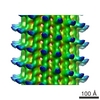
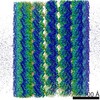
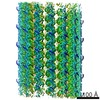
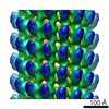
| Title | Cryo-EM structure of GMPCPP-microtubule (14 protofilaments) decorated with kinesin | |||||||||
 Map data Map data | 3D reconstruction of kinesin-bound GMPCPP microtubule (14 protofilaments) with pseudo-helical symmetry imposed | |||||||||
 Sample Sample |
| |||||||||
 Keywords Keywords | microtubule / GMPCPP / kinesin | |||||||||
| Function / homology |  Function and homology information Function and homology informationMicrotubule-dependent trafficking of connexons from Golgi to the plasma membrane / Resolution of Sister Chromatid Cohesion / Hedgehog 'off' state / Cilium Assembly / Intraflagellar transport / COPI-dependent Golgi-to-ER retrograde traffic / Mitotic Prometaphase / Carboxyterminal post-translational modifications of tubulin / RHOH GTPase cycle / EML4 and NUDC in mitotic spindle formation ...Microtubule-dependent trafficking of connexons from Golgi to the plasma membrane / Resolution of Sister Chromatid Cohesion / Hedgehog 'off' state / Cilium Assembly / Intraflagellar transport / COPI-dependent Golgi-to-ER retrograde traffic / Mitotic Prometaphase / Carboxyterminal post-translational modifications of tubulin / RHOH GTPase cycle / EML4 and NUDC in mitotic spindle formation / Sealing of the nuclear envelope (NE) by ESCRT-III / Kinesins / PKR-mediated signaling / Separation of Sister Chromatids / The role of GTSE1 in G2/M progression after G2 checkpoint / Aggrephagy / RHO GTPases activate IQGAPs / RHO GTPases Activate Formins / HSP90 chaperone cycle for steroid hormone receptors (SHR) in the presence of ligand / MHC class II antigen presentation / Recruitment of NuMA to mitotic centrosomes / COPI-mediated anterograde transport / structural constituent of cytoskeleton / microtubule cytoskeleton organization / neuron migration / mitotic cell cycle / microtubule cytoskeleton / Hydrolases; Acting on acid anhydrides; Acting on GTP to facilitate cellular and subcellular movement / microtubule / GTPase activity / GTP binding / metal ion binding / cytoplasm Similarity search - Function | |||||||||
| Biological species |   Homo sapiens (human) Homo sapiens (human) | |||||||||
| Method | helical reconstruction / cryo EM / Resolution: 3.5 Å | |||||||||
 Authors Authors | Zhang R / Alushin GM / Brown A / Nogales E | |||||||||
 Citation Citation | Journal: Cell / Year: 2015 Title: Mechanistic Origin of Microtubule Dynamic Instability and Its Modulation by EB Proteins. Authors: Rui Zhang / Gregory M Alushin / Alan Brown / Eva Nogales /   Abstract: Microtubule (MT) dynamic instability is driven by GTP hydrolysis and regulated by microtubule-associated proteins, including the plus-end tracking end-binding protein (EB) family. We report six cryo- ...Microtubule (MT) dynamic instability is driven by GTP hydrolysis and regulated by microtubule-associated proteins, including the plus-end tracking end-binding protein (EB) family. We report six cryo-electron microscopy (cryo-EM) structures of MTs, at 3.5 Å or better resolution, bound to GMPCPP, GTPγS, or GDP, either decorated with kinesin motor domain after polymerization or copolymerized with EB3. Subtle changes around the E-site nucleotide during hydrolysis trigger conformational changes in α-tubulin around an "anchor point," leading to global lattice rearrangements and strain generation. Unlike the extended lattice of the GMPCPP-MT, the EB3-bound GTPγS-MT has a compacted lattice that differs in lattice twist from that of the also compacted GDP-MT. These results and the observation that EB3 promotes rapid hydrolysis of GMPCPP suggest that EB proteins modulate structural transitions at growing MT ends by recognizing and promoting an intermediate state generated during GTP hydrolysis. Our findings explain both EBs end-tracking behavior and their effect on microtubule dynamics. | |||||||||
| History |
|
- Structure visualization
| Movie |
Movie viewer |
|---|---|
| Structure viewer | EM map: SurfViewMolmilJmol/JSmol |
| Supplemental images |

- Downloads & links
-EMDB archive
| Map data | emd_6352.map.gz | 155.9 MB | EMDB map data format | |
|---|---|---|---|---|
| Header (meta data) | emd-6352-v30.xmlemd-6352.xml | 12.9 KB 12.9 KB | Display Display | EMDB header |
| Images | 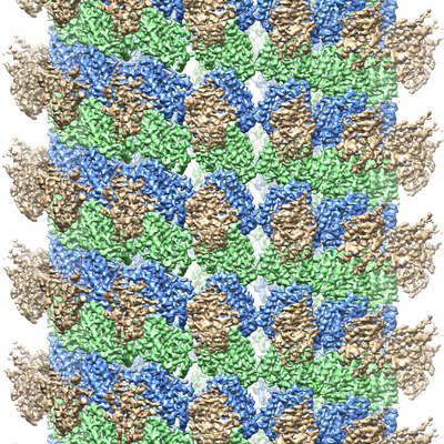 400_6352.gif 400_6352.gif 80_6352.gif 80_6352.gif | 165.8 KB 8.1 KB | ||
| Archive directory |  http://ftp.pdbj.org/pub/emdb/structures/EMD-6352ftp://ftp.pdbj.org/pub/emdb/structures/EMD-6352 http://ftp.pdbj.org/pub/emdb/structures/EMD-6352ftp://ftp.pdbj.org/pub/emdb/structures/EMD-6352 | HTTPS FTP |
-Related structure data
| Related structure data |  3jatMC 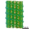 6347C 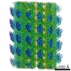 6348C  6349C  6350C  6351C  6353C 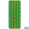 6354C 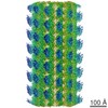 6355C  3jakC  3jalC  3jarC  3jasC  3jawC M: atomic model generated by this map C: citing same article ( |
|---|---|
| Similar structure data |
-Links
| EMDB pages | EMDB (EBI/PDBe) / EMDataResource |
|---|---|
| Related items in Molecule of the Month |


-Map
| File | Download / File: emd_6352.map.gz / Format: CCP4 / Size: 500 MB / Type: IMAGE STORED AS FLOATING POINT NUMBER (4 BYTES) | ||||||||||||||||||||||||||||||||||||||||||||||||||||||||||||
|---|---|---|---|---|---|---|---|---|---|---|---|---|---|---|---|---|---|---|---|---|---|---|---|---|---|---|---|---|---|---|---|---|---|---|---|---|---|---|---|---|---|---|---|---|---|---|---|---|---|---|---|---|---|---|---|---|---|---|---|---|---|
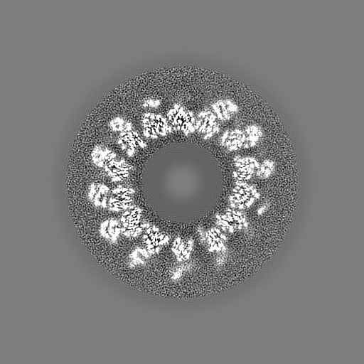
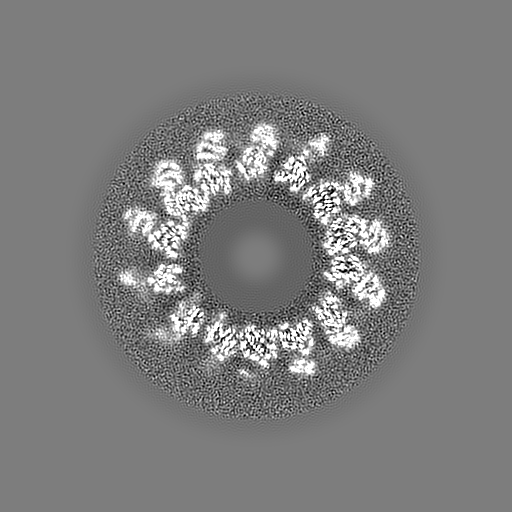
| Annotation | 3D reconstruction of kinesin-bound GMPCPP microtubule (14 protofilaments) with pseudo-helical symmetry imposed | ||||||||||||||||||||||||||||||||||||||||||||||||||||||||||||
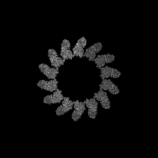
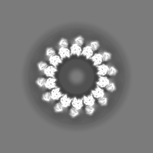
| Projections & slices | Image control
Images are generated by Spider. | ||||||||||||||||||||||||||||||||||||||||||||||||||||||||||||
| Voxel size | X=Y=Z: 1.32 Å | ||||||||||||||||||||||||||||||||||||||||||||||||||||||||||||
| Density |
| ||||||||||||||||||||||||||||||||||||||||||||||||||||||||||||
| Symmetry | Space group: 1 | ||||||||||||||||||||||||||||||||||||||||||||||||||||||||||||
| Details | EMDB XML:
CCP4 map header:
| ||||||||||||||||||||||||||||||||||||||||||||||||||||||||||||
 Z (Sec.)
 Y (Row.)
 X (Col.)


-Supplemental data
- Sample components
-Entire : kinesin-bound GMPCPP microtubule
| Entire | Name: kinesin-bound GMPCPP microtubule |
|---|---|
| Components |
|
-Supramolecule #1000: kinesin-bound GMPCPP microtubule
| Supramolecule | Name: kinesin-bound GMPCPP microtubule / type: sample / ID: 1000 / Oligomeric state: helical assembly / Number unique components: 3 |
|---|
-Macromolecule #1: Alpha tubulin
| Macromolecule | Name: Alpha tubulin / type: protein_or_peptide / ID: 1 Details: Porcine tubulin powder was purchased from Cytoskeleton Inc. Oligomeric state: Helical assembly / Recombinant expression: No / Database: NCBI |
|---|---|
| Source (natural) | Organism: |
| Molecular weight | Theoretical: 55 KDa |
| Sequence | UniProtKB: Tubulin alpha-1B chain |
-Macromolecule #2: Beta tubulin
| Macromolecule | Name: Beta tubulin / type: protein_or_peptide / ID: 2 Details: Porcine tubulin powder was purchased from Cytoskeleton Inc. Oligomeric state: Helical assembly / Recombinant expression: No / Database: NCBI |
|---|---|
| Source (natural) | Organism: |
| Molecular weight | Theoretical: 55 KDa |
| Sequence | UniProtKB: Tubulin beta chain |
-Macromolecule #3: kinesin
| Macromolecule | Name: kinesin / type: protein_or_peptide / ID: 3 Details: Human monomeric kinesin K349 cys-lite described in: Rice, S., Lin, A.W., Safer, D., Hart, C.L., Naber, N., Carragher, B.O., Cain, S.M., Pechatnikova, E., Wilson-Kubalek, E.M., Whittaker, M., ...Details: Human monomeric kinesin K349 cys-lite described in: Rice, S., Lin, A.W., Safer, D., Hart, C.L., Naber, N., Carragher, B.O., Cain, S.M., Pechatnikova, E., Wilson-Kubalek, E.M., Whittaker, M., et al. (1999). A structural change in the kinesin motor protein that drives motility. Nature 402, 778-784. Oligomeric state: monomer / Recombinant expression: Yes |
|---|---|
| Source (natural) | Organism: Homo sapiens (human) / synonym: Human / Organelle: Cytoplasm / Location in cell: Cytoskeleton |
| Molecular weight | Theoretical: 36 KDa |
| Recombinant expression | Organism:  |
-Experimental details
-Structure determination
| Method | cryo EM |
|---|---|
 Processing Processing | helical reconstruction |
| Aggregation state | helical array |
-Sample preparation
| Concentration | 3 mg/mL |
|---|---|
| Buffer | pH: 6.8 Details: 80 mM PIPES, 1 mM EGTA, 1 mM MgCl2, 1 mM DTT, 0.05% Nonidet P-40 |
| Grid | Details: 400 mesh C-flat 1.2/1.3 EM grid, glow discharged in Ar/O2 gas (Solarus, Gatan Inc) |
| Vitrification | Cryogen name: ETHANE / Chamber humidity: 95 % / Chamber temperature: 90.4 K / Instrument: FEI VITROBOT MARK II / Method: Blot once for 4 seconds before plunging. |
- Electron microscopy
| Microscope | FEI TITAN |
|---|---|
| Temperature | Average: 90 K |
| Alignment procedure | Legacy - Astigmatism: Objective lens astigmatism was corrected at 27,500 times magnification. |
| Details | The camera was operated in counting mode, with a dose rate of ~8 electrons/pixel/s on the camera. A total exposure time of 6 s was fractionated into 20 movie frames. |
| Date | Mar 2, 2014 |
| Image recording | Category: CCD / Film or detector model: GATAN K2 (4k x 4k) / Number real images: 695 / Average electron dose: 27.6 e/Å2 Details: The camera was operated in counting mode, with a dose rate of ~8 electrons/pixel/s on the camera. A total exposure time of 6 s was fractionated into 20 movie frames. |
| Electron beam | Acceleration voltage: 300 kV / Electron source:  FIELD EMISSION GUN FIELD EMISSION GUN |
| Electron optics | Calibrated magnification: 27500 / Illumination mode: FLOOD BEAM / Imaging mode: BRIGHT FIELD / Cs: 2.7 mm / Nominal defocus max: 3.5 µm / Nominal defocus min: 1.0 µm / Nominal magnification: 27500 |
| Sample stage | Specimen holder: Gatan 626 holder / Specimen holder model: GATAN LIQUID NITROGEN |
-Image processing
| Details | IHRSR algorithm with microtubule-specific pseudo-helical symmetry applied |
|---|---|
| Final reconstruction | Applied symmetry - Helical parameters - Δz: 8.92 Å Applied symmetry - Helical parameters - Δ&Phi: 25.75 ° Applied symmetry - Helical parameters - Axial symmetry: C1 (asymmetric) Algorithm: OTHER / Resolution.type: BY AUTHOR / Resolution: 3.5 Å / Resolution method: OTHER / Software - Name: EMAN1, FREALIGN Details: Pseudo-helical symmetry was applied during the reconstruction step. |
| CTF correction | Details: CTFFIND4, each particle |
