 Movie
Movie Controller

[English] 日本語
 Yorodumi
- EMDB-5888: Electron cryo-microscopy of nanobody AB29 in complex with poliovi... -
+ Open data

- Basic information
| Entry | Database: EMDB / ID: EMD-5888 | |||||||||
|---|---|---|---|---|---|---|---|---|---|---|
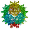
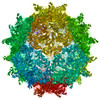
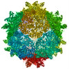
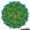
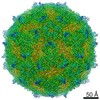
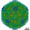
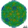
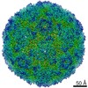
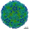
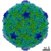
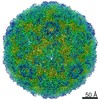
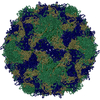
| Title | Electron cryo-microscopy of nanobody AB29 in complex with poliovirus P1/Mahoney | |||||||||
 Map data Map data | reconstruction of nanobody AB29 in complex with poliovirus P1/Mahoney | |||||||||
 Sample Sample |
| |||||||||
 Keywords Keywords | picornavirus / nanobody / antibody / VHH / poliovirus / mechanism of neutralization | |||||||||
| Function / homology |  Function and homology information Function and homology informationsymbiont-mediated suppression of host translation initiation / symbiont-mediated suppression of host cytoplasmic pattern recognition receptor signaling pathway via inhibition of RIG-I activity / symbiont-mediated suppression of host cytoplasmic pattern recognition receptor signaling pathway via inhibition of MDA-5 activity / symbiont-mediated suppression of host cytoplasmic pattern recognition receptor signaling pathway via inhibition of MAVS activity / picornain 2A / symbiont-mediated suppression of host mRNA export from nucleus / ribonucleoside triphosphate phosphatase activity / symbiont genome entry into host cell via pore formation in plasma membrane / picornain 3C / T=pseudo3 icosahedral viral capsid ...symbiont-mediated suppression of host translation initiation / symbiont-mediated suppression of host cytoplasmic pattern recognition receptor signaling pathway via inhibition of RIG-I activity / symbiont-mediated suppression of host cytoplasmic pattern recognition receptor signaling pathway via inhibition of MDA-5 activity / symbiont-mediated suppression of host cytoplasmic pattern recognition receptor signaling pathway via inhibition of MAVS activity / picornain 2A / symbiont-mediated suppression of host mRNA export from nucleus / ribonucleoside triphosphate phosphatase activity / symbiont genome entry into host cell via pore formation in plasma membrane / picornain 3C / T=pseudo3 icosahedral viral capsid / host cell cytoplasmic vesicle membrane / endocytosis involved in viral entry into host cell / channel activity / nucleoside-triphosphate phosphatase / monoatomic ion transmembrane transport / RNA helicase activity / induction by virus of host autophagy / RNA-directed RNA polymerase / viral RNA genome replication / cysteine-type endopeptidase activity / RNA-dependent RNA polymerase activity / virus-mediated perturbation of host defense response / DNA-templated transcription / host cell nucleus / virion attachment to host cell / structural molecule activity / proteolysis / RNA binding / ATP binding / membrane / metal ion binding Similarity search - Function | |||||||||
| Biological species |   Human poliovirus 1 Human poliovirus 1 | |||||||||
| Method | single particle reconstruction / cryo EM / Resolution: 6.5 Å | |||||||||
 Authors Authors | Schotte L / Strauss M / Thys B / Halewyck H / Filman DJ / Bostina M / Hogle JM / Rombaut B | |||||||||
 Citation Citation | Journal: J Virol / Year: 2016 Title: Five of Five VHHs Neutralizing Poliovirus Bind the Receptor-Binding Site. Authors: Mike Strauss / Lise Schotte / Bert Thys / David J Filman / James M Hogle /   Abstract: Nanobodies, or VHHs, that recognize poliovirus type 1 have previously been selected and characterized as candidates for antiviral agents or reagents for standardization of vaccine quality control. In ...Nanobodies, or VHHs, that recognize poliovirus type 1 have previously been selected and characterized as candidates for antiviral agents or reagents for standardization of vaccine quality control. In this study, we present high-resolution cryo-electron microscopy reconstructions of poliovirus with five neutralizing VHHs. All VHHs bind the capsid in the canyon at sites that extensively overlap the poliovirus receptor-binding site. In contrast, the interaction involves a unique (and surprisingly extensive) surface for each of the five VHHs. Five regions of the capsid were found to participate in binding with all five VHHs. Four of these five regions are known to alter during the expansion of the capsid associated with viral entry. Interestingly, binding of one of the VHHs, PVSS21E, resulted in significant changes of the capsid structure and thus seems to trap the virus in an early stage of expansion. IMPORTANCE: We describe the cryo-electron microscopy structures of complexes of five neutralizing VHHs with the Mahoney strain of type 1 poliovirus at resolutions ranging from 3.8 to 6.3Å. All five ...IMPORTANCE: We describe the cryo-electron microscopy structures of complexes of five neutralizing VHHs with the Mahoney strain of type 1 poliovirus at resolutions ranging from 3.8 to 6.3Å. All five VHHs bind deep in the virus canyon at similar sites that overlap extensively with the binding site for the receptor (CD155). The binding surfaces on the VHHs are surprisingly extensive, but despite the use of similar binding surfaces on the virus, the binding surface on the VHHs is unique for each VHH. In four of the five complexes, the virus remains essentially unchanged, but for the fifth there are significant changes reminiscent of but smaller in magnitude than the changes associated with cell entry, suggesting that this VHH traps the virus in a previously undescribed early intermediate state. The neutralizing mechanisms of the VHHs and their potential use as quality control agents for the end game of poliovirus eradication are discussed. | |||||||||
| History |
|
- Structure visualization
| Movie |
Movie viewer |
|---|---|
| Structure viewer | EM map: SurfViewMolmilJmol/JSmol |
| Supplemental images |

- Downloads & links
-EMDB archive
| Map data | emd_5888.map.gz | 111.2 MB | EMDB map data format | |
|---|---|---|---|---|
| Header (meta data) | emd-5888-v30.xmlemd-5888.xml | 14.5 KB 14.5 KB | Display Display | EMDB header |
| Images | 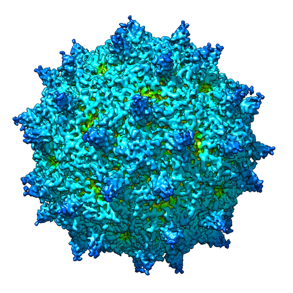 emd_5888_1.jpg emd_5888_1.jpg | 393.3 KB | ||
| Archive directory |  http://ftp.pdbj.org/pub/emdb/structures/EMD-5888ftp://ftp.pdbj.org/pub/emdb/structures/EMD-5888 http://ftp.pdbj.org/pub/emdb/structures/EMD-5888ftp://ftp.pdbj.org/pub/emdb/structures/EMD-5888 | HTTPS FTP |
-Validation report
| Summary document | emd_5888_validation.pdf.gz | 370.7 KB | Display | EMDB validaton report |
|---|---|---|---|---|
| Full document | emd_5888_full_validation.pdf.gz | 370.2 KB | Display | |
| Data in XML | emd_5888_validation.xml.gz | 6.5 KB | Display | |
| Arichive directory | https://ftp.pdbj.org/pub/emdb/validation_reports/EMD-5888ftp://ftp.pdbj.org/pub/emdb/validation_reports/EMD-5888 | HTTPS FTP |
-Related structure data
| Related structure data |  3jbcM 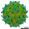 5886C C: citing same article ( M: atomic model generated by this map |
|---|---|
| Similar structure data |


-Links
| EMDB pages | EMDB (EBI/PDBe) / EMDataResource |
|---|---|
| Related items in Molecule of the Month |


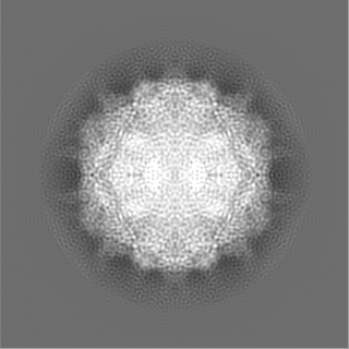
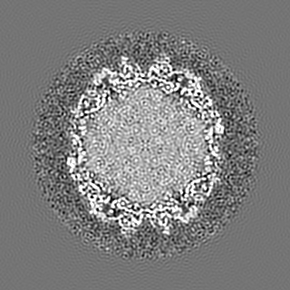
-Map
| File | Download / File: emd_5888.map.gz / Format: CCP4 / Size: 122.1 MB / Type: IMAGE STORED AS FLOATING POINT NUMBER (4 BYTES) | ||||||||||||||||||||||||||||||||||||||||||||||||||||||||||||||||||||
|---|---|---|---|---|---|---|---|---|---|---|---|---|---|---|---|---|---|---|---|---|---|---|---|---|---|---|---|---|---|---|---|---|---|---|---|---|---|---|---|---|---|---|---|---|---|---|---|---|---|---|---|---|---|---|---|---|---|---|---|---|---|---|---|---|---|---|---|---|---|
| Annotation | reconstruction of nanobody AB29 in complex with poliovirus P1/Mahoney | ||||||||||||||||||||||||||||||||||||||||||||||||||||||||||||||||||||
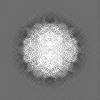
| Projections & slices | Image control
Images are generated by Spider. | ||||||||||||||||||||||||||||||||||||||||||||||||||||||||||||||||||||
| Voxel size | X=Y=Z: 1.681 Å | ||||||||||||||||||||||||||||||||||||||||||||||||||||||||||||||||||||
| Density |
| ||||||||||||||||||||||||||||||||||||||||||||||||||||||||||||||||||||
| Symmetry | Space group: 1 | ||||||||||||||||||||||||||||||||||||||||||||||||||||||||||||||||||||
| Details | EMDB XML:
CCP4 map header:
| ||||||||||||||||||||||||||||||||||||||||||||||||||||||||||||||||||||
 Z (Sec.)
 Y (Row.)
 X (Col.)


-Supplemental data
- Sample components
-Entire : nanobody AB29 in complex with poliovirus P1/Mahoney
| Entire | Name: nanobody AB29 in complex with poliovirus P1/Mahoney |
|---|---|
| Components |
|
-Supramolecule #1000: nanobody AB29 in complex with poliovirus P1/Mahoney
| Supramolecule | Name: nanobody AB29 in complex with poliovirus P1/Mahoney / type: sample / ID: 1000 / Details: 1 Oligomeric state: 60 nanobody VHH monomers bind to each poliovirion Number unique components: 2 |
|---|---|
| Molecular weight | Experimental: 9.0 MDa / Theoretical: 9.0 MDa / Method: 1 |
-Supramolecule #1: Human poliovirus 1
| Supramolecule | Name: Human poliovirus 1 / type: virus / ID: 1 / NCBI-ID: 12080 / Sci species name: Human poliovirus 1 / Database: NCBI / Virus type: VIRION / Virus isolate: SEROTYPE / Virus enveloped: No / Virus empty: No / Sci species serotype: Mahoney |
|---|---|
| Host (natural) | Organism:  Homo sapiens (human) / synonym: VERTEBRATES Homo sapiens (human) / synonym: VERTEBRATES |
| Molecular weight | Experimental: 9 MDa / Theoretical: 9 MDa |
| Virus shell | Shell ID: 1 / Diameter: 350 Å / T number (triangulation number): 1 |
-Macromolecule #1: nanobody PVSP29F
| Macromolecule | Name: nanobody PVSP29F / type: protein_or_peptide / ID: 1 / Name.synonym: nanobody AB29 Details: Each virus is decorated with 60 copies of nanobody PVSP29F. Recombinant expression: Yes |
|---|---|
| Source (natural) | Organism: |
| Recombinant expression | Organism:  |
-Experimental details
-Structure determination
| Method | cryo EM |
|---|---|
 Processing Processing | single particle reconstruction |
| Aggregation state | particle |
-Sample preparation
| Concentration | 1 mg/mL |
|---|---|
| Buffer | pH: 7.4 / Details: 145 mM NaCl, 50 mM Na2HPO4.12H2O |
| Grid | Details: Quantifoil R2/2 |
| Vitrification | Cryogen name: ETHANE / Chamber temperature: 154 K / Instrument: HOMEMADE PLUNGER |
- Electron microscopy
| Microscope | FEI TITAN KRIOS |
|---|---|
| Date | Oct 23, 2012 |
| Image recording | Category: CCD / Film or detector model: GATAN ULTRASCAN 4000 (4k x 4k) / Digitization - Sampling interval: 15 µm / Number real images: 1503 / Average electron dose: 25 e/Å2 / Bits/pixel: 16 |
| Electron beam | Acceleration voltage: 200 kV / Electron source:  FIELD EMISSION GUN FIELD EMISSION GUN |
| Electron optics | Calibrated magnification: 89232 / Illumination mode: FLOOD BEAM / Imaging mode: BRIGHT FIELD / Cs: 2.7 mm / Nominal defocus max: 3.0 µm / Nominal defocus min: 1.0 µm / Nominal magnification: 96000 |
| Sample stage | Specimen holder model: FEI TITAN KRIOS AUTOGRID HOLDER |
| Experimental equipment |  Model: Titan Krios / Image courtesy: FEI Company |
-Image processing
| Details | The particles were processed using Frealign. |
|---|---|
| CTF correction | Details: per particle |
| Final reconstruction | Resolution.type: BY AUTHOR / Resolution: 6.5 Å / Resolution method: OTHER / Software - Name: Frealign / Number images used: 9764 |
-Atomic model buiding 1
| Initial model | PDB ID: |
|---|---|
| Software | Name: REFMAC5, COOT, SPDBV |
| Details | Rigid body with some flexible loops and side chains. LSQKAB was applied after each refinement cycle to re-impose exact icosahedral operators and rigid body constraints. |
| Refinement | Space: RECIPROCAL / Protocol: RIGID BODY FIT Target criteria: Stereochemically restrained maximum likelihood refinement of both Fourier phase and amplitude agreement |
| Output model | PDB-3jbc: |
-Atomic model buiding 2
| Initial model | PDB ID: |
|---|---|
| Software | Name: REFMAC5, COOT, SPDBV |
| Details | Rigid body with some flexible loops and side chains. LSQKAB was applied after each refinement cycle to re-impose exact icosahedral operators and rigid body constraints. |
| Refinement | Space: RECIPROCAL / Protocol: RIGID BODY FIT Target criteria: Stereochemically restrained maximum likelihood refinement of both Fourier phase and amplitude agreement |
| Output model | PDB-3jbc: |
-Atomic model buiding 3
| Initial model | PDB ID: |
|---|---|
| Software | Name: REFMAC5, COOT, SPDBV |
| Details | Rigid body with some flexible loops and side chains. LSQKAB was applied after each refinement cycle to re-impose exact icosahedral operators and rigid body constraints. |
| Refinement | Space: RECIPROCAL / Protocol: RIGID BODY FIT Target criteria: Stereochemically restrained maximum likelihood refinement of both Fourier phase and amplitude agreement |
| Output model | PDB-3jbc: |

