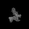
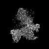
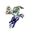
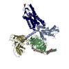
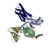
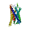
登録情報 データベース : EMDB / ID : EMD-35444タイトル Cryo-EM structure of HCA2-Gi complex with LUF6283 map 複合体 : Multiprotein complexタンパク質・ペプチド : Guanine nucleotide-binding protein G(i) subunit alpha-1タンパク質・ペプチド : Guanine nucleotide-binding protein G(I)/G(S)/G(T) subunit beta-1タンパク質・ペプチド : Guanine nucleotide-binding protein G(I)/G(S)/G(O) subunit gamma-2タンパク質・ペプチド : Soluble cytochrome b562,Hydroxycarboxylic acid receptor 2タンパク質・ペプチド : scFv16リガンド : 2-acetamido-2-deoxy-beta-D-glucopyranoseリガンド : 5-butyl-1~{H}-pyrazole-3-carboxylic acid / 機能・相同性 分子機能 ドメイン・相同性 構成要素
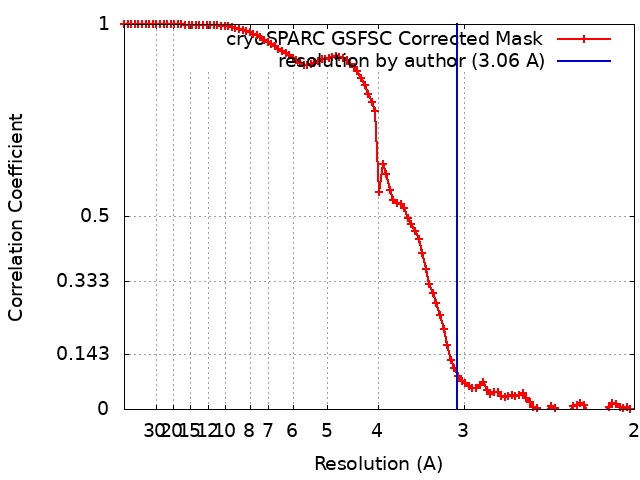
/ / / / / / / / / / / / / / / / / / / / / / / / / / / / / / / / / / / / / / / / / / / / / / / / / / / / / / / / / / / / / / / / / / / / / / / / / / / / / / / / / / / / / / / / / / / / / / / / / / / / / / / / / / / / / / / / / / / / / / / / / / / / / / / / / / / / / / / / / / / / / / / / / / / / / / / / / / / 生物種 Homo sapiens (ヒト) / Mus musculus (ハツカネズミ) / synthetic construct (人工物) 手法 / / 解像度 : 3.06 Å Suzuki S / Nishikawa K / Suzuki H / Fujiyoshi Y 資金援助 Organization Grant number 国 Japan Society for the Promotion of Science (JSPS) 20H00451
ジャーナル : Nat Commun / 年 : 2023タイトル : Structural basis of hydroxycarboxylic acid receptor signaling mechanisms through ligand binding.著者 : Shota Suzuki / Kotaro Tanaka / Kouki Nishikawa / Hiroshi Suzuki / Atsunori Oshima / Yoshinori Fujiyoshi / 要旨 : Hydroxycarboxylic acid receptors (HCA) are expressed in various tissues and immune cells. HCA2 and its agonist are thus important targets for treating inflammatory and metabolic disorders. Only ... Hydroxycarboxylic acid receptors (HCA) are expressed in various tissues and immune cells. HCA2 and its agonist are thus important targets for treating inflammatory and metabolic disorders. Only limited information is available, however, on the active-state binding of HCAs with agonists. Here, we present cryo-EM structures of human HCA2-Gi and HCA3-Gi signaling complexes binding with multiple compounds bound. Agonists were revealed to form a salt bridge with arginine, which is conserved in the HCA family, to activate these receptors. Extracellular regions of the receptors form a lid-like structure that covers the ligand-binding pocket. Although transmembrane (TM) 6 in HCAs undergoes dynamic conformational changes, ligands do not directly interact with amino acids in TM6, suggesting that indirect signaling induces a slight shift in TM6 to activate Gi proteins. Structural analyses of agonist-bound HCA2 and HCA3 together with mutagenesis and molecular dynamics simulation provide molecular insights into HCA ligand recognition and activation mechanisms. 履歴 登録 2023年2月22日 - ヘッダ(付随情報) 公開 2023年8月30日 - マップ公開 2023年8月30日 - 更新 2025年7月2日 - 現状 2025年7月2日 処理サイト : PDBj / 状態 : 公開
すべて表示 表示を減らす
 ムービー
ムービー コントローラー


 データを開く
 基本情報
基本情報
 マップデータ
 試料
試料 キーワード
キーワード 機能・相同性情報
 Homo sapiens (ヒト) /
 データ登録者
 日本, 1件
  引用
引用 構造の表示
構造の表示
 ダウンロードとリンク
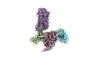 emd_35444.png
 http://ftp.pdbj.org/pub/emdb/structures/EMD-35444
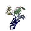


 Z (Sec.)
 Y (Row.)
 X (Col.)


 試料の構成要素
 Spodoptera frugiperda (ツマジロクサヨトウ)

 解析
解析 電子顕微鏡法
 FIELD EMISSION GUN