Movie
Movie Controller


+ Open data

- Basic information
| Entry |  | |||||||||||||||||||||
|---|---|---|---|---|---|---|---|---|---|---|---|---|---|---|---|---|---|---|---|---|---|---|
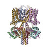
| Title | human KCNQ1-CaM in apo state | |||||||||||||||||||||
 Map data Map data | ||||||||||||||||||||||
 Sample Sample |
| |||||||||||||||||||||
 Keywords Keywords | potassium voltage-gated channel / MEMBRANE PROTEIN | |||||||||||||||||||||
| Function / homology |  Function and homology information Function and homology informationgastrin-induced gastric acid secretion / corticosterone secretion / voltage-gated potassium channel activity involved in atrial cardiac muscle cell action potential repolarization / negative regulation of voltage-gated potassium channel activity / basolateral part of cell / lumenal side of membrane / negative regulation of delayed rectifier potassium channel activity / rhythmic behavior / stomach development / regulation of gastric acid secretion ...gastrin-induced gastric acid secretion / corticosterone secretion / voltage-gated potassium channel activity involved in atrial cardiac muscle cell action potential repolarization / negative regulation of voltage-gated potassium channel activity / basolateral part of cell / lumenal side of membrane / negative regulation of delayed rectifier potassium channel activity / rhythmic behavior / stomach development / regulation of gastric acid secretion / voltage-gated potassium channel activity involved in cardiac muscle cell action potential repolarization / Phase 3 - rapid repolarisation / membrane repolarization during action potential / membrane repolarization during atrial cardiac muscle cell action potential / Phase 2 - plateau phase / iodide transport / regulation of atrial cardiac muscle cell membrane repolarization / membrane repolarization during ventricular cardiac muscle cell action potential / intracellular chloride ion homeostasis / membrane repolarization during cardiac muscle cell action potential / potassium ion export across plasma membrane / voltage-gated potassium channel activity involved in ventricular cardiac muscle cell action potential repolarization / atrial cardiac muscle cell action potential / renal sodium ion absorption / regulation of membrane repolarization / auditory receptor cell development / protein phosphatase 1 binding / detection of mechanical stimulus involved in sensory perception of sound / transporter inhibitor activity / delayed rectifier potassium channel activity / : / ventricular cardiac muscle cell action potential / potassium ion homeostasis / Voltage gated Potassium channels / positive regulation of potassium ion transmembrane transport / regulation of ventricular cardiac muscle cell membrane repolarization / type 3 metabotropic glutamate receptor binding / non-motile cilium assembly / outward rectifier potassium channel activity / intestinal absorption / cardiac muscle cell contraction / inner ear morphogenesis / negative regulation of high voltage-gated calcium channel activity / adrenergic receptor signaling pathway / response to corticosterone / renal absorption / regulation of synaptic vesicle exocytosis / ciliary base / negative regulation of calcium ion export across plasma membrane / regulation of cardiac muscle cell action potential / protein kinase A regulatory subunit binding / protein kinase A catalytic subunit binding / presynaptic endocytosis / regulation of heart contraction / inner ear development / potassium ion import across plasma membrane / regulation of heart rate by cardiac conduction / calcineurin-mediated signaling / monoatomic ion channel complex / nitric-oxide synthase binding / regulation of cell communication by electrical coupling involved in cardiac conduction / adenylate cyclase binding / protein phosphatase activator activity / action potential / cochlea development / voltage-gated potassium channel activity / regulation of synaptic vesicle endocytosis / social behavior / detection of calcium ion / regulation of cardiac muscle contraction / postsynaptic cytosol / positive regulation of heart rate / catalytic complex / phosphatidylinositol 3-kinase binding / transport vesicle / presynaptic cytosol / regulation of release of sequestered calcium ion into cytosol by sarcoplasmic reticulum / cardiac muscle contraction / titin binding / phosphatidylinositol-4,5-bisphosphate binding / regulation of calcium-mediated signaling / regulation of cardiac muscle contraction by regulation of the release of sequestered calcium ion / voltage-gated potassium channel complex / positive regulation of cardiac muscle contraction / cellular response to epinephrine stimulus / potassium ion transmembrane transport / calcium channel complex / substantia nigra development / regulation of heart rate / cytoplasmic vesicle membrane / cellular response to cAMP / calyx of Held / nitric-oxide synthase regulator activity / adenylate cyclase activator activity / response to amphetamine / regulation of cytokinesis / sarcomere / protein serine/threonine kinase activator activity / erythrocyte differentiation / spindle microtubule Similarity search - Function | |||||||||||||||||||||
| Biological species |  Homo sapiens (human) Homo sapiens (human) | |||||||||||||||||||||
| Method | single particle reconstruction / cryo EM / Resolution: 3.5 Å | |||||||||||||||||||||
 Authors Authors | Ma D / Guo J | |||||||||||||||||||||
| Funding support |  China, 6 items China, 6 items
| |||||||||||||||||||||
 Citation Citation | Journal: Proc Natl Acad Sci U S A / Year: 2022 Title: Structural mechanisms for the activation of human cardiac KCNQ1 channel by electro-mechanical coupling enhancers. Authors: Demin Ma / Ling Zhong / Zhenzhen Yan / Jing Yao / Yan Zhang / Fan Ye / Yuan Huang / Dongwu Lai / Wei Yang / Panpan Hou / Jiangtao Guo / Abstract: The cardiac KCNQ1 potassium channel carries the important current and controls the heart rhythm. Hundreds of mutations in KCNQ1 can cause life-threatening cardiac arrhythmia. Although KCNQ1 ...The cardiac KCNQ1 potassium channel carries the important current and controls the heart rhythm. Hundreds of mutations in KCNQ1 can cause life-threatening cardiac arrhythmia. Although KCNQ1 structures have been recently resolved, the structural basis for the dynamic electro-mechanical coupling, also known as the voltage sensor domain-pore domain (VSD-PD) coupling, remains largely unknown. In this study, utilizing two VSD-PD coupling enhancers, namely, the membrane lipid phosphatidylinositol 4,5-bisphosphate (PIP) and a small-molecule ML277, we determined 2.5-3.5 Å resolution cryo-electron microscopy structures of full-length human KCNQ1-calmodulin (CaM) complex in the apo closed, ML277-bound open, and ML277-PIP-bound open states. ML277 binds at the "elbow" pocket above the S4-S5 linker and directly induces an upward movement of the S4-S5 linker and the opening of the activation gate without affecting the C-terminal domain (CTD) of KCNQ1. PIP binds at the cleft between the VSD and the PD and brings a large structural rearrangement of the CTD together with the CaM to activate the PD. These findings not only elucidate the structural basis for the dynamic VSD-PD coupling process during KCNQ1 gating but also pave the way to develop new therapeutics for anti-arrhythmia. | |||||||||||||||||||||
| History |
|
- Structure visualization
| Supplemental images |
|---|

- Downloads & links
-EMDB archive
| Map data | emd_33316.map.gz | 43.8 MB | EMDB map data format | |
|---|---|---|---|---|
| Header (meta data) | emd-33316-v30.xmlemd-33316.xml | 16.2 KB 16.2 KB | Display Display | EMDB header |
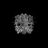
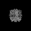
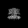
| Images | 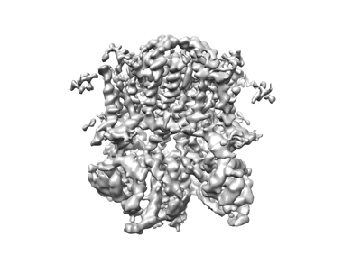 emd_33316.png emd_33316.png | 61.9 KB | ||
| Filedesc metadata | emd-33316.cif.gz | 5.7 KB | ||
| Others | emd_33316_half_map_1.map.gzemd_33316_half_map_2.map.gz | 37 MB 37 MB | ||
| Archive directory |  http://ftp.pdbj.org/pub/emdb/structures/EMD-33316ftp://ftp.pdbj.org/pub/emdb/structures/EMD-33316 http://ftp.pdbj.org/pub/emdb/structures/EMD-33316ftp://ftp.pdbj.org/pub/emdb/structures/EMD-33316 | HTTPS FTP |
-Related structure data
| Related structure data |  7xniMC  7xnkC 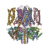 7xnlC  7xnnC M: atomic model generated by this map C: citing same article ( |
|---|---|
| Similar structure data |
-Links
| EMDB pages | EMDB (EBI/PDBe) / EMDataResource |
|---|---|
| Related items in Molecule of the Month |

-Map
| File | Download / File: emd_33316.map.gz / Format: CCP4 / Size: 52.7 MB / Type: IMAGE STORED AS FLOATING POINT NUMBER (4 BYTES) | ||||||||||||||||||||||||||||||||||||
|---|---|---|---|---|---|---|---|---|---|---|---|---|---|---|---|---|---|---|---|---|---|---|---|---|---|---|---|---|---|---|---|---|---|---|---|---|---|
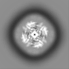
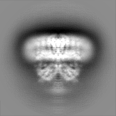
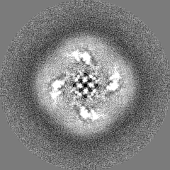
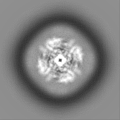
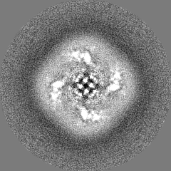
| Projections & slices | Image control
Images are generated by Spider. | ||||||||||||||||||||||||||||||||||||
| Voxel size | X=Y=Z: 1.014 Å | ||||||||||||||||||||||||||||||||||||
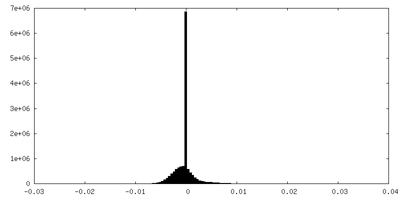
| Density |
| ||||||||||||||||||||||||||||||||||||
| Symmetry | Space group: 1 | ||||||||||||||||||||||||||||||||||||
| Details | EMDB XML:
|
 Z (Sec.)
 Y (Row.)
 X (Col.)


-Supplemental data
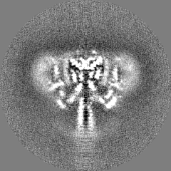
-Half map: #1
| File | emd_33316_half_map_1.map | ||||||||||||
|---|---|---|---|---|---|---|---|---|---|---|---|---|---|
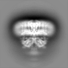
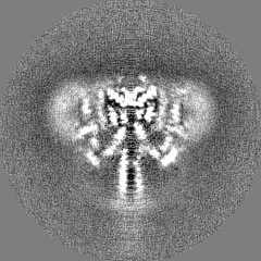
| Projections & Slices |
| ||||||||||||
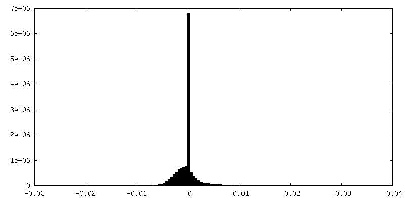
| Density Histograms |

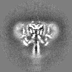

-Half map: #2
| File | emd_33316_half_map_2.map | ||||||||||||
|---|---|---|---|---|---|---|---|---|---|---|---|---|---|
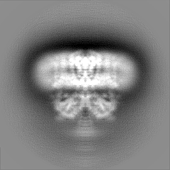
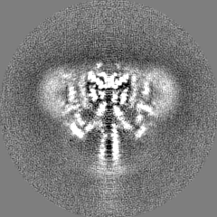
| Projections & Slices |
| ||||||||||||
| Density Histograms |
- Sample components
-Entire : KCNQ1-CaM complex
| Entire | Name: KCNQ1-CaM complex |
|---|---|
| Components |
|
-Supramolecule #1: KCNQ1-CaM complex
| Supramolecule | Name: KCNQ1-CaM complex / type: organelle_or_cellular_component / ID: 1 / Parent: 0 / Macromolecule list: all |
|---|---|
| Source (natural) | Organism: Homo sapiens (human) |
-Macromolecule #1: Potassium voltage-gated channel subfamily KQT member 1
| Macromolecule | Name: Potassium voltage-gated channel subfamily KQT member 1 type: protein_or_peptide / ID: 1 / Number of copies: 4 / Enantiomer: LEVO |
|---|---|
| Source (natural) | Organism: Homo sapiens (human) |
| Molecular weight | Theoretical: 76.487297 KDa |
| Recombinant expression | Organism: Homo sapiens (human) |
| Sequence | String: MAAASSPPRA ERKRWGWGRL PGARRGSAGL AKKCPFSLEL AEGGPAGGAL YAPIAPGAPG PAPPASPAAP AAPPVASDLG PRPPVSLDP RVSIYSTRRP VLARTHVQGR VYNFLERPTG WKCFVYHFAV FLIVLVCLIF SVLSTIEQYA ALATGTLFWM E IVLVVFFG ...String: MAAASSPPRA ERKRWGWGRL PGARRGSAGL AKKCPFSLEL AEGGPAGGAL YAPIAPGAPG PAPPASPAAP AAPPVASDLG PRPPVSLDP RVSIYSTRRP VLARTHVQGR VYNFLERPTG WKCFVYHFAV FLIVLVCLIF SVLSTIEQYA ALATGTLFWM E IVLVVFFG TEYVVRLWSA GCRSKYVGLW GRLRFARKPI SIIDLIVVVA SMVVLCVGSK GQVFATSAIR GIRFLQILRM LH VDRQGGT WRLLGSVVFI HRQELITTLY IGFLGLIFSS YFVYLAEKDA VNESGRVEFG SYADALWWGV VTVTTIGYGD KVP QTWVGK TIASCFSVFA ISFFALPAGI LGSGFALKVQ QKQRQKHFNR QIPAAASLIQ TAWRCYAAEN PDSSTWKIYI RKAP RSHTL LSPSPKPKKS VVVKKKKFKL DKDNGVTPGE KMLTVPHITC DPPEERRLDH FSVDGYDSSV RKSPTLLEVS MPHFM RTNS FAEDLDLEGE TLLTPITHIS QLREHHRATI KVIRRMQYFV AKKKFQQARK PYDVRDVIEQ YSQGHLNLMV RIKELQ RRL DQSIGKPSLF ISVSEKSKDR GSNTIGARLN RVEDKVTQLD QRLALITDML HQLLSLHGGS TPGSGGPPRE GGAHITQ PC GSGGSVDPEL FLPSNTLPTY EQLTVPRRGP DEGSLEGGSS GGWSHPQFEK UniProtKB: Potassium voltage-gated channel subfamily KQT member 1 |
-Macromolecule #2: Calmodulin-3
| Macromolecule | Name: Calmodulin-3 / type: protein_or_peptide / ID: 2 / Number of copies: 4 / Enantiomer: LEVO |
|---|---|
| Source (natural) | Organism: Homo sapiens (human) |
| Molecular weight | Theoretical: 19.615445 KDa |
| Recombinant expression | Organism: Homo sapiens (human) |
| Sequence | String: MADQLTEEQI AEFKEAFSLF DKDGDGTITT KELGTVMRSL GQNPTEAELQ DMINEVDADG NGTIDFPEFL TMMARKMKDT DSEEEIREA FRVFDKDGNG YISAAELRHV MTNLGEKLTD EEVDEMIREA DIDGDGQVNY EEFVQMMTAK LEGGSSGGLV P RGSGGSSG GHHHHHHHH UniProtKB: Calmodulin-3 |
-Experimental details
-Structure determination
| Method | cryo EM |
|---|---|
 Processing Processing | single particle reconstruction |
| Aggregation state | particle |
-Sample preparation
| Buffer | pH: 8 |
|---|---|
| Vitrification | Cryogen name: ETHANE |
- Electron microscopy
| Microscope | FEI TITAN KRIOS |
|---|---|
| Image recording | Film or detector model: GATAN K2 SUMMIT (4k x 4k) / Average electron dose: 64.0 e/Å2 |
| Electron beam | Acceleration voltage: 300 kV / Electron source:  FIELD EMISSION GUN FIELD EMISSION GUN |
| Electron optics | Illumination mode: FLOOD BEAM / Imaging mode: BRIGHT FIELD / Nominal defocus max: -1.3 µm / Nominal defocus min: -1.1 µm |
| Experimental equipment |  Model: Titan Krios / Image courtesy: FEI Company |
-Image processing
| Startup model | Type of model: PDB ENTRY PDB model - PDB ID: |
|---|---|
| Final reconstruction | Resolution.type: BY AUTHOR / Resolution: 3.5 Å / Resolution method: FSC 0.143 CUT-OFF / Number images used: 169344 |
| Initial angle assignment | Type: MAXIMUM LIKELIHOOD |
| Final angle assignment | Type: MAXIMUM LIKELIHOOD |