National Institutes of Health/National Institute Of Allergy and Infectious Diseases (NIH/NIAID)
AI081726
United States
National Science Foundation (NSF, United States)
MCB-0923873
United States
Citation
Journal: Proc Natl Acad Sci U S A / Year: 2022 Title: Structures of a large prolate virus capsid in unexpanded and expanded states generate insights into the icosahedral virus assembly. Authors: Qianglin Fang / Wei-Chun Tang / Andrei Fokine / Marthandan Mahalingam / Qianqian Shao / Michael G Rossmann / Venigalla B Rao / Abstract: Many icosahedral viruses assemble proteinaceous precursors called proheads or procapsids. Proheads are metastable structures that undergo a profound structural transition known as expansion that ...Many icosahedral viruses assemble proteinaceous precursors called proheads or procapsids. Proheads are metastable structures that undergo a profound structural transition known as expansion that transforms an immature unexpanded head into a mature genome-packaging head. Bacteriophage T4 is a model virus, well studied genetically and biochemically, but its structure determination has been challenging because of its large size and unusually prolate-shaped, ∼1,200-Å-long and ∼860-Å-wide capsid. Here, we report the cryogenic electron microscopy (cryo-EM) structures of T4 capsid in both of its major conformational states: unexpanded at a resolution of 5.1 Å and expanded at a resolution of 3.4 Å. These are among the largest structures deposited in Protein Data Bank to date and provide insights into virus assembly, head length determination, and shell expansion. First, the structures illustrate major domain movements and ∼70% additional gain in inner capsid volume, an essential transformation to contain the entire viral genome. Second, intricate intracapsomer interactions involving a unique insertion domain dramatically change, allowing the capsid subunits to rotate and twist while the capsomers remain fastened at quasi-threefold axes. Third, high-affinity binding sites emerge for a capsid decoration protein that clamps adjacent capsomers, imparting extraordinary structural stability. Fourth, subtle conformational changes at capsomers' periphery modulate intercapsomer angles between capsomer planes that control capsid length. Finally, conformational changes were observed at the symmetry-mismatched portal vertex, which might be involved in triggering head expansion. These analyses illustrate how small changes in local capsid subunit interactions lead to profound shifts in viral capsid morphology, stability, and volume.
In the structure databanks used in Yorodumi, some data are registered as the other names, "COVID-19 virus" and "2019-nCoV". Here are the details of the virus and the list of structure data.
Jan 31, 2019. EMDB accession codes are about to change! (news from PDBe EMDB page)
EMDB accession codes are about to change! (news from PDBe EMDB page)
The allocation of 4 digits for EMDB accession codes will soon come to an end. Whilst these codes will remain in use, new EMDB accession codes will include an additional digit and will expand incrementally as the available range of codes is exhausted. The current 4-digit format prefixed with “EMD-” (i.e. EMD-XXXX) will advance to a 5-digit format (i.e. EMD-XXXXX), and so on. It is currently estimated that the 4-digit codes will be depleted around Spring 2019, at which point the 5-digit format will come into force.
The EM Navigator/Yorodumi systems omit the EMD- prefix.
Related info.:Q: What is EMD? / ID/Accession-code notation in Yorodumi/EM Navigator
Yorodumi is a browser for structure data from EMDB, PDB, SASBDB, etc.
This page is also the successor to EM Navigator detail page, and also detail information page/front-end page for Omokage search.
The word "yorodu" (or yorozu) is an old Japanese word meaning "ten thousand". "mi" (miru) is to see.
Related info.:EMDB / PDB / SASBDB / Comparison of 3 databanks / Yorodumi Search / Aug 31, 2016. New EM Navigator & Yorodumi / Yorodumi Papers / Jmol/JSmol / Function and homology information / Changes in new EM Navigator and Yorodumi
 Movie
Movie Controller


 Open data
 Basic information
 Map data
 Sample
 Keywords
 Function and homology information
 Enterobacteria phage T4 (virus) /
  Authors
 United States, 2 items
  Citation
 Structure visualization
 Downloads & links
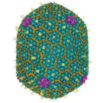 emd_32109.png
 http://ftp.pdbj.org/pub/emdb/structures/EMD-32109


 Z (Sec.)
 Y (Row.)
 X (Col.)


 Sample components
 Processing
 Electron microscopy
 FIELD EMISSION GUN
