 Movie
Movie Controller


+ Open data

- Basic information
| Entry | 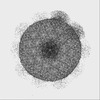 | |||||||||
|---|---|---|---|---|---|---|---|---|---|---|
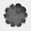
| Title | The unexpanded head structure of phage T4 | |||||||||
 Map data Map data | ||||||||||
 Sample Sample |
| |||||||||
 Keywords Keywords | virus assembly / capsid expansion / capsid length control / prolate virus structure / virus decoration proteins / capsid stabilization / VIRUS | |||||||||
| Function / homology | Capsid vertex protein / Major capsid protein, Myoviridae / Major capsid protein Gp23 / Capsid protein, T4-like bacteriophage-like / T=13 icosahedral viral capsid / viral capsid / Major capsid protein / Capsid vertex protein Function and homology information Function and homology information | |||||||||
| Biological species |  Enterobacteria phage T4 (virus) / Escherichia virus T4 Enterobacteria phage T4 (virus) / Escherichia virus T4 | |||||||||
| Method | single particle reconstruction / cryo EM / Resolution: 5.1 Å | |||||||||
 Authors Authors | Fang Q / Tang W | |||||||||
| Funding support |  United States, 2 items United States, 2 items
| |||||||||
 Citation Citation | Journal: Proc Natl Acad Sci U S A / Year: 2022 Title: Structures of a large prolate virus capsid in unexpanded and expanded states generate insights into the icosahedral virus assembly. Authors: Qianglin Fang / Wei-Chun Tang / Andrei Fokine / Marthandan Mahalingam / Qianqian Shao / Michael G Rossmann / Venigalla B Rao /  Abstract: Many icosahedral viruses assemble proteinaceous precursors called proheads or procapsids. Proheads are metastable structures that undergo a profound structural transition known as expansion that ...Many icosahedral viruses assemble proteinaceous precursors called proheads or procapsids. Proheads are metastable structures that undergo a profound structural transition known as expansion that transforms an immature unexpanded head into a mature genome-packaging head. Bacteriophage T4 is a model virus, well studied genetically and biochemically, but its structure determination has been challenging because of its large size and unusually prolate-shaped, ∼1,200-Å-long and ∼860-Å-wide capsid. Here, we report the cryogenic electron microscopy (cryo-EM) structures of T4 capsid in both of its major conformational states: unexpanded at a resolution of 5.1 Å and expanded at a resolution of 3.4 Å. These are among the largest structures deposited in Protein Data Bank to date and provide insights into virus assembly, head length determination, and shell expansion. First, the structures illustrate major domain movements and ∼70% additional gain in inner capsid volume, an essential transformation to contain the entire viral genome. Second, intricate intracapsomer interactions involving a unique insertion domain dramatically change, allowing the capsid subunits to rotate and twist while the capsomers remain fastened at quasi-threefold axes. Third, high-affinity binding sites emerge for a capsid decoration protein that clamps adjacent capsomers, imparting extraordinary structural stability. Fourth, subtle conformational changes at capsomers' periphery modulate intercapsomer angles between capsomer planes that control capsid length. Finally, conformational changes were observed at the symmetry-mismatched portal vertex, which might be involved in triggering head expansion. These analyses illustrate how small changes in local capsid subunit interactions lead to profound shifts in viral capsid morphology, stability, and volume. | |||||||||
| History |
|
- Structure visualization
| Supplemental images |
|---|

- Downloads & links
-EMDB archive
| Map data | emd_32103.map.gz | 1.6 GB | EMDB map data format | |
|---|---|---|---|---|
| Header (meta data) | emd-32103-v30.xmlemd-32103.xml | 11.3 KB 11.3 KB | Display Display | EMDB header |
| Images |  emd_32103.png emd_32103.png | 258.4 KB | ||
| Filedesc metadata | emd-32103.cif.gz | 5.5 KB | ||
| Archive directory |  http://ftp.pdbj.org/pub/emdb/structures/EMD-32103ftp://ftp.pdbj.org/pub/emdb/structures/EMD-32103 http://ftp.pdbj.org/pub/emdb/structures/EMD-32103ftp://ftp.pdbj.org/pub/emdb/structures/EMD-32103 | HTTPS FTP |
-Related structure data
| Related structure data | 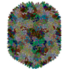 7vrtMC 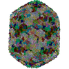 7vs5C M: atomic model generated by this map C: citing same article ( |
|---|---|
| Similar structure data |
-Links
| EMDB pages | EMDB (EBI/PDBe) / EMDataResource |
|---|---|
| Related items in Molecule of the Month |

-Map
| File | Download / File: emd_32103.map.gz / Format: CCP4 / Size: 1.7 GB / Type: IMAGE STORED AS FLOATING POINT NUMBER (4 BYTES) | ||||||||||||||||||||||||||||||||||||
|---|---|---|---|---|---|---|---|---|---|---|---|---|---|---|---|---|---|---|---|---|---|---|---|---|---|---|---|---|---|---|---|---|---|---|---|---|---|
| Projections & slices | Image control
Images are generated by Spider. | ||||||||||||||||||||||||||||||||||||
| Voxel size | X=Y=Z: 1.62 Å | ||||||||||||||||||||||||||||||||||||
| Density |
| ||||||||||||||||||||||||||||||||||||
| Symmetry | Space group: 1 | ||||||||||||||||||||||||||||||||||||
| Details | EMDB XML:
|
 Z (Sec.)
 Y (Row.)
 X (Col.)


-Supplemental data
- Sample components
-Entire : Escherichia virus T4
| Entire | Name: Escherichia virus T4 |
|---|---|
| Components |
|
-Supramolecule #1: Escherichia virus T4
| Supramolecule | Name: Escherichia virus T4 / type: virus / ID: 1 / Parent: 0 / Macromolecule list: all / NCBI-ID: 10665 / Sci species name: Escherichia virus T4 / Virus type: VIRION / Virus isolate: STRAIN / Virus enveloped: No / Virus empty: Yes |
|---|---|
| Host (natural) | Organism:  |
-Macromolecule #1: Major capsid protein
| Macromolecule | Name: Major capsid protein / type: protein_or_peptide / ID: 1 / Number of copies: 180 / Enantiomer: LEVO |
|---|---|
| Source (natural) | Organism: Enterobacteria phage T4 (virus) |
| Molecular weight | Theoretical: 56.074242 KDa |
| Sequence | String: MTIKTKAELL NKWKPLLEGE GLPEIANSKQ AIIAKIFENQ EKDFQTAPEY KDEKIAQAFG SFLTEAEIGG DHGYNATNIA AGQTSGAVT QIGPAVMGMV RRAIPNLIAF DICGVQPMNS PTGQVFALRA VYGKDPVAAG AKEAFHPMYG PDAMFSGQGA A KKFPALAA ...String: MTIKTKAELL NKWKPLLEGE GLPEIANSKQ AIIAKIFENQ EKDFQTAPEY KDEKIAQAFG SFLTEAEIGG DHGYNATNIA AGQTSGAVT QIGPAVMGMV RRAIPNLIAF DICGVQPMNS PTGQVFALRA VYGKDPVAAG AKEAFHPMYG PDAMFSGQGA A KKFPALAA STQTTVGDIY THFFQETGTV YLQASVQVTI DAGATDAAKL DAEIKKQMEA GALVEIAEGM ATSIAELQEG FN GSTDNPW NEMGFRIDKQ VIEAKSRQLK AAYSIELAQD LRAVHGMDAD AELSGILATE IMLEINREVV DWINYSAQVG KSG MTLTPG SKAGVFDFQD PIDIRGARWA GESFKALLFQ IDKEAVEIAR QTGRGEGNFI IASRNVVNVL ASVDTGISYA AQGL ATGFS TDTTKSVFAG VLGGKYRVYI DQYAKQDYFT VGYKGPNEMD AGIYYAPYVA LTPLRGSDPK NFQPVMGFKT RYGIG INPF AESAAQAPAS RIQSGMPSIL NSLGKNAYFR RVYVKGI UniProtKB: Major capsid protein |
-Macromolecule #2: Capsid vertex protein
| Macromolecule | Name: Capsid vertex protein / type: protein_or_peptide / ID: 2 / Number of copies: 11 / Enantiomer: LEVO |
|---|---|
| Source (natural) | Organism: Enterobacteria phage T4 (virus) |
| Molecular weight | Theoretical: 47.039023 KDa |
| Sequence | String: MAKINELLRE STTTNSNSIG RPNLVALTRA TTKLIYSDIV ATQRTNQPVA AFYGIKYLNP DNEFTFKTGA TYAGEAGYVD REQITELTE ESKLTLNKGD LFKYNNIVYK VLEDTPFATI EESDLELALQ IAIVLLKVRL FSDAASTSKF ESSDSEIADA R FQINKWQT ...String: MAKINELLRE STTTNSNSIG RPNLVALTRA TTKLIYSDIV ATQRTNQPVA AFYGIKYLNP DNEFTFKTGA TYAGEAGYVD REQITELTE ESKLTLNKGD LFKYNNIVYK VLEDTPFATI EESDLELALQ IAIVLLKVRL FSDAASTSKF ESSDSEIADA R FQINKWQT AVKSRKLKTG ITVELAQDLE ANGFDAPNFL EDLLATEMAD EINKDILQSL ITVSKRYKVT GITDSGFIDL SY ASAPEAG RSLYRMVCEM VSHIQKESTY TATFCVASAR AAAILAASGW LKHKPEDDKY LSQNAYGFLA NGLPLYCDTN SPL DYVIVG VVENIGEKEI VGSIFYAPYT EGLDLDDPEH VGAFKVVVDP ESLQPSIGLL VRYALSANPY TVAKDEKEAR IIDG GDMDK MAGRSDLSVL LGVKLPKIII DE UniProtKB: Capsid vertex protein |
-Experimental details
-Structure determination
| Method | cryo EM |
|---|---|
 Processing Processing | single particle reconstruction |
| Aggregation state | particle |
-Sample preparation
| Buffer | pH: 7.5 |
|---|---|
| Vitrification | Cryogen name: NITROGEN / Instrument: GATAN CRYOPLUNGE 3 |
- Electron microscopy
| Microscope | FEI TITAN KRIOS |
|---|---|
| Image recording | Film or detector model: GATAN K2 SUMMIT (4k x 4k) / Average electron dose: 25.3 e/Å2 |
| Electron beam | Acceleration voltage: 300 kV / Electron source:  FIELD EMISSION GUN FIELD EMISSION GUN |
| Electron optics | Illumination mode: FLOOD BEAM / Imaging mode: BRIGHT FIELD |
| Experimental equipment |  Model: Titan Krios / Image courtesy: FEI Company |
-Image processing
| Startup model | Type of model: OTHER |
|---|---|
| Final reconstruction | Applied symmetry - Point group: C5 (5 fold cyclic) / Resolution.type: BY AUTHOR / Resolution: 5.1 Å / Resolution method: FSC 0.143 CUT-OFF / Number images used: 28706 |
| Initial angle assignment | Type: PROJECTION MATCHING |
| Final angle assignment | Type: PROJECTION MATCHING |
