Capsid vertex protein / Small outer capsid protein / Small outer capsid protein superfamily / Small outer capsid protein / Major capsid protein, Myoviridae / Major capsid protein Gp23 / Capsid protein, T4-like bacteriophage-like Similarity search - Domain/homology
National Institutes of Health/National Institute Of Allergy and Infectious Diseases (NIH/NIAID)
AI081726
United States
National Science Foundation (NSF, United States)
MCB-0923873
United States
Citation
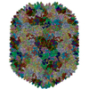
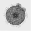
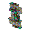
Journal: Proc Natl Acad Sci U S A / Year: 2022 Title: Structures of a large prolate virus capsid in unexpanded and expanded states generate insights into the icosahedral virus assembly. Authors: Qianglin Fang / Wei-Chun Tang / Andrei Fokine / Marthandan Mahalingam / Qianqian Shao / Michael G Rossmann / Venigalla B Rao / Abstract: Many icosahedral viruses assemble proteinaceous precursors called proheads or procapsids. Proheads are metastable structures that undergo a profound structural transition known as expansion that ...Many icosahedral viruses assemble proteinaceous precursors called proheads or procapsids. Proheads are metastable structures that undergo a profound structural transition known as expansion that transforms an immature unexpanded head into a mature genome-packaging head. Bacteriophage T4 is a model virus, well studied genetically and biochemically, but its structure determination has been challenging because of its large size and unusually prolate-shaped, ∼1,200-Å-long and ∼860-Å-wide capsid. Here, we report the cryogenic electron microscopy (cryo-EM) structures of T4 capsid in both of its major conformational states: unexpanded at a resolution of 5.1 Å and expanded at a resolution of 3.4 Å. These are among the largest structures deposited in Protein Data Bank to date and provide insights into virus assembly, head length determination, and shell expansion. First, the structures illustrate major domain movements and ∼70% additional gain in inner capsid volume, an essential transformation to contain the entire viral genome. Second, intricate intracapsomer interactions involving a unique insertion domain dramatically change, allowing the capsid subunits to rotate and twist while the capsomers remain fastened at quasi-threefold axes. Third, high-affinity binding sites emerge for a capsid decoration protein that clamps adjacent capsomers, imparting extraordinary structural stability. Fourth, subtle conformational changes at capsomers' periphery modulate intercapsomer angles between capsomer planes that control capsid length. Finally, conformational changes were observed at the symmetry-mismatched portal vertex, which might be involved in triggering head expansion. These analyses illustrate how small changes in local capsid subunit interactions lead to profound shifts in viral capsid morphology, stability, and volume.
#200 - Aug 2016 Quasisymmetry in Icosahedral Viruses similarity (1)
-
Assembly
Deposited unit
aa: Major capsid protein ab: Major capsid protein ac: Major capsid protein ad: Major capsid protein ae: Major capsid protein af: Major capsid protein ag: Major capsid protein ah: Major capsid protein ai: Major capsid protein aj: Major capsid protein ak: Major capsid protein al: Major capsid protein am: Major capsid protein an: Major capsid protein ao: Major capsid protein ap: Major capsid protein aq: Major capsid protein ar: Major capsid protein as: Major capsid protein at: Major capsid protein au: Major capsid protein av: Major capsid protein aw: Major capsid protein ax: Major capsid protein ay: Major capsid protein az: Major capsid protein ba: Major capsid protein bb: Major capsid protein bc: Major capsid protein bd: Major capsid protein be: Major capsid protein bf: Major capsid protein bg: Major capsid protein bh: Major capsid protein bi: Major capsid protein bj: Major capsid protein bk: Major capsid protein bl: Major capsid protein bm: Major capsid protein bn: Major capsid protein bo: Major capsid protein bp: Major capsid protein bq: Major capsid protein br: Major capsid protein bs: Major capsid protein bt: Major capsid protein bu: Major capsid protein bv: Major capsid protein bw: Major capsid protein bx: Major capsid protein by: Major capsid protein bz: Major capsid protein ca: Major capsid protein cb: Major capsid protein cc: Major capsid protein cd: Major capsid protein ce: Major capsid protein cf: Major capsid protein cg: Major capsid protein ch: Major capsid protein ci: Major capsid protein cj: Major capsid protein ck: Major capsid protein cl: Major capsid protein cm: Major capsid protein cn: Major capsid protein co: Major capsid protein cp: Major capsid protein cq: Major capsid protein cr: Major capsid protein cs: Major capsid protein ct: Major capsid protein cu: Major capsid protein cv: Major capsid protein cw: Major capsid protein cx: Major capsid protein cy: Major capsid protein cz: Major capsid protein da: Major capsid protein db: Major capsid protein dc: Major capsid protein dd: Major capsid protein de: Major capsid protein df: Major capsid protein dg: Major capsid protein dh: Major capsid protein di: Major capsid protein dj: Major capsid protein dk: Major capsid protein dl: Major capsid protein dm: Major capsid protein dn: Major capsid protein do: Major capsid protein dp: Major capsid protein dq: Major capsid protein dr: Major capsid protein ds: Major capsid protein dt: Major capsid protein du: Major capsid protein dv: Major capsid protein dw: Major capsid protein dx: Major capsid protein dy: Major capsid protein dz: Major capsid protein ea: Major capsid protein eb: Major capsid protein ec: Major capsid protein ed: Major capsid protein ee: Major capsid protein ef: Major capsid protein eg: Major capsid protein eh: Major capsid protein ei: Major capsid protein ej: Major capsid protein ek: Major capsid protein el: Major capsid protein em: Major capsid protein en: Major capsid protein eo: Major capsid protein ep: Major capsid protein eq: Major capsid protein er: Major capsid protein es: Major capsid protein et: Major capsid protein eu: Major capsid protein ev: Major capsid protein ew: Major capsid protein ex: Major capsid protein ey: Major capsid protein ez: Major capsid protein fa: Major capsid protein fb: Major capsid protein fc: Major capsid protein fd: Major capsid protein fe: Major capsid protein ff: Major capsid protein fg: Major capsid protein fh: Major capsid protein fi: Major capsid protein fj: Major capsid protein fk: Major capsid protein fl: Major capsid protein fm: Major capsid protein fn: Major capsid protein fo: Major capsid protein fp: Major capsid protein fq: Major capsid protein fr: Major capsid protein fs: Major capsid protein ft: Major capsid protein fu: Major capsid protein fv: Major capsid protein fw: Major capsid protein fx: Major capsid protein fy: Major capsid protein fz: Major capsid protein ga: Major capsid protein gb: Major capsid protein gc: Major capsid protein gd: Major capsid protein ge: Major capsid protein gf: Major capsid protein gg: Major capsid protein gh: Major capsid protein gi: Major capsid protein gj: Major capsid protein gk: Major capsid protein gl: Major capsid protein gm: Major capsid protein gn: Major capsid protein go: Major capsid protein gp: Major capsid protein gq: Major capsid protein gr: Major capsid protein gs: Major capsid protein gt: Major capsid protein gu: Major capsid protein gv: Major capsid protein gw: Major capsid protein gx: Major capsid protein gy: Major capsid protein gz: Major capsid protein ha: Major capsid protein hb: Major capsid protein hc: Major capsid protein hd: Major capsid protein he: Capsid vertex protein hf: Capsid vertex protein hg: Capsid vertex protein hh: Capsid vertex protein hi: Capsid vertex protein hj: Capsid vertex protein hk: Capsid vertex protein hl: Capsid vertex protein hm: Capsid vertex protein hn: Capsid vertex protein ho: Capsid vertex protein hp: Small outer capsid protein hq: Small outer capsid protein hr: Small outer capsid protein hs: Small outer capsid protein ht: Small outer capsid protein hu: Small outer capsid protein hv: Small outer capsid protein hw: Small outer capsid protein hx: Small outer capsid protein hy: Small outer capsid protein hz: Small outer capsid protein ia: Small outer capsid protein ib: Small outer capsid protein ic: Small outer capsid protein id: Small outer capsid protein ie: Small outer capsid protein if: Small outer capsid protein ig: Small outer capsid protein ih: Small outer capsid protein ii: Small outer capsid protein ij: Small outer capsid protein ik: Small outer capsid protein il: Small outer capsid protein im: Small outer capsid protein in: Small outer capsid protein io: Small outer capsid protein ip: Small outer capsid protein iq: Small outer capsid protein ir: Small outer capsid protein is: Small outer capsid protein it: Small outer capsid protein iu: Small outer capsid protein iv: Small outer capsid protein iw: Small outer capsid protein ix: Small outer capsid protein iy: Small outer capsid protein iz: Small outer capsid protein ja: Small outer capsid protein jb: Small outer capsid protein jc: Small outer capsid protein jd: Small outer capsid protein je: Small outer capsid protein jf: Small outer capsid protein jg: Small outer capsid protein jh: Small outer capsid protein ji: Small outer capsid protein jj: Small outer capsid protein jk: Small outer capsid protein jl: Small outer capsid protein jm: Small outer capsid protein jn: Small outer capsid protein jo: Small outer capsid protein jp: Small outer capsid protein jq: Small outer capsid protein jr: Small outer capsid protein js: Small outer capsid protein jt: Small outer capsid protein ju: Small outer capsid protein jv: Small outer capsid protein jw: Small outer capsid protein jx: Small outer capsid protein jy: Small outer capsid protein jz: Small outer capsid protein ka: Small outer capsid protein kb: Small outer capsid protein kc: Small outer capsid protein kd: Small outer capsid protein ke: Small outer capsid protein kf: Small outer capsid protein kg: Small outer capsid protein kh: Small outer capsid protein ki: Small outer capsid protein kj: Small outer capsid protein kk: Small outer capsid protein kl: Small outer capsid protein km: Small outer capsid protein kn: Small outer capsid protein ko: Small outer capsid protein kp: Small outer capsid protein kq: Small outer capsid protein kr: Small outer capsid protein ks: Small outer capsid protein kt: Small outer capsid protein ku: Small outer capsid protein kv: Small outer capsid protein kw: Small outer capsid protein kx: Small outer capsid protein ky: Small outer capsid protein kz: Small outer capsid protein la: Small outer capsid protein lb: Small outer capsid protein lc: Small outer capsid protein ld: Small outer capsid protein le: Small outer capsid protein lf: Small outer capsid protein lg: Small outer capsid protein lh: Small outer capsid protein li: Small outer capsid protein lj: Small outer capsid protein lk: Small outer capsid protein ll: Small outer capsid protein lm: Small outer capsid protein ln: Small outer capsid protein lo: Small outer capsid protein lp: Small outer capsid protein lq: Small outer capsid protein lr: Small outer capsid protein ls: Small outer capsid protein lt: Small outer capsid protein lu: Small outer capsid protein lv: Small outer capsid protein lw: Small outer capsid protein lx: Small outer capsid protein ly: Small outer capsid protein lz: Small outer capsid protein ma: Small outer capsid protein mb: Small outer capsid protein mc: Small outer capsid protein md: Small outer capsid protein me: Small outer capsid protein mf: Small outer capsid protein mg: Small outer capsid protein mh: Small outer capsid protein mi: Small outer capsid protein mj: Small outer capsid protein mk: Small outer capsid protein ml: Small outer capsid protein mm: Small outer capsid protein mn: Small outer capsid protein mo: Small outer capsid protein mp: Small outer capsid protein mq: Small outer capsid protein ms: Small outer capsid protein mt: Small outer capsid protein mu: Small outer capsid protein mv: Small outer capsid protein mw: Small outer capsid protein mx: Small outer capsid protein my: Small outer capsid protein mz: Small outer capsid protein na: Small outer capsid protein nb: Small outer capsid protein nc: Small outer capsid protein nd: Small outer capsid protein ne: Small outer capsid protein nf: Small outer capsid protein ng: Small outer capsid protein nh: Small outer capsid protein ni: Small outer capsid protein nj: Small outer capsid protein nk: Small outer capsid protein nl: Small outer capsid protein nm: Small outer capsid protein nn: Small outer capsid protein no: Small outer capsid protein np: Small outer capsid protein nq: Small outer capsid protein ns: Small outer capsid protein nt: Small outer capsid protein nu: Small outer capsid protein nv: Small outer capsid protein nw: Small outer capsid protein nx: Small outer capsid protein ny: Small outer capsid protein nz: Small outer capsid protein oa: Small outer capsid protein ob: Small outer capsid protein oc: Small outer capsid protein od: Small outer capsid protein oe: Small outer capsid protein of: Small outer capsid protein og: Small outer capsid protein
aa: Major capsid protein ab: Major capsid protein ac: Major capsid protein ad: Major capsid protein ae: Major capsid protein af: Major capsid protein ag: Major capsid protein ah: Major capsid protein ai: Major capsid protein aj: Major capsid protein ak: Major capsid protein al: Major capsid protein am: Major capsid protein an: Major capsid protein ao: Major capsid protein ap: Major capsid protein aq: Major capsid protein ar: Major capsid protein as: Major capsid protein at: Major capsid protein au: Major capsid protein av: Major capsid protein aw: Major capsid protein ax: Major capsid protein ay: Major capsid protein az: Major capsid protein ba: Major capsid protein bb: Major capsid protein bc: Major capsid protein bd: Major capsid protein be: Major capsid protein bf: Major capsid protein bg: Major capsid protein bh: Major capsid protein bi: Major capsid protein bj: Major capsid protein bk: Major capsid protein bl: Major capsid protein bm: Major capsid protein bn: Major capsid protein bo: Major capsid protein bp: Major capsid protein bq: Major capsid protein br: Major capsid protein bs: Major capsid protein bt: Major capsid protein bu: Major capsid protein bv: Major capsid protein bw: Major capsid protein bx: Major capsid protein by: Major capsid protein bz: Major capsid protein ca: Major capsid protein cb: Major capsid protein cc: Major capsid protein cd: Major capsid protein ce: Major capsid protein cf: Major capsid protein cg: Major capsid protein ch: Major capsid protein ci: Major capsid protein cj: Major capsid protein ck: Major capsid protein cl: Major capsid protein cm: Major capsid protein cn: Major capsid protein co: Major capsid protein cp: Major capsid protein cq: Major capsid protein cr: Major capsid protein cs: Major capsid protein ct: Major capsid protein cu: Major capsid protein cv: Major capsid protein cw: Major capsid protein cx: Major capsid protein cy: Major capsid protein cz: Major capsid protein da: Major capsid protein db: Major capsid protein dc: Major capsid protein dd: Major capsid protein de: Major capsid protein df: Major capsid protein dg: Major capsid protein dh: Major capsid protein di: Major capsid protein dj: Major capsid protein dk: Major capsid protein dl: Major capsid protein dm: Major capsid protein dn: Major capsid protein do: Major capsid protein dp: Major capsid protein dq: Major capsid protein dr: Major capsid protein ds: Major capsid protein dt: Major capsid protein du: Major capsid protein dv: Major capsid protein dw: Major capsid protein dx: Major capsid protein dy: Major capsid protein dz: Major capsid protein ea: Major capsid protein eb: Major capsid protein ec: Major capsid protein ed: Major capsid protein ee: Major capsid protein ef: Major capsid protein eg: Major capsid protein eh: Major capsid protein ei: Major capsid protein ej: Major capsid protein ek: Major capsid protein el: Major capsid protein em: Major capsid protein en: Major capsid protein eo: Major capsid protein ep: Major capsid protein eq: Major capsid protein er: Major capsid protein es: Major capsid protein et: Major capsid protein eu: Major capsid protein ev: Major capsid protein ew: Major capsid protein ex: Major capsid protein ey: Major capsid protein ez: Major capsid protein fa: Major capsid protein fb: Major capsid protein fc: Major capsid protein fd: Major capsid protein fe: Major capsid protein ff: Major capsid protein fg: Major capsid protein fh: Major capsid protein fi: Major capsid protein fj: Major capsid protein fk: Major capsid protein fl: Major capsid protein fm: Major capsid protein fn: Major capsid protein fo: Major capsid protein fp: Major capsid protein fq: Major capsid protein fr: Major capsid protein fs: Major capsid protein ft: Major capsid protein fu: Major capsid protein fv: Major capsid protein fw: Major capsid protein fx: Major capsid protein fy: Major capsid protein fz: Major capsid protein ga: Major capsid protein gb: Major capsid protein gc: Major capsid protein gd: Major capsid protein ge: Major capsid protein gf: Major capsid protein gg: Major capsid protein gh: Major capsid protein gi: Major capsid protein gj: Major capsid protein gk: Major capsid protein gl: Major capsid protein gm: Major capsid protein gn: Major capsid protein go: Major capsid protein gp: Major capsid protein gq: Major capsid protein gr: Major capsid protein gs: Major capsid protein gt: Major capsid protein gu: Major capsid protein gv: Major capsid protein gw: Major capsid protein gx: Major capsid protein gy: Major capsid protein gz: Major capsid protein ha: Major capsid protein hb: Major capsid protein hc: Major capsid protein hd: Major capsid protein he: Capsid vertex protein hf: Capsid vertex protein hg: Capsid vertex protein hh: Capsid vertex protein hi: Capsid vertex protein hj: Capsid vertex protein hk: Capsid vertex protein hl: Capsid vertex protein hm: Capsid vertex protein hn: Capsid vertex protein ho: Capsid vertex protein hp: Small outer capsid protein hq: Small outer capsid protein hr: Small outer capsid protein hs: Small outer capsid protein ht: Small outer capsid protein hu: Small outer capsid protein hv: Small outer capsid protein hw: Small outer capsid protein hx: Small outer capsid protein hy: Small outer capsid protein hz: Small outer capsid protein ia: Small outer capsid protein ib: Small outer capsid protein ic: Small outer capsid protein id: Small outer capsid protein ie: Small outer capsid protein if: Small outer capsid protein ig: Small outer capsid protein ih: Small outer capsid protein ii: Small outer capsid protein ij: Small outer capsid protein ik: Small outer capsid protein il: Small outer capsid protein im: Small outer capsid protein in: Small outer capsid protein io: Small outer capsid protein ip: Small outer capsid protein iq: Small outer capsid protein ir: Small outer capsid protein is: Small outer capsid protein it: Small outer capsid protein iu: Small outer capsid protein iv: Small outer capsid protein iw: Small outer capsid protein ix: Small outer capsid protein iy: Small outer capsid protein iz: Small outer capsid protein ja: Small outer capsid protein jb: Small outer capsid protein jc: Small outer capsid protein jd: Small outer capsid protein je: Small outer capsid protein jf: Small outer capsid protein jg: Small outer capsid protein jh: Small outer capsid protein ji: Small outer capsid protein jj: Small outer capsid protein jk: Small outer capsid protein jl: Small outer capsid protein jm: Small outer capsid protein jn: Small outer capsid protein jo: Small outer capsid protein jp: Small outer capsid protein jq: Small outer capsid protein jr: Small outer capsid protein js: Small outer capsid protein jt: Small outer capsid protein ju: Small outer capsid protein jv: Small outer capsid protein jw: Small outer capsid protein jx: Small outer capsid protein jy: Small outer capsid protein jz: Small outer capsid protein ka: Small outer capsid protein kb: Small outer capsid protein kc: Small outer capsid protein kd: Small outer capsid protein ke: Small outer capsid protein kf: Small outer capsid protein kg: Small outer capsid protein kh: Small outer capsid protein ki: Small outer capsid protein kj: Small outer capsid protein kk: Small outer capsid protein kl: Small outer capsid protein km: Small outer capsid protein kn: Small outer capsid protein ko: Small outer capsid protein kp: Small outer capsid protein kq: Small outer capsid protein kr: Small outer capsid protein ks: Small outer capsid protein kt: Small outer capsid protein ku: Small outer capsid protein kv: Small outer capsid protein kw: Small outer capsid protein kx: Small outer capsid protein ky: Small outer capsid protein kz: Small outer capsid protein la: Small outer capsid protein lb: Small outer capsid protein lc: Small outer capsid protein ld: Small outer capsid protein le: Small outer capsid protein lf: Small outer capsid protein lg: Small outer capsid protein lh: Small outer capsid protein li: Small outer capsid protein lj: Small outer capsid protein lk: Small outer capsid protein ll: Small outer capsid protein lm: Small outer capsid protein ln: Small outer capsid protein lo: Small outer capsid protein lp: Small outer capsid protein lq: Small outer capsid protein lr: Small outer capsid protein ls: Small outer capsid protein lt: Small outer capsid protein lu: Small outer capsid protein lv: Small outer capsid protein lw: Small outer capsid protein lx: Small outer capsid protein ly: Small outer capsid protein lz: Small outer capsid protein ma: Small outer capsid protein mb: Small outer capsid protein mc: Small outer capsid protein md: Small outer capsid protein me: Small outer capsid protein mf: Small outer capsid protein mg: Small outer capsid protein mh: Small outer capsid protein mi: Small outer capsid protein mj: Small outer capsid protein mk: Small outer capsid protein ml: Small outer capsid protein mm: Small outer capsid protein mn: Small outer capsid protein mo: Small outer capsid protein mp: Small outer capsid protein mq: Small outer capsid protein ms: Small outer capsid protein mt: Small outer capsid protein mu: Small outer capsid protein mv: Small outer capsid protein mw: Small outer capsid protein mx: Small outer capsid protein my: Small outer capsid protein mz: Small outer capsid protein na: Small outer capsid protein nb: Small outer capsid protein nc: Small outer capsid protein nd: Small outer capsid protein ne: Small outer capsid protein nf: Small outer capsid protein ng: Small outer capsid protein nh: Small outer capsid protein ni: Small outer capsid protein nj: Small outer capsid protein nk: Small outer capsid protein nl: Small outer capsid protein nm: Small outer capsid protein nn: Small outer capsid protein no: Small outer capsid protein np: Small outer capsid protein nq: Small outer capsid protein ns: Small outer capsid protein nt: Small outer capsid protein nu: Small outer capsid protein nv: Small outer capsid protein nw: Small outer capsid protein nx: Small outer capsid protein ny: Small outer capsid protein nz: Small outer capsid protein oa: Small outer capsid protein ob: Small outer capsid protein oc: Small outer capsid protein od: Small outer capsid protein oe: Small outer capsid protein of: Small outer capsid protein og: Small outer capsid protein
In the structure databanks used in Yorodumi, some data are registered as the other names, "COVID-19 virus" and "2019-nCoV". Here are the details of the virus and the list of structure data.
Jan 31, 2019. EMDB accession codes are about to change! (news from PDBe EMDB page)
EMDB accession codes are about to change! (news from PDBe EMDB page)
The allocation of 4 digits for EMDB accession codes will soon come to an end. Whilst these codes will remain in use, new EMDB accession codes will include an additional digit and will expand incrementally as the available range of codes is exhausted. The current 4-digit format prefixed with “EMD-” (i.e. EMD-XXXX) will advance to a 5-digit format (i.e. EMD-XXXXX), and so on. It is currently estimated that the 4-digit codes will be depleted around Spring 2019, at which point the 5-digit format will come into force.
The EM Navigator/Yorodumi systems omit the EMD- prefix.
Related info.:Q: What is EMD? / ID/Accession-code notation in Yorodumi/EM Navigator
Yorodumi is a browser for structure data from EMDB, PDB, SASBDB, etc.
This page is also the successor to EM Navigator detail page, and also detail information page/front-end page for Omokage search.
The word "yorodu" (or yorozu) is an old Japanese word meaning "ten thousand". "mi" (miru) is to see.
Related info.:EMDB / PDB / SASBDB / Comparison of 3 databanks / Yorodumi Search / Aug 31, 2016. New EM Navigator & Yorodumi / Yorodumi Papers / Jmol/JSmol / Function and homology information / Changes in new EM Navigator and Yorodumi
 Movie
Movie Controller


 Open data
 Basic information
 Components
 Keywords
 Function and homology information
 Enterobacteria phage T4 (virus)
 Authors
 United States, 2items
  Citation
 Structure visualization
 Downloads & links
 Other downloads
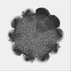
 PDBj
PDBj
 Assembly

 Sample preparation
 Electron microscopy imaging
 FIELD EMISSION GUN / Accelerating voltage: 300 kV / Illumination mode: FLOOD BEAM
 Processing