 Movie
Movie Controller


+ Open data

- Basic information
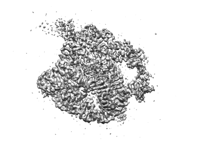
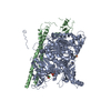
| Entry | Database: EMDB / ID: EMD-31386 | |||||||||||||||||||||
|---|---|---|---|---|---|---|---|---|---|---|---|---|---|---|---|---|---|---|---|---|---|---|
| Title | Holo L-16 ScaI Tetrahymena ribozyme | |||||||||||||||||||||
 Map data Map data | ||||||||||||||||||||||
 Sample Sample |
| |||||||||||||||||||||
 Keywords Keywords | RNA structure / Tetrahymena ribozyme / RNA | |||||||||||||||||||||
| Biological species |   Tetrahymena thermophila (eukaryote) Tetrahymena thermophila (eukaryote) | |||||||||||||||||||||
| Method | single particle reconstruction / cryo EM / Resolution: 3.05 Å | |||||||||||||||||||||
 Authors Authors | Su Z / Zhang K | |||||||||||||||||||||
| Funding support |  China, China,  United States, 6 items United States, 6 items
| |||||||||||||||||||||
 Citation Citation | Journal: Nature / Year: 2021 Title: Cryo-EM structures of full-length Tetrahymena ribozyme at 3.1 Å resolution. Authors: Zhaoming Su / Kaiming Zhang / Kalli Kappel / Shanshan Li / Michael Z Palo / Grigore D Pintilie / Ramya Rangan / Bingnan Luo / Yuquan Wei / Rhiju Das / Wah Chiu / Abstract: Single-particle cryogenic electron microscopy (cryo-EM) has become a standard technique for determining protein structures at atomic resolution. However, cryo-EM studies of protein-free RNA are in ...Single-particle cryogenic electron microscopy (cryo-EM) has become a standard technique for determining protein structures at atomic resolution. However, cryo-EM studies of protein-free RNA are in their early days. The Tetrahymena thermophila group I self-splicing intron was the first ribozyme to be discovered and has been a prominent model system for the study of RNA catalysis and structure-function relationships, but its full structure remains unknown. Here we report cryo-EM structures of the full-length Tetrahymena ribozyme in substrate-free and bound states at a resolution of 3.1 Å. Newly resolved peripheral regions form two coaxially stacked helices; these are interconnected by two kissing loop pseudoknots that wrap around the catalytic core and include two previously unforeseen (to our knowledge) tertiary interactions. The global architecture is nearly identical in both states; only the internal guide sequence and guanosine binding site undergo a large conformational change and a localized shift, respectively, upon binding of RNA substrates. These results provide a long-sought structural view of a paradigmatic RNA enzyme and signal a new era for the cryo-EM-based study of structure-function relationships in ribozymes. | |||||||||||||||||||||
| History |
|
- Structure visualization
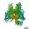
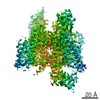
| Movie |
 Movie viewer Movie viewer |
|---|---|
| Structure viewer | EM map: SurfViewMolmilJmol/JSmol |
| Supplemental images |
- Downloads & links
-EMDB archive
| Map data | emd_31386.map.gz | 59.8 MB | EMDB map data format | |
|---|---|---|---|---|
| Header (meta data) | emd-31386-v30.xmlemd-31386.xml | 15.6 KB 15.6 KB | Display Display | EMDB header |
| Images |  emd_31386.png emd_31386.png | 73.6 KB | ||
| Filedesc metadata | emd-31386.cif.gz | 5.4 KB | ||
| Archive directory |  http://ftp.pdbj.org/pub/emdb/structures/EMD-31386ftp://ftp.pdbj.org/pub/emdb/structures/EMD-31386 http://ftp.pdbj.org/pub/emdb/structures/EMD-31386ftp://ftp.pdbj.org/pub/emdb/structures/EMD-31386 | HTTPS FTP |
-Validation report
| Summary document | emd_31386_validation.pdf.gz | 565.6 KB | Display | EMDB validaton report |
|---|---|---|---|---|
| Full document | emd_31386_full_validation.pdf.gz | 565.2 KB | Display | |
| Data in XML | emd_31386_validation.xml.gz | 6 KB | Display | |
| Data in CIF | emd_31386_validation.cif.gz | 6.8 KB | Display | |
| Arichive directory | https://ftp.pdbj.org/pub/emdb/validation_reports/EMD-31386ftp://ftp.pdbj.org/pub/emdb/validation_reports/EMD-31386 | HTTPS FTP |
-Related structure data
| Related structure data |  7ez2MC 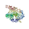 7ez0C C: citing same article ( M: atomic model generated by this map |
|---|---|
| Similar structure data |


-Links
| EMDB pages | EMDB (EBI/PDBe) / EMDataResource |
|---|
-Map
| File | Download / File: emd_31386.map.gz / Format: CCP4 / Size: 64 MB / Type: IMAGE STORED AS FLOATING POINT NUMBER (4 BYTES) | ||||||||||||||||||||||||||||||||||||||||||||||||||||||||||||||||||||
|---|---|---|---|---|---|---|---|---|---|---|---|---|---|---|---|---|---|---|---|---|---|---|---|---|---|---|---|---|---|---|---|---|---|---|---|---|---|---|---|---|---|---|---|---|---|---|---|---|---|---|---|---|---|---|---|---|---|---|---|---|---|---|---|---|---|---|---|---|---|
| Projections & slices | Image control
Images are generated by Spider. | ||||||||||||||||||||||||||||||||||||||||||||||||||||||||||||||||||||
| Voxel size | X=Y=Z: 0.86 Å | ||||||||||||||||||||||||||||||||||||||||||||||||||||||||||||||||||||
| Density |
| ||||||||||||||||||||||||||||||||||||||||||||||||||||||||||||||||||||
| Symmetry | Space group: 1 | ||||||||||||||||||||||||||||||||||||||||||||||||||||||||||||||||||||
| Details | EMDB XML:
CCP4 map header:
| ||||||||||||||||||||||||||||||||||||||||||||||||||||||||||||||||||||
 Z (Sec.)
 Y (Row.)
 X (Col.)


-Supplemental data
- Sample components
-Entire : Holo L-16 ScaI Tetrahymena ribozyme with substrates S1 and S2, an...
| Entire | Name: Holo L-16 ScaI Tetrahymena ribozyme with substrates S1 and S2, and metal ions. |
|---|---|
| Components |
|
-Supramolecule #1: Holo L-16 ScaI Tetrahymena ribozyme with substrates S1 and S2, an...
| Supramolecule | Name: Holo L-16 ScaI Tetrahymena ribozyme with substrates S1 and S2, and metal ions. type: complex / ID: 1 / Parent: 0 / Macromolecule list: #1-#3 |
|---|---|
| Source (natural) | Organism: Tetrahymena thermophila (eukaryote) |
-Macromolecule #1: Holo L-16 ScaI Tetrahymena ribozyme S1
| Macromolecule | Name: Holo L-16 ScaI Tetrahymena ribozyme S1 / type: rna / ID: 1 / Number of copies: 1 |
|---|---|
| Source (natural) | Organism: Tetrahymena thermophila (eukaryote) |
| Molecular weight | Theoretical: 2.502603 KDa |
| Sequence | String: UCG(SSU)AACC |
-Macromolecule #2: Holo L-16 ScaI Tetrahymena ribozyme S2
| Macromolecule | Name: Holo L-16 ScaI Tetrahymena ribozyme S2 / type: rna / ID: 2 / Number of copies: 1 |
|---|---|
| Source (natural) | Organism: Tetrahymena thermophila (eukaryote) |
| Molecular weight | Theoretical: 1.788101 KDa |
| Sequence | String: CCCUCU |
-Macromolecule #3: Holo L-16 ScaI Tetrahymena ribozyme
| Macromolecule | Name: Holo L-16 ScaI Tetrahymena ribozyme / type: rna / ID: 3 / Number of copies: 1 |
|---|---|
| Source (natural) | Organism: Tetrahymena thermophila (eukaryote) |
| Molecular weight | Theoretical: 126.705711 KDa |
| Sequence | String: GGUUUGGAGG GAAAAGUUAU CAGGCAUGCA CCUGGUAGCU AGUCUUUAAA CCAAUAGAUU GCAUCGGUUU AAAAGGCAAG ACCGUCAAA UUGCGGGAAA GGGGUCAACA GCCGUUCAGU ACCAAGUCUC AGGGGAAACU UUGAGAUGGC CUUGCAAAGG G UAUGGUAA ...String: GGUUUGGAGG GAAAAGUUAU CAGGCAUGCA CCUGGUAGCU AGUCUUUAAA CCAAUAGAUU GCAUCGGUUU AAAAGGCAAG ACCGUCAAA UUGCGGGAAA GGGGUCAACA GCCGUUCAGU ACCAAGUCUC AGGGGAAACU UUGAGAUGGC CUUGCAAAGG G UAUGGUAA UAAGCUGACG GACAUGGUCC UAACCACGCA GCCAAGUCCU AAGUCAACAG AUCUUCUGUU GAUAUGGAUG CA GUUCACA GACUAAAUGU CGGUCGGGGA AGAUGUAUUC UUCUCAUAAG AUAUAGUCGG ACCUCUCCUU AAUGGGAGCU AGC GGAUGA AGUGAUGCAA CACUGGAGCC GCUGGGAACU AAUUUGUAUG CGAAAGUAUA UUGAUUAGUU UUGGAG |
-Macromolecule #4: MAGNESIUM ION
| Macromolecule | Name: MAGNESIUM ION / type: ligand / ID: 4 / Number of copies: 31 / Formula: MG |
|---|---|
| Molecular weight | Theoretical: 24.305 Da |
-Experimental details
-Structure determination
| Method | cryo EM |
|---|---|
 Processing Processing | single particle reconstruction |
| Aggregation state | particle |
-Sample preparation
| Concentration | 3 mg/mL |
|---|---|
| Buffer | pH: 8 |
| Grid | Model: Quantifoil R2/1 / Material: COPPER / Mesh: 200 / Pretreatment - Type: GLOW DISCHARGE / Pretreatment - Time: 40 sec. / Pretreatment - Atmosphere: AIR / Pretreatment - Pressure: 0.36 kPa |
| Vitrification | Cryogen name: ETHANE / Chamber humidity: 100 % / Chamber temperature: 278 K / Instrument: FEI VITROBOT MARK IV |
- Electron microscopy
| Microscope | FEI TITAN KRIOS |
|---|---|
| Temperature | Min: 78.2 K |
| Specialist optics | Energy filter - Name: GIF Bioquantum / Energy filter - Slit width: 20 eV |
| Image recording | Film or detector model: GATAN K3 (6k x 4k) / Number real images: 5559 / Average electron dose: 50.0 e/Å2 |
| Electron beam | Acceleration voltage: 300 kV / Electron source:  FIELD EMISSION GUN FIELD EMISSION GUN |
| Electron optics | C2 aperture diameter: 70.0 µm / Illumination mode: FLOOD BEAM / Imaging mode: BRIGHT FIELD / Cs: 2.7 mm / Nominal defocus max: 2.0 µm / Nominal defocus min: 0.8 µm / Nominal magnification: 105000 |
| Sample stage | Specimen holder model: FEI TITAN KRIOS AUTOGRID HOLDER / Cooling holder cryogen: NITROGEN |
| Experimental equipment |  Model: Titan Krios / Image courtesy: FEI Company |
+Image processing
-Atomic model buiding 1
| Refinement | Space: REAL / Protocol: AB INITIO MODEL |
|---|---|
| Output model | PDB-7ez2: |
