 Movie
Movie Controller

[English] 日本語
 Yorodumi
- EMDB-2831: Electron cryotomography, subtomogram averaging and classification... -
+ Open data

- Basic information
| Entry | Database: EMDB / ID: EMD-2831 | |||||||||
|---|---|---|---|---|---|---|---|---|---|---|
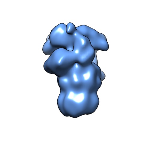
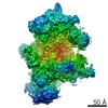
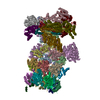
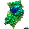
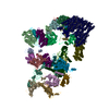
| Title | Electron cryotomography, subtomogram averaging and classification of 26S proteasomes in situ in intact hippocampal neurons | |||||||||
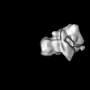
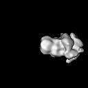
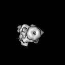
 Map data Map data | 26S Proteasome from mammal hippocampus neurons in substrate-processing state (SPS) | |||||||||
 Sample Sample |
| |||||||||
 Keywords Keywords | Proteasome / 26S / 26S Proteasome / Ubiquitin / Ubiquitin-Proteasome Pathway / UPP / Ubiquitin-Proteasome System / UPS / Protease | |||||||||
| Biological species |  | |||||||||
| Method | subtomogram averaging / cryo EM / Resolution: 31.0 Å | |||||||||
 Authors Authors | Asano S / Fukuda Y / Beck F / Aufderheide A / Foerster F / Danev R / Baumeister W | |||||||||
 Citation Citation | Journal: Science / Year: 2015 Title: Proteasomes. A molecular census of 26S proteasomes in intact neurons. Authors: Shoh Asano / Yoshiyuki Fukuda / Florian Beck / Antje Aufderheide / Friedrich Förster / Radostin Danev / Wolfgang Baumeister /  Abstract: The 26S proteasome is a key player in eukaryotic protein quality control and in the regulation of numerous cellular processes. Here, we describe quantitative in situ structural studies of this highly ...The 26S proteasome is a key player in eukaryotic protein quality control and in the regulation of numerous cellular processes. Here, we describe quantitative in situ structural studies of this highly dynamic molecular machine in intact hippocampal neurons. We used electron cryotomography with the Volta phase plate, which allowed high fidelity and nanometer precision localization of 26S proteasomes. We undertook a molecular census of single- and double-capped proteasomes and assessed the conformational states of individual complexes. Under the conditions of the experiment—that is, in the absence of proteotoxic stress—only 20% of the 26S proteasomes were engaged in substrate processing. The remainder was in the substrate-accepting ground state. These findings suggest that in the absence of stress, the capacity of the proteasome system is not fully used. | |||||||||
| History |
|
- Structure visualization
| Movie |
 Movie viewer Movie viewer |
|---|---|
| Structure viewer | EM map: SurfViewMolmilJmol/JSmol |
| Supplemental images |
- Downloads & links
-EMDB archive
| Map data | emd_2831.map.gz | 7.5 MB | EMDB map data format | |
|---|---|---|---|---|
| Header (meta data) | emd-2831-v30.xmlemd-2831.xml | 8.3 KB 8.3 KB | Display Display | EMDB header |
| Images | emd_2831.tif | 1.2 MB | ||
| Archive directory |  http://ftp.pdbj.org/pub/emdb/structures/EMD-2831ftp://ftp.pdbj.org/pub/emdb/structures/EMD-2831 http://ftp.pdbj.org/pub/emdb/structures/EMD-2831ftp://ftp.pdbj.org/pub/emdb/structures/EMD-2831 | HTTPS FTP |
-Validation report
| Summary document | emd_2831_validation.pdf.gz | 210 KB | Display | EMDB validaton report |
|---|---|---|---|---|
| Full document | emd_2831_full_validation.pdf.gz | 209.1 KB | Display | |
| Data in XML | emd_2831_validation.xml.gz | 5.4 KB | Display | |
| Arichive directory | https://ftp.pdbj.org/pub/emdb/validation_reports/EMD-2831ftp://ftp.pdbj.org/pub/emdb/validation_reports/EMD-2831 | HTTPS FTP |
-Related structure data

-Links
| EMDB pages | EMDB (EBI/PDBe) / EMDataResource |
|---|
-Map
| File | Download / File: emd_2831.map.gz / Format: CCP4 / Size: 7.8 MB / Type: IMAGE STORED AS FLOATING POINT NUMBER (4 BYTES) | ||||||||||||||||||||||||||||||||||||||||||||||||||||||||||||
|---|---|---|---|---|---|---|---|---|---|---|---|---|---|---|---|---|---|---|---|---|---|---|---|---|---|---|---|---|---|---|---|---|---|---|---|---|---|---|---|---|---|---|---|---|---|---|---|---|---|---|---|---|---|---|---|---|---|---|---|---|---|
| Annotation | 26S Proteasome from mammal hippocampus neurons in substrate-processing state (SPS) | ||||||||||||||||||||||||||||||||||||||||||||||||||||||||||||
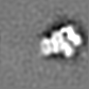
| Projections & slices | Image control
Images are generated by Spider. | ||||||||||||||||||||||||||||||||||||||||||||||||||||||||||||
| Voxel size | X=Y=Z: 4.21 Å | ||||||||||||||||||||||||||||||||||||||||||||||||||||||||||||
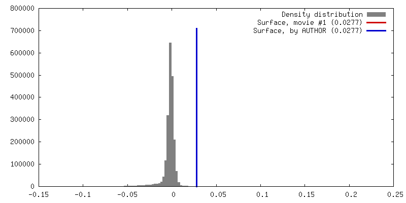
| Density |
| ||||||||||||||||||||||||||||||||||||||||||||||||||||||||||||
| Symmetry | Space group: 1 | ||||||||||||||||||||||||||||||||||||||||||||||||||||||||||||
| Details | EMDB XML:
CCP4 map header:
| ||||||||||||||||||||||||||||||||||||||||||||||||||||||||||||
 Z (Sec.)
 Y (Row.)
 X (Col.)


-Supplemental data
- Sample components
-Entire : 26S Proteasome from mammal hippocampus neurons in substrate-proce...
| Entire | Name: 26S Proteasome from mammal hippocampus neurons in substrate-processing state (SPS) |
|---|---|
| Components |
|
-Supramolecule #1000: 26S Proteasome from mammal hippocampus neurons in substrate-proce...
| Supramolecule | Name: 26S Proteasome from mammal hippocampus neurons in substrate-processing state (SPS) type: sample / ID: 1000 / Number unique components: 1 |
|---|
-Macromolecule #1: 26S Proteasome
| Macromolecule | Name: 26S Proteasome / type: protein_or_peptide / ID: 1 / Recombinant expression: No |
|---|---|
| Source (natural) | Organism: |
-Experimental details
-Structure determination
| Method | cryo EM |
|---|---|
 Processing Processing | subtomogram averaging |
| Aggregation state | particle |
-Sample preparation
| Grid | Details: Quantifoil Au 200 mesh grids R1/4 |
|---|---|
| Vitrification | Cryogen name: ETHANE-PROPANE MIXTURE / Chamber humidity: 90 % / Instrument: FEI VITROBOT MARK III Method: 37 deg C, waiting time 5 sec, and 10 seconds blot time |
- Electron microscopy
| Microscope | FEI TITAN KRIOS |
|---|---|
| Specialist optics | Energy filter - Name: Gatan |
| Details | Volta Phase Plate |
| Date | Oct 24, 2013 |
| Image recording | Category: CCD / Film or detector model: GATAN K2 (4k x 4k) / Average electron dose: 110 e/Å2 |
| Electron beam | Acceleration voltage: 300 kV / Electron source:  FIELD EMISSION GUN FIELD EMISSION GUN |
| Electron optics | Illumination mode: FLOOD BEAM / Imaging mode: BRIGHT FIELD / Nominal defocus min: 1.0 µm / Nominal magnification: 33000 |
| Sample stage | Specimen holder model: FEI TITAN KRIOS AUTOGRID HOLDER / Tilt series - Axis1 - Min angle: -60 ° / Tilt series - Axis1 - Max angle: 60 ° |
| Experimental equipment |  Model: Titan Krios / Image courtesy: FEI Company |
-Image processing
| Final reconstruction | Applied symmetry - Point group: C1 (asymmetric) / Resolution.type: BY AUTHOR / Resolution: 31.0 Å / Resolution method: OTHER / Software - Name: etomo, TOM, PYTOM / Number subtomograms used: 339 |
|---|
