 Movie
Movie Controller

[English] 日本語
 Yorodumi
- EMDB-2626: The Cryo-Electron Microscopy Structure of the CorA channel from M... -
+ Open data

- Basic information
| Entry | Database: EMDB / ID: EMD-2626 | |||||||||
|---|---|---|---|---|---|---|---|---|---|---|
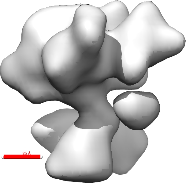
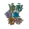
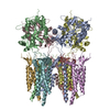
| Title | The Cryo-Electron Microscopy Structure of the CorA channel from Methanocaldococcus jannaschii at 21.6 Angstrom in low magnesium. | |||||||||
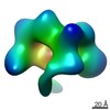
 Map data Map data | Single particle cryo-electron microscopy map of the CorA channel from Methanocaldococcus jannaschii | |||||||||
 Sample Sample |
| |||||||||
 Keywords Keywords | membrane protein / magnesium ion channel | |||||||||
| Function / homology |  Function and homology information Function and homology informationcobalt ion transmembrane transporter activity / magnesium ion transmembrane transporter activity / cobalt ion binding / magnesium ion binding / identical protein binding / plasma membrane Similarity search - Function | |||||||||
| Biological species |   Methanocaldococcus jannaschii (archaea) Methanocaldococcus jannaschii (archaea) | |||||||||
| Method | single particle reconstruction / cryo EM / Resolution: 21.6 Å | |||||||||
 Authors Authors | Cleverley RM / Kean J / Shintre CA / Baldock C / Derrick JP / Ford RC / Prince SM | |||||||||
 Citation Citation | Journal: Biochim Biophys Acta / Year: 2015 Title: The Cryo-EM structure of the CorA channel from Methanocaldococcus jannaschii in low magnesium conditions. Authors: Robert M Cleverley / James Kean / Chitra A Shintre / Clair Baldock / Jeremy P Derrick / Robert C Ford / Stephen M Prince /  Abstract: CorA channels are responsible for the uptake of essential magnesium ions by bacteria. X-ray crystal structures have been resolved for two full-length CorA channels, each in a non-conducting state ...CorA channels are responsible for the uptake of essential magnesium ions by bacteria. X-ray crystal structures have been resolved for two full-length CorA channels, each in a non-conducting state with magnesium ions bound to the protein: These structures reveal a homo-pentameric quaternary structure with approximate 5-fold rotational symmetry about a central pore axis. We report the structure of the detergent solubilized Methanocaldococcus jannaschii CorA channel determined by Cryo-Electron Microscopy and Single Particle Averaging, supported by Small Angle X-ray Scattering and X-ray crystallography. This structure also shows a pentameric channel but with a highly asymmetric domain structure. The asymmetry of the domains includes differential separations between the trans-membrane segments, which reflects mechanical coupling of the cytoplasmic domain to the trans-membrane domain. This structure therefore reveals an important aspect of the gating mechanism of CorA channels by providing an indication of how the absence of magnesium ions leads to major structural changes. | |||||||||
| History |
|
- Structure visualization
| Movie |
Movie viewer |
|---|---|
| Structure viewer | EM map: SurfViewMolmilJmol/JSmol |
| Supplemental images |
- Downloads & links
-EMDB archive
| Map data | emd_2626.map.gz | 430.5 KB | EMDB map data format | |
|---|---|---|---|---|
| Header (meta data) | emd-2626-v30.xmlemd-2626.xml | 11.3 KB 11.3 KB | Display Display | EMDB header |
| FSC (resolution estimation) | emd_2626_fsc.xml | 2.1 KB | Display | FSC data file |
| Images |  emd_2626.jpg emd_2626.jpg | 85.7 KB | ||
| Archive directory |  http://ftp.pdbj.org/pub/emdb/structures/EMD-2626ftp://ftp.pdbj.org/pub/emdb/structures/EMD-2626 http://ftp.pdbj.org/pub/emdb/structures/EMD-2626ftp://ftp.pdbj.org/pub/emdb/structures/EMD-2626 | HTTPS FTP |
-Related structure data
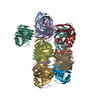
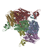
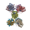
| Related structure data |  4cy4MC M: atomic model generated by this map C: citing same article ( |
|---|---|
| Similar structure data |
-Links
| EMDB pages | EMDB (EBI/PDBe) / EMDataResource |
|---|
-Map
| File | Download / File: emd_2626.map.gz / Format: CCP4 / Size: 450.2 KB / Type: IMAGE STORED AS FLOATING POINT NUMBER (4 BYTES) | ||||||||||||||||||||||||||||||||||||||||||||||||||||||||||||||||||||
|---|---|---|---|---|---|---|---|---|---|---|---|---|---|---|---|---|---|---|---|---|---|---|---|---|---|---|---|---|---|---|---|---|---|---|---|---|---|---|---|---|---|---|---|---|---|---|---|---|---|---|---|---|---|---|---|---|---|---|---|---|---|---|---|---|---|---|---|---|---|
| Annotation | Single particle cryo-electron microscopy map of the CorA channel from Methanocaldococcus jannaschii | ||||||||||||||||||||||||||||||||||||||||||||||||||||||||||||||||||||
| Projections & slices | Image control
Images are generated by Spider. | ||||||||||||||||||||||||||||||||||||||||||||||||||||||||||||||||||||
| Voxel size | X=Y=Z: 3.5 Å | ||||||||||||||||||||||||||||||||||||||||||||||||||||||||||||||||||||
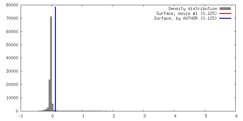
| Density |
| ||||||||||||||||||||||||||||||||||||||||||||||||||||||||||||||||||||
| Symmetry | Space group: 1 | ||||||||||||||||||||||||||||||||||||||||||||||||||||||||||||||||||||
| Details | EMDB XML:
CCP4 map header:
| ||||||||||||||||||||||||||||||||||||||||||||||||||||||||||||||||||||
 Y (Sec.)
 X (Row.)
 Z (Col.)


-Supplemental data
- Sample components
-Entire : CorA channel from Methanocaldococcus jannaschii
| Entire | Name: CorA channel from Methanocaldococcus jannaschii |
|---|---|
| Components |
|
-Supramolecule #1000: CorA channel from Methanocaldococcus jannaschii
| Supramolecule | Name: CorA channel from Methanocaldococcus jannaschii / type: sample / ID: 1000 Details: Single peak in DDM detergent in size exclusion, analysed by PFO-PAGE electrophoresis Oligomeric state: Pentamer / Number unique components: 1 |
|---|---|
| Molecular weight | Theoretical: 200 KDa |
-Macromolecule #1: Magnesium transport protein CorA
| Macromolecule | Name: Magnesium transport protein CorA / type: protein_or_peptide / ID: 1 / Name.synonym: MjCorA Details: Recombinantly over-expressed protein, solubilized from membrane fraction. Purified by affinity (His-tag), size exclusion chromatography. Proteolytic tag cleaveage. Number of copies: 5 / Oligomeric state: Pentamer / Recombinant expression: Yes |
|---|---|
| Source (natural) | Organism: Methanocaldococcus jannaschii (archaea) / Strain: ATCC 43067 / Location in cell: Plasma membrane |
| Molecular weight | Theoretical: 37 KDa |
| Recombinant expression | Organism:  |
| Sequence | UniProtKB: Cobalt/magnesium transport protein CorA / InterPro: Magnesium/cobalt transport protein CorA |
-Experimental details
-Structure determination
| Method | cryo EM |
|---|---|
 Processing Processing | single particle reconstruction |
| Aggregation state | particle |
-Sample preparation
| Concentration | 1 mg/mL |
|---|---|
| Buffer | pH: 8 / Details: 20mM Tris/HCl,200mM NaCl,0.04% DodecylMaltoside |
| Grid | Details: Quantifoil R 1.3/2 holey carbon-coated EM grids |
| Vitrification | Cryogen name: ETHANE / Chamber humidity: 90 % / Instrument: FEI VITROBOT MARK I Method: Blotted twice with Whatman No.1 filter paper for 1s each time |
- Electron microscopy
| Microscope | FEI TECNAI F20 |
|---|---|
| Temperature | Max: 104 K |
| Details | Low dose mode |
| Date | Jun 5, 2009 |
| Image recording | Category: CCD / Film or detector model: GATAN ULTRASCAN 4000 (4k x 4k) / Number real images: 28 Details: 4k by 4k pixel each pixel maps to 3.5 by 3.5 Angstroms |
| Electron beam | Acceleration voltage: 200 kV / Electron source:  FIELD EMISSION GUN FIELD EMISSION GUN |
| Electron optics | Illumination mode: FLOOD BEAM / Imaging mode: BRIGHT FIELD / Nominal defocus max: 4.8 µm / Nominal defocus min: 3.3 µm |
| Sample stage | Specimen holder model: SIDE ENTRY, EUCENTRIC |
| Experimental equipment |  Model: Tecnai F20 / Image courtesy: FEI Company |
+Image processing

-Atomic model buiding 1
| Initial model | PDB ID: Chain - #0 - Chain ID: A / Chain - #1 - Chain ID: B / Chain - #2 - Chain ID: C / Chain - #3 - Chain ID: D / Chain - #4 - Chain ID: E |
|---|---|
| Software | Name: UCSF Chimera |
| Details | The coordinates of the MjCorA pentamer from PDB entry 4ev6 were positioned manually within the envelope. The fit was adjusted automatically by maximizing the correlation of the EM map with a map calculated from 4ev6 at a resolution of 21.6 Angstroms. |
| Refinement | Space: REAL / Protocol: RIGID BODY FIT Target criteria: Maximum correlation of map calculated from coordinates |
| Output model | PDB-4cy4: |

