 Movie
Movie Controller

[English] 日本語
 Yorodumi
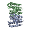
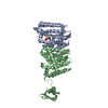
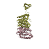
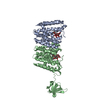
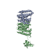
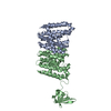
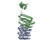
- PDB-5b0l: Structure of MoeN5-Sso7d fusion protein in complex with beta-nony... -
+ Open data

- Basic information
| Entry | Database: PDB / ID: 5b0l | ||||||
|---|---|---|---|---|---|---|---|
| Title | Structure of MoeN5-Sso7d fusion protein in complex with beta-nonyl glucoside | ||||||
 Components Components | MoeN5,DNA-binding protein 7d | ||||||
 Keywords Keywords | TRANSFERASE / DNA BINDING PROTEIN / prenyltransferase / alpha-helical fold / complex / fusion tag | ||||||
| Function / homology |  Function and homology information Function and homology information | ||||||
| Biological species |  Streptomyces ghanaensis (bacteria) Streptomyces ghanaensis (bacteria)  Sulfolobus solfataricus (archaea) Sulfolobus solfataricus (archaea) | ||||||
| Method |  X-RAY DIFFRACTION / SYNCHROTRON / MOLECULAR REPLACEMENT / Resolution: 2.8 Å X-RAY DIFFRACTION / SYNCHROTRON / MOLECULAR REPLACEMENT / Resolution: 2.8 Å | ||||||
 Authors Authors | Ko, T.-P. / Zhang, L. / Chen, C.-C. / Guo, R.-T. / Oldfield, E.O. | ||||||
 Citation Citation | Journal: Angew.Chem.Int.Ed.Engl. / Year: 2016 Title: Moenomycin Biosynthesis: Structure and Mechanism of Action of the Prenyltransferase MoeN5. Authors: Zhang, L. / Chen, C.C. / Ko, T.P. / Huang, J.W. / Zheng, Y. / Liu, W. / Wang, I. / Malwal, S.R. / Feng, X. / Wang, K. / Huang, C.H. / Hsu, S.T. / Wang, A.H. / Oldfield, E. / Guo, R.T. | ||||||
| History |
|
- Structure visualization
| Structure viewer | Molecule: MolmilJmol/JSmol |
|---|
- Downloads & links
-Download
| PDBx/mmCIF format | 5b0l.cif.gz | 483.9 KB | Display | PDBx/mmCIF format |
|---|---|---|---|---|
| PDB format | pdb5b0l.ent.gz | 396.1 KB | Display | PDB format |
| PDBx/mmJSON format | 5b0l.json.gz | Tree view | PDBx/mmJSON format | |
| Others |  Other downloads Other downloads |
-Validation report
| Arichive directory | https://data.pdbj.org/pub/pdb/validation_reports/b0/5b0lftp://data.pdbj.org/pub/pdb/validation_reports/b0/5b0l | HTTPS FTP |
|---|
-Related structure data
| Related structure data | 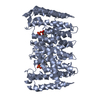 5b00C 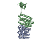 5b02SC  5b03C 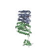 5b0iC 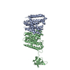 5b0jC  5b0kC 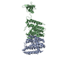 5b0mC S: Starting model for refinement C: citing same article ( |
|---|---|
| Similar structure data |


-Links
 PDBj
PDBj
- Assembly
| Deposited unit | 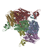
| ||||||||||||
|---|---|---|---|---|---|---|---|---|---|---|---|---|---|
| 1 | 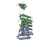
| ||||||||||||
| 2 | 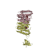
| ||||||||||||
| 3 | 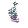
| ||||||||||||
| 4 | 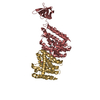
| ||||||||||||
| Unit cell |
| ||||||||||||
| Components on special symmetry positions |
|
-Components
| #1: Protein | Mass: 37581.781 Da / Num. of mol.: 8 Source method: isolated from a genetically manipulated source Details: The fusion protein of prenyltransferase (RESIDUES 1-260), linker AGAGA (RESIDUES 261-265) and Sso7d (RESIDUES 266-329) Source: (gene. exp.) Streptomyces ghanaensis (bacteria), (gene. exp.) Sulfolobus solfataricus (strain ATCC 35092 / DSM 1617 / JCM 11322 / P2) (archaea)Gene: moeN5, sso7d, sso7d-1, SSO10610 / Strain: ATCC 35092 / DSM 1617 / JCM 11322 / P2 / Production host: #2: Sugar | ChemComp-BNG / 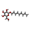  Type: D-saccharide / Mass: 306.395 Da / Num. of mol.: 13 Type: D-saccharide / Mass: 306.395 Da / Num. of mol.: 13Source method: isolated from a genetically manipulated source Formula: C15H30O6 / Comment: detergent*YM #3: Water | ChemComp-HOH / |  Mass: 18.015 Da / Num. of mol.: 1083 / Source method: isolated from a natural source / Formula: H2O Mass: 18.015 Da / Num. of mol.: 1083 / Source method: isolated from a natural source / Formula: H2O |
|---|
-Experimental details
-Experiment
| Experiment | Method: X-RAY DIFFRACTION |
|---|
- Sample preparation
| Crystal | Density Matthews: 2.68 Å3/Da / Density % sol: 54.06 % |
|---|---|
| Crystal grow | Temperature: 298 K / Method: vapor diffusion, sitting drop / pH: 8.1 Details: 0.2M Li3-citrate, 0.3M NaCl, 25% PEG 3350, 1% beta-nonyl-D-glucoside |
-Data collection
| Diffraction | Mean temperature: 100 K |
|---|---|
| Diffraction source | Source: SYNCHROTRON / Site: NSRRC  / Beamline: BL13B1 / Wavelength: 1 Å / Beamline: BL13B1 / Wavelength: 1 Å |
| Detector | Type: ADSC QUANTUM 315r / Detector: CCD / Date: Jul 14, 2015 |
| Radiation | Protocol: SINGLE WAVELENGTH / Monochromatic (M) / Laue (L): M / Scattering type: x-ray |
| Radiation wavelength | Wavelength: 1 Å / Relative weight: 1 |
| Reflection | Resolution: 2.8→25 Å / Num. obs: 79481 / % possible obs: 100 % / Redundancy: 7.1 % / Rmerge(I) obs: 0.081 / Net I/σ(I): 20.6 |
| Reflection shell | Resolution: 2.8→2.9 Å / Redundancy: 7.3 % / Rmerge(I) obs: 0.415 / Mean I/σ(I) obs: 3.5 / % possible all: 100 |
- Processing
| Software |
| ||||||||||||||||
|---|---|---|---|---|---|---|---|---|---|---|---|---|---|---|---|---|---|
| Refinement | Method to determine structure: MOLECULAR REPLACEMENT Starting model: 5B02 Resolution: 2.8→25 Å / Cross valid method: THROUGHOUT
| ||||||||||||||||
| Refine analyze | Luzzati coordinate error obs: 0.27 Å / Luzzati d res low obs: 5 Å / Luzzati sigma a obs: 0.34 Å | ||||||||||||||||
| Refinement step | Cycle: LAST / Resolution: 2.8→25 Å
| ||||||||||||||||
| LS refinement shell | Resolution: 2.8→2.9 Å / Rfactor Rfree: 0.322 / Rfactor Rwork: 0.247 |
