 Movie
Movie Controller

[English] 日本語
 Yorodumi
- PDB-5b00: Structure of the prenyltransferase MoeN5 in complex with geranyl ... -
+ Open data

- Basic information
| Entry | Database: PDB / ID: 5b00 | ||||||
|---|---|---|---|---|---|---|---|
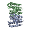
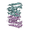
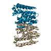
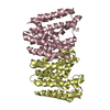
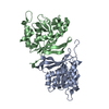
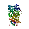
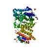
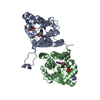
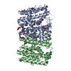
| Title | Structure of the prenyltransferase MoeN5 in complex with geranyl pyrophosphate | ||||||
 Components Components | MoeN5 | ||||||
 Keywords Keywords | TRANSFERASE / prenyltransferase / alpha-helical fold | ||||||
| Function / homology | Farnesyl Diphosphate Synthase / Farnesyl Diphosphate Synthase / Isoprenoid synthase domain superfamily / Orthogonal Bundle / Mainly Alpha / GERANYL DIPHOSPHATE / MoeN5 Function and homology information Function and homology information | ||||||
| Biological species |  Streptomyces ghanaensis (bacteria) Streptomyces ghanaensis (bacteria) | ||||||
| Method |  X-RAY DIFFRACTION / SYNCHROTRON / SAD / Resolution: 2.95 Å X-RAY DIFFRACTION / SYNCHROTRON / SAD / Resolution: 2.95 Å | ||||||
 Authors Authors | Ko, T.-P. / Zhang, L. / Chen, C.-C. / Guo, R.-T. | ||||||
 Citation Citation | Journal: Angew.Chem.Int.Ed.Engl. / Year: 2016 Title: Moenomycin Biosynthesis: Structure and Mechanism of Action of the Prenyltransferase MoeN5. Authors: Zhang, L. / Chen, C.C. / Ko, T.P. / Huang, J.W. / Zheng, Y. / Liu, W. / Wang, I. / Malwal, S.R. / Feng, X. / Wang, K. / Huang, C.H. / Hsu, S.T. / Wang, A.H. / Oldfield, E. / Guo, R.T. | ||||||
| History |
|
- Structure visualization
| Structure viewer | Molecule: MolmilJmol/JSmol |
|---|
- Downloads & links
-Download
| PDBx/mmCIF format | 5b00.cif.gz | 164.9 KB | Display | PDBx/mmCIF format |
|---|---|---|---|---|
| PDB format | pdb5b00.ent.gz | 130.7 KB | Display | PDB format |
| PDBx/mmJSON format | 5b00.json.gz | Tree view | PDBx/mmJSON format | |
| Others |  Other downloads Other downloads |
-Validation report
| Arichive directory | https://data.pdbj.org/pub/pdb/validation_reports/b0/5b00ftp://data.pdbj.org/pub/pdb/validation_reports/b0/5b00 | HTTPS FTP |
|---|
-Related structure data
| Related structure data |  5b02C  5b03C  5b0iC  5b0jC  5b0kC 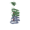 5b0lC 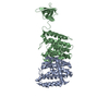 5b0mC C: citing same article ( |
|---|---|
| Similar structure data |


-Links
 PDBj
PDBj
- Assembly
| Deposited unit | 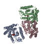
| ||||||||
|---|---|---|---|---|---|---|---|---|---|
| 1 | 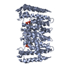
| ||||||||
| 2 | 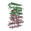
| ||||||||
| Unit cell |
|
-Components
| #1: Protein | Mass: 32258.320 Da / Num. of mol.: 3 Source method: isolated from a genetically manipulated source Source: (gene. exp.) Streptomyces ghanaensis (bacteria) / Gene: moeN5 / Production host: #2: Chemical |   Mass: 314.209 Da / Num. of mol.: 3 / Source method: obtained synthetically / Formula: C10H20O7P2 Mass: 314.209 Da / Num. of mol.: 3 / Source method: obtained synthetically / Formula: C10H20O7P2#3: Water | ChemComp-HOH / |  Mass: 18.015 Da / Num. of mol.: 191 / Source method: isolated from a natural source / Formula: H2O Mass: 18.015 Da / Num. of mol.: 191 / Source method: isolated from a natural source / Formula: H2O |
|---|
-Experimental details
-Experiment
| Experiment | Method: X-RAY DIFFRACTION |
|---|
- Sample preparation
| Crystal | Density Matthews: 2.9 Å3/Da / Density % sol: 57.57 % / Description: hicup uppsala |
|---|---|
| Crystal grow | Temperature: 298 K / Method: vapor diffusion, sitting drop / pH: 9.1 / Details: 0.2 M Na2HPO4, 16% PEG 3350 |
-Data collection
| Diffraction | Mean temperature: 100 K |
|---|---|
| Diffraction source | Source: SYNCHROTRON / Site: NSRRC  / Beamline: BL13B1 / Wavelength: 1 Å / Beamline: BL13B1 / Wavelength: 1 Å |
| Detector | Type: ADSC QUANTUM 315r / Detector: CCD / Date: Jan 14, 2015 |
| Radiation | Protocol: SINGLE WAVELENGTH / Monochromatic (M) / Laue (L): M / Scattering type: x-ray |
| Radiation wavelength | Wavelength: 1 Å / Relative weight: 1 |
| Reflection | Resolution: 2.95→25 Å / Num. obs: 23648 / % possible obs: 99 % / Redundancy: 4.5 % / Rmerge(I) obs: 0.087 / Net I/σ(I): 19.9 |
| Reflection shell | Resolution: 2.95→3.06 Å / Redundancy: 4.3 % / Rmerge(I) obs: 0.453 / Mean I/σ(I) obs: 3.5 / % possible all: 93.4 |
- Processing
| Software |
| ||||||||||||||||||||
|---|---|---|---|---|---|---|---|---|---|---|---|---|---|---|---|---|---|---|---|---|---|
| Refinement | Method to determine structure: SAD / Resolution: 2.95→25 Å / Cross valid method: THROUGHOUT
| ||||||||||||||||||||
| Refine analyze | Luzzati coordinate error obs: 0.67 Å / Luzzati d res low obs: 5 Å / Luzzati sigma a obs: 1.16 Å | ||||||||||||||||||||
| Refinement step | Cycle: LAST / Resolution: 2.95→25 Å
|
