Movie
Movie Controller

[English] 日本語
 Yorodumi
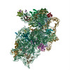
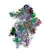
- EMDB-25543: Structure of the wt IRES eIF5B-containing 48S initiation complex,... -
+ Open data

- Basic information
| Entry | 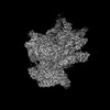 | |||||||||||||||
|---|---|---|---|---|---|---|---|---|---|---|---|---|---|---|---|---|
| Title | Structure of the wt IRES eIF5B-containing 48S initiation complex, closed conformation. Structure 15(wt) | |||||||||||||||
 Map data Map data | Post processed map | |||||||||||||||
 Sample Sample |
| |||||||||||||||
 Keywords Keywords | HCV / IRES / 40S / RIBOSOME | |||||||||||||||
| Function / homology |  Function and homology information Function and homology informationmulti-eIF complex / eukaryotic 43S preinitiation complex / translation factor activity, RNA binding / eukaryotic 48S preinitiation complex / protein-synthesizing GTPase / Formation of the ternary complex, and subsequently, the 43S complex / laminin receptor activity / Ribosomal scanning and start codon recognition / Translation initiation complex formation / Formation of a pool of free 40S subunits ...multi-eIF complex / eukaryotic 43S preinitiation complex / translation factor activity, RNA binding / eukaryotic 48S preinitiation complex / protein-synthesizing GTPase / Formation of the ternary complex, and subsequently, the 43S complex / laminin receptor activity / Ribosomal scanning and start codon recognition / Translation initiation complex formation / Formation of a pool of free 40S subunits / 90S preribosome / ubiquitin ligase inhibitor activity / positive regulation of signal transduction by p53 class mediator / GTP hydrolysis and joining of the 60S ribosomal subunit / L13a-mediated translational silencing of Ceruloplasmin expression / phagocytic cup / translation regulator activity / rough endoplasmic reticulum / ribosomal small subunit export from nucleus / laminin binding / translation initiation factor activity / gastrulation / MDM2/MDM4 family protein binding / cytosolic ribosome / class I DNA-(apurinic or apyrimidinic site) endonuclease activity / DNA-(apurinic or apyrimidinic site) lyase / ribosome assembly / positive regulation of apoptotic signaling pathway / maturation of SSU-rRNA from tricistronic rRNA transcript (SSU-rRNA, 5.8S rRNA, LSU-rRNA) / maturation of SSU-rRNA / translational initiation / small-subunit processome / spindle / rRNA processing / regulation of translation / rhythmic process / positive regulation of canonical Wnt signaling pathway / ribosomal small subunit assembly / virus receptor activity / ribosome binding / ribosomal small subunit biogenesis / small ribosomal subunit / small ribosomal subunit rRNA binding / cytosolic small ribosomal subunit / perikaryon / cell differentiation / cytoplasmic translation / tRNA binding / mitochondrial inner membrane / postsynaptic density / rRNA binding / structural constituent of ribosome / ribosome / translation / ribonucleoprotein complex / cell division / DNA repair / GTPase activity / apoptotic process / centrosome / synapse / dendrite / GTP binding / nucleolus / perinuclear region of cytoplasm / Golgi apparatus / mitochondrion / DNA binding / RNA binding / zinc ion binding / membrane / metal ion binding / nucleus / plasma membrane / cytoplasm / cytosol Similarity search - Function | |||||||||||||||
| Biological species |   Hepatitis C virus (isolate 1) / Hepatitis C virus (isolate 1) /  Homo sapiens (human) Homo sapiens (human) | |||||||||||||||
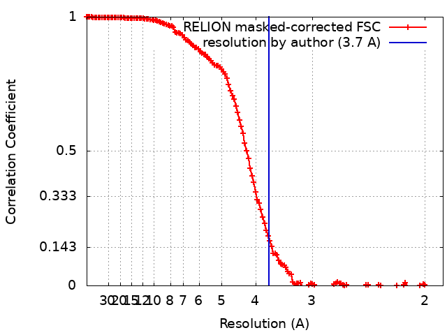
| Method | single particle reconstruction / cryo EM / Resolution: 3.7 Å | |||||||||||||||
 Authors Authors | Brown ZP / Abaeva IS / De S / Hellen CUT / Pestova TV / Frank J | |||||||||||||||
| Funding support |  United States, 4 items United States, 4 items
| |||||||||||||||
 Citation Citation | Journal: EMBO J / Year: 2022 Title: Molecular architecture of 40S translation initiation complexes on the hepatitis C virus IRES. Authors: Zuben P Brown / Irina S Abaeva / Swastik De / Christopher U T Hellen / Tatyana V Pestova / Joachim Frank / Abstract: Hepatitis C virus mRNA contains an internal ribosome entry site (IRES) that mediates end-independent translation initiation, requiring a subset of eukaryotic initiation factors (eIFs). Biochemical ...Hepatitis C virus mRNA contains an internal ribosome entry site (IRES) that mediates end-independent translation initiation, requiring a subset of eukaryotic initiation factors (eIFs). Biochemical studies revealed that direct binding of the IRES to the 40S ribosomal subunit places the initiation codon into the P site, where it base pairs with eIF2-bound Met-tRNAiMet forming a 48S initiation complex. Subsequently, eIF5 and eIF5B mediate subunit joining, yielding an elongation-competent 80S ribosome. Initiation can also proceed without eIF2, in which case Met-tRNAiMet is recruited directly by eIF5B. However, the structures of initiation complexes assembled on the HCV IRES, the transitions between different states, and the accompanying conformational changes have remained unknown. To fill these gaps, we now obtained cryo-EM structures of IRES initiation complexes, at resolutions up to 3.5 Å, that cover all major stages from the initial ribosomal association, through eIF2-containing 48S initiation complexes, to eIF5B-containing complexes immediately prior to subunit joining. These structures provide insights into the dynamic network of 40S/IRES contacts, highlight the role of IRES domain II, and reveal conformational changes that occur during the transition from eIF2- to eIF5B-containing 48S complexes and prepare them for subunit joining. | |||||||||||||||
| History |
|
- Structure visualization
| Supplemental images |
|---|

- Downloads & links
-EMDB archive
| Map data | emd_25543.map.gz | 222.1 MB | EMDB map data format | |
|---|---|---|---|---|
| Header (meta data) | emd-25543-v30.xmlemd-25543.xml | 65.5 KB 65.5 KB | Display Display | EMDB header |
| FSC (resolution estimation) | emd_25543_fsc.xml | 14.3 KB | Display | FSC data file |
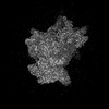
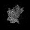
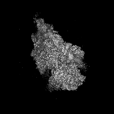
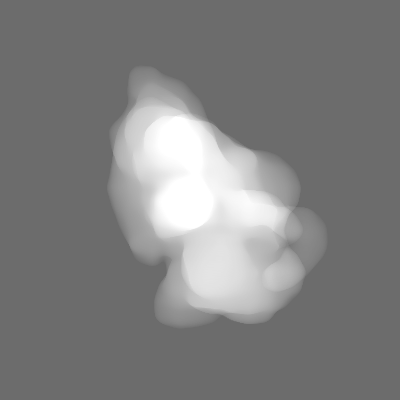
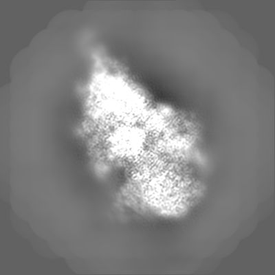
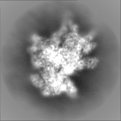
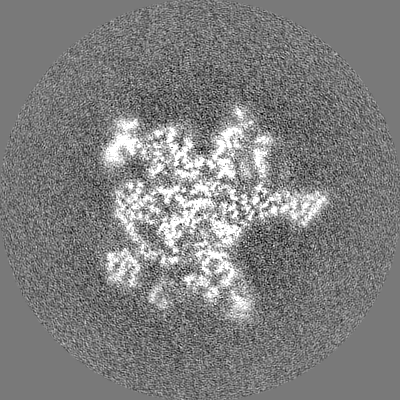
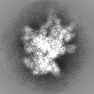
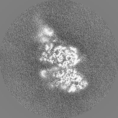
| Images |  emd_25543.png emd_25543.png | 33.6 KB | ||
| Masks | emd_25543_msk_1.map | 244.1 MB | Mask map | |
| Filedesc metadata | emd-25543.cif.gz | 13.8 KB | ||
| Others | emd_25543_additional_1.map.gzemd_25543_additional_2.map.gzemd_25543_additional_3.map.gzemd_25543_half_map_1.map.gzemd_25543_half_map_2.map.gz | 92.6 MB 86.8 MB 192.3 MB 194 MB 192.9 MB | ||
| Archive directory |  http://ftp.pdbj.org/pub/emdb/structures/EMD-25543ftp://ftp.pdbj.org/pub/emdb/structures/EMD-25543 http://ftp.pdbj.org/pub/emdb/structures/EMD-25543ftp://ftp.pdbj.org/pub/emdb/structures/EMD-25543 | HTTPS FTP |
-Related structure data
| Related structure data |  7sywMC 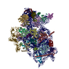 7syiC  7syjC  7sykC  7sylC  7syoC  7sypC  7syqC 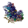 7syrC  7sysC  7sytC  7syuC  7syvC  7syxC M: atomic model generated by this map C: citing same article ( |
|---|---|
| Similar structure data |


-Links
| EMDB pages | EMDB (EBI/PDBe) / EMDataResource |
|---|---|
| Related items in Molecule of the Month |


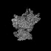
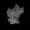
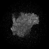
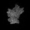
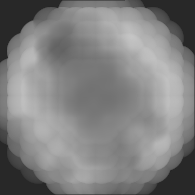
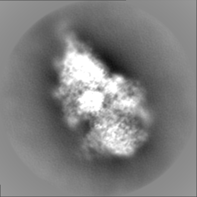
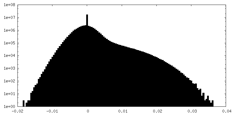
-Map
| File | Download / File: emd_25543.map.gz / Format: CCP4 / Size: 244.1 MB / Type: IMAGE STORED AS FLOATING POINT NUMBER (4 BYTES) | ||||||||||||||||||||||||||||||||||||
|---|---|---|---|---|---|---|---|---|---|---|---|---|---|---|---|---|---|---|---|---|---|---|---|---|---|---|---|---|---|---|---|---|---|---|---|---|---|
| Annotation | Post processed map | ||||||||||||||||||||||||||||||||||||
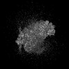
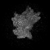
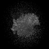
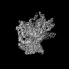
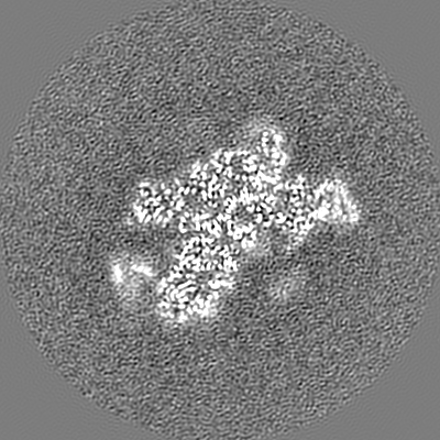
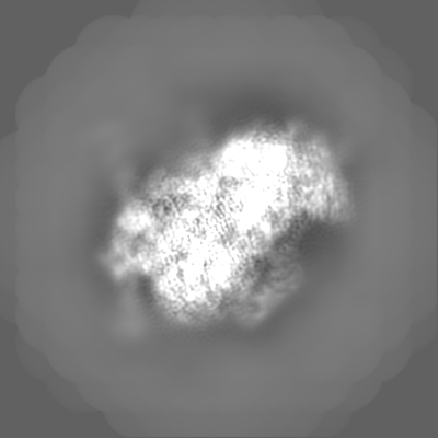
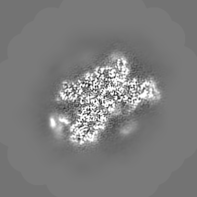
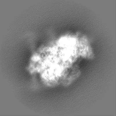
| Projections & slices | Image control
Images are generated by Spider. | ||||||||||||||||||||||||||||||||||||
| Voxel size | X=Y=Z: 0.95 Å | ||||||||||||||||||||||||||||||||||||
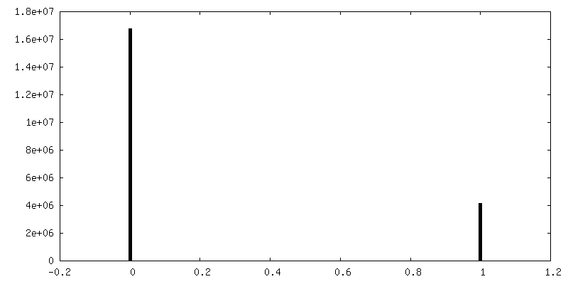
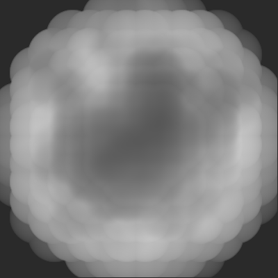
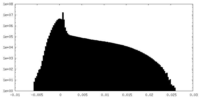
| Density |
| ||||||||||||||||||||||||||||||||||||
| Symmetry | Space group: 1 | ||||||||||||||||||||||||||||||||||||
| Details | EMDB XML:
|
 Z (Sec.)
 Y (Row.)
 X (Col.)


-Supplemental data
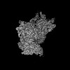
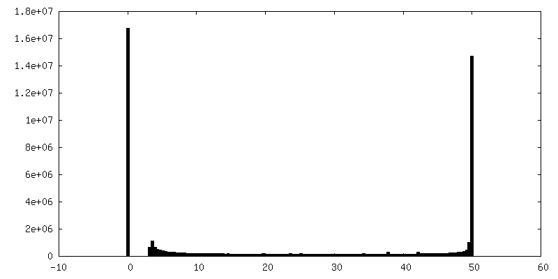
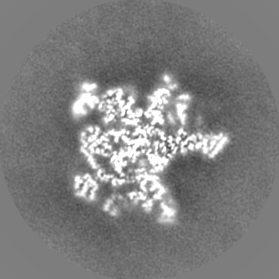
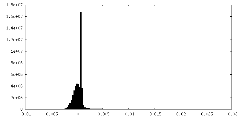
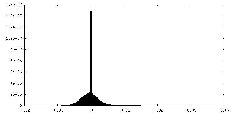
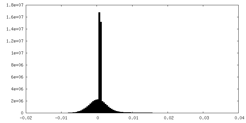
-Mask #1
| File | emd_25543_msk_1.map | ||||||||||||
|---|---|---|---|---|---|---|---|---|---|---|---|---|---|
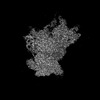
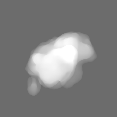
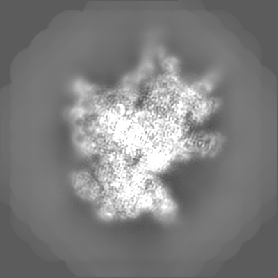
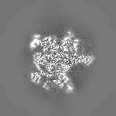
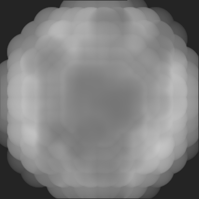
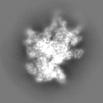
| Projections & Slices |
| ||||||||||||
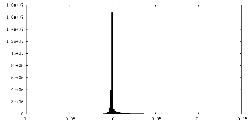
| Density Histograms |


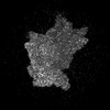
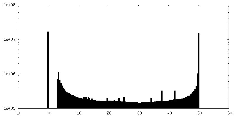
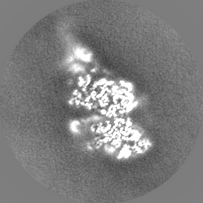
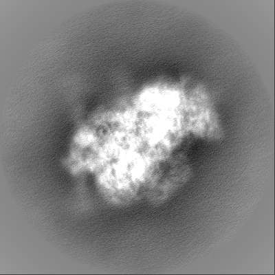
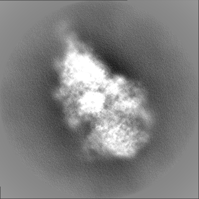
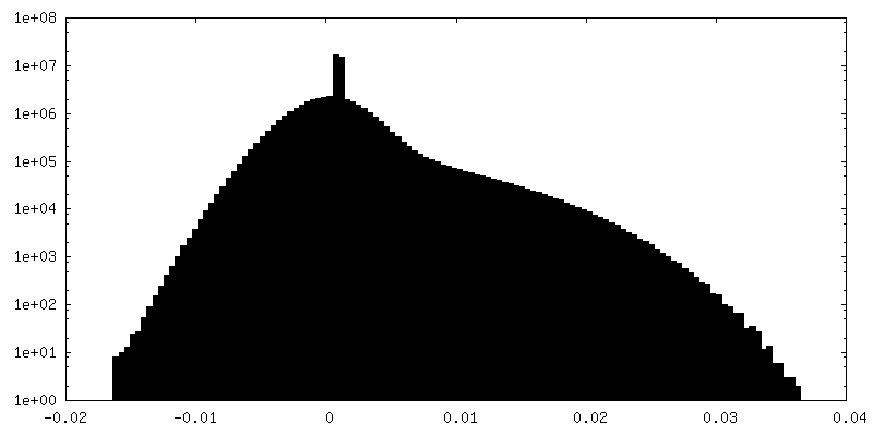
-Additional map: Local resolution map filtered at local resolution
| File | emd_25543_additional_1.map | ||||||||||||
|---|---|---|---|---|---|---|---|---|---|---|---|---|---|
| Annotation | Local resolution map filtered at local resolution | ||||||||||||
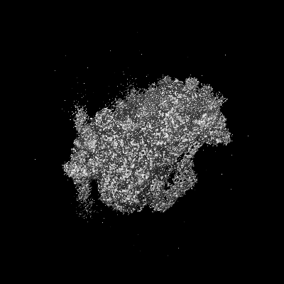
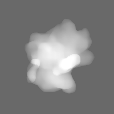
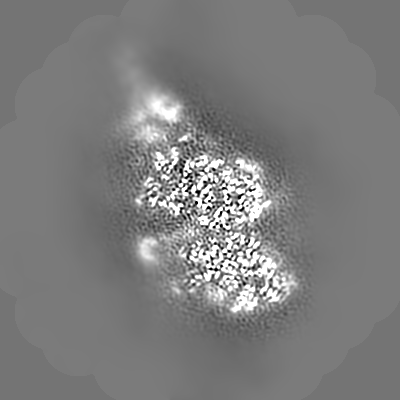
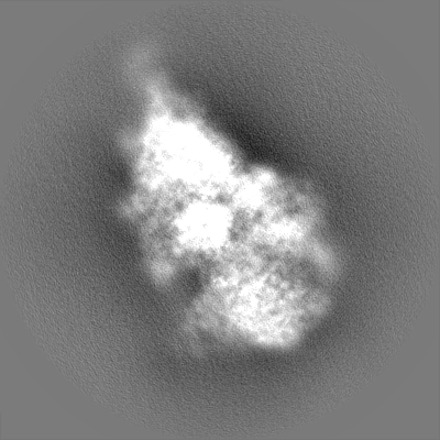
| Projections & Slices |
| ||||||||||||
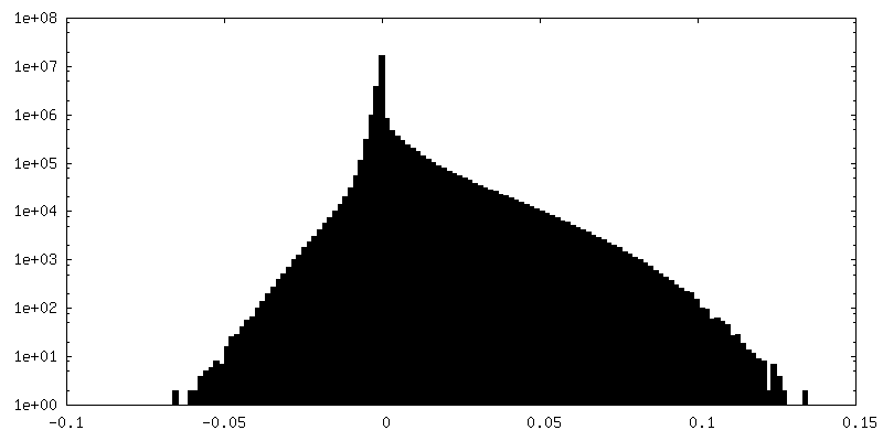
| Density Histograms |
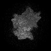
-Additional map: Local resolution values
| File | emd_25543_additional_2.map | ||||||||||||
|---|---|---|---|---|---|---|---|---|---|---|---|---|---|
| Annotation | Local resolution values | ||||||||||||
| Projections & Slices |
| ||||||||||||
| Density Histograms |


-Additional map: Unsharpened map
| File | emd_25543_additional_3.map | ||||||||||||
|---|---|---|---|---|---|---|---|---|---|---|---|---|---|
| Annotation | Unsharpened map | ||||||||||||
| Projections & Slices |
| ||||||||||||
| Density Histograms |


-Half map: Half map
| File | emd_25543_half_map_1.map | ||||||||||||
|---|---|---|---|---|---|---|---|---|---|---|---|---|---|
| Annotation | Half map | ||||||||||||
| Projections & Slices |
| ||||||||||||
| Density Histograms |


-Half map: Half map
| File | emd_25543_half_map_2.map | ||||||||||||
|---|---|---|---|---|---|---|---|---|---|---|---|---|---|
| Annotation | Half map | ||||||||||||
| Projections & Slices |
| ||||||||||||
| Density Histograms |
- Sample components
+Entire : 40S ribosomal small subunit with HCV IRES
+Supramolecule #1: 40S ribosomal small subunit with HCV IRES
+Macromolecule #1: 18S rRNA
+Macromolecule #36: Met-tRNA-i-Met
+Macromolecule #39: HCV IRES
+Macromolecule #2: Eukaryotic translation initiation factor 1A, X-chromosomal

+Macromolecule #3: 40S ribosomal protein SA
+Macromolecule #4: eS1
+Macromolecule #5: 40S ribosomal protein S2
+Macromolecule #6: uS3
+Macromolecule #7: 40S ribosomal protein S4
+Macromolecule #8: uS7
+Macromolecule #9: eS6
+Macromolecule #10: 40S ribosomal protein S7
+Macromolecule #11: eS8
+Macromolecule #12: uS4
+Macromolecule #13: eS10
+Macromolecule #14: uS17
+Macromolecule #15: eS12
+Macromolecule #16: uS15
+Macromolecule #17: uS11
+Macromolecule #18: uS19
+Macromolecule #19: uS9
+Macromolecule #20: eS17
+Macromolecule #21: uS13
+Macromolecule #22: eS19
+Macromolecule #23: uS10
+Macromolecule #24: 40S ribosomal protein S21
+Macromolecule #25: uS8
+Macromolecule #26: uS12
+Macromolecule #27: 40S ribosomal protein S24
+Macromolecule #28: 40S ribosomal protein S25
+Macromolecule #29: 40S ribosomal protein S26
+Macromolecule #30: eS27
+Macromolecule #31: eS28
+Macromolecule #32: eS29
+Macromolecule #33: eS30
+Macromolecule #34: 40S ribosomal protein S27a
+Macromolecule #35: RACK1
+Macromolecule #37: 60s ribosomal protein l41
+Macromolecule #38: Eukaryotic translation initiation factor 5B
+Macromolecule #40: ZINC ION
+Macromolecule #41: GUANOSINE-5'-TRIPHOSPHATE

+Macromolecule #42: MAGNESIUM ION
+Macromolecule #43: SODIUM ION
-Experimental details
-Structure determination
| Method | cryo EM |
|---|---|
 Processing Processing | single particle reconstruction |
| Aggregation state | particle |
-Sample preparation
| Concentration | 0.000075 mg/mL |
|---|---|
| Buffer | pH: 7.5 |
| Grid | Model: Quantifoil R0.6/1 / Material: GOLD / Mesh: 300 / Support film - Material: GOLD / Support film - topology: HOLEY / Support film - Film thickness: 50 / Pretreatment - Type: PLASMA CLEANING / Pretreatment - Time: 25 sec. / Pretreatment - Atmosphere: OTHER / Details: H2/O2 mixture for 25 seconds at 25W power |
| Vitrification | Cryogen name: ETHANE-PROPANE / Chamber humidity: 100 % / Chamber temperature: 277.15 K / Instrument: FEI VITROBOT MARK IV / Details: 4 second blot time, force 3. |
- Electron microscopy
| Microscope | FEI TECNAI F30 |
|---|---|
| Image recording | Film or detector model: GATAN K3 (6k x 4k) / Average exposure time: 4.0 sec. / Average electron dose: 70.9 e/Å2 |
| Electron beam | Acceleration voltage: 300 kV / Electron source:  FIELD EMISSION GUN FIELD EMISSION GUN |
| Electron optics | Illumination mode: FLOOD BEAM / Imaging mode: BRIGHT FIELD / Cs: 2.26 mm / Nominal defocus max: 2.0 µm / Nominal defocus min: 1.0 µm / Nominal magnification: 52000 |
| Sample stage | Cooling holder cryogen: NITROGEN |
| Experimental equipment |  Model: Tecnai F30 / Image courtesy: FEI Company |
+Image processing
-Atomic model buiding 1
| Initial model |
| ||||||||||
|---|---|---|---|---|---|---|---|---|---|---|---|
| Output model | PDB-7syw: |