+ Open data
- Basic information
| Entry | Database: EMDB / ID: EMD-23405 | ||||||||||||||||||||||||||||||||||||
|---|---|---|---|---|---|---|---|---|---|---|---|---|---|---|---|---|---|---|---|---|---|---|---|---|---|---|---|---|---|---|---|---|---|---|---|---|---|
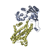
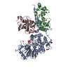
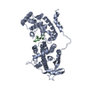
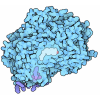
| Title | WT Chicken Scap L1-L7 / Fab 4G10 complex focused refinement | ||||||||||||||||||||||||||||||||||||
 Map data Map data | Focused classification map of WT Scap L1-L7/Fab | ||||||||||||||||||||||||||||||||||||
 Sample Sample |
| ||||||||||||||||||||||||||||||||||||
 Keywords Keywords | Cholesterol / LIPID BINDING PROTEIN / SREBP | ||||||||||||||||||||||||||||||||||||
| Function / homology |  Function and homology information Function and homology informationRegulation of cholesterol biosynthesis by SREBP (SREBF) / SREBP-SCAP complex / regulation of cholesterol biosynthetic process / sterol binding / SREBP signaling pathway / regulation of fatty acid biosynthetic process / cholesterol metabolic process / response to insulin / positive regulation of cholesterol biosynthetic process / ER to Golgi transport vesicle membrane ...Regulation of cholesterol biosynthesis by SREBP (SREBF) / SREBP-SCAP complex / regulation of cholesterol biosynthetic process / sterol binding / SREBP signaling pathway / regulation of fatty acid biosynthetic process / cholesterol metabolic process / response to insulin / positive regulation of cholesterol biosynthetic process / ER to Golgi transport vesicle membrane / response to hypoxia / immune response / Golgi membrane / endoplasmic reticulum membrane Similarity search - Function | ||||||||||||||||||||||||||||||||||||
| Biological species |   | ||||||||||||||||||||||||||||||||||||
| Method | single particle reconstruction / cryo EM / Resolution: 2.9 Å | ||||||||||||||||||||||||||||||||||||
 Authors Authors | Kober DL / Radhakrishnan A | ||||||||||||||||||||||||||||||||||||
| Funding support |  United States, United States,  France, 11 items France, 11 items
| ||||||||||||||||||||||||||||||||||||
 Citation Citation |  Journal: Cell / Year: 2021 Journal: Cell / Year: 2021Title: Scap structures highlight key role for rotation of intertwined luminal loops in cholesterol sensing. Authors: Daniel L Kober / Arun Radhakrishnan / Joseph L Goldstein / Michael S Brown / Lindsay D Clark / Xiao-Chen Bai / Daniel M Rosenbaum /  Abstract: The cholesterol-sensing protein Scap induces cholesterol synthesis by transporting membrane-bound transcription factors called sterol regulatory element-binding proteins (SREBPs) from the endoplasmic ...The cholesterol-sensing protein Scap induces cholesterol synthesis by transporting membrane-bound transcription factors called sterol regulatory element-binding proteins (SREBPs) from the endoplasmic reticulum (ER) to the Golgi apparatus for proteolytic activation. Transport requires interaction between Scap's two ER luminal loops (L1 and L7), which flank an intramembrane sterol-sensing domain (SSD). Cholesterol inhibits Scap transport by binding to L1, which triggers Scap's binding to Insig, an ER retention protein. Here we used cryoelectron microscopy (cryo-EM) to elucidate two structures of full-length chicken Scap: (1) a wild-type free of Insigs and (2) mutant Scap bound to chicken Insig without cholesterol. Strikingly, L1 and L7 intertwine tightly to form a globular domain that acts as a luminal platform connecting the SSD to the rest of Scap. In the presence of Insig, this platform undergoes a large rotation accompanied by rearrangement of Scap's transmembrane helices. We postulate that this conformational change halts Scap transport of SREBPs and inhibits cholesterol synthesis. | ||||||||||||||||||||||||||||||||||||
| History |
|
- Structure visualization
| Movie |
 Movie viewer Movie viewer |
|---|---|
| Structure viewer | EM map:  SurfView SurfView Molmil Molmil Jmol/JSmol Jmol/JSmol |
| Supplemental images |
- Downloads & links
-EMDB archive
| Map data |  emd_23405.map.gz emd_23405.map.gz | 70.3 MB |  EMDB map data format EMDB map data format | |
|---|---|---|---|---|
| Header (meta data) |  emd-23405-v30.xml emd-23405-v30.xml emd-23405.xml emd-23405.xml | 17.5 KB 17.5 KB | Display Display |  EMDB header EMDB header |
| FSC (resolution estimation) |  emd_23405_fsc.xml emd_23405_fsc.xml | 9.6 KB | Display |  FSC data file FSC data file |
| Images |  emd_23405.png emd_23405.png | 33.1 KB | ||
| Filedesc metadata |  emd-23405.cif.gz emd-23405.cif.gz | 7.1 KB | ||
| Archive directory |  http://ftp.pdbj.org/pub/emdb/structures/EMD-23405 http://ftp.pdbj.org/pub/emdb/structures/EMD-23405 ftp://ftp.pdbj.org/pub/emdb/structures/EMD-23405 ftp://ftp.pdbj.org/pub/emdb/structures/EMD-23405 | HTTPS FTP |
-Validation report
| Summary document |  emd_23405_validation.pdf.gz emd_23405_validation.pdf.gz | 542.6 KB | Display |  EMDB validaton report EMDB validaton report |
|---|---|---|---|---|
| Full document |  emd_23405_full_validation.pdf.gz emd_23405_full_validation.pdf.gz | 542.2 KB | Display | |
| Data in XML |  emd_23405_validation.xml.gz emd_23405_validation.xml.gz | 11 KB | Display | |
| Data in CIF |  emd_23405_validation.cif.gz emd_23405_validation.cif.gz | 14.6 KB | Display | |
| Arichive directory |  https://ftp.pdbj.org/pub/emdb/validation_reports/EMD-23405 https://ftp.pdbj.org/pub/emdb/validation_reports/EMD-23405 ftp://ftp.pdbj.org/pub/emdb/validation_reports/EMD-23405 ftp://ftp.pdbj.org/pub/emdb/validation_reports/EMD-23405 | HTTPS FTP |
-Related structure data
| Related structure data |  7lkfMC  7lkhC M: atomic model generated by this map C: citing same article ( |
|---|---|
| Similar structure data |
- Links
Links
| EMDB pages |  EMDB (EBI/PDBe) / EMDB (EBI/PDBe) /  EMDataResource EMDataResource |
|---|---|
| Related items in Molecule of the Month |
- Map
Map
| File |  Download / File: emd_23405.map.gz / Format: CCP4 / Size: 75.1 MB / Type: IMAGE STORED AS FLOATING POINT NUMBER (4 BYTES) Download / File: emd_23405.map.gz / Format: CCP4 / Size: 75.1 MB / Type: IMAGE STORED AS FLOATING POINT NUMBER (4 BYTES) | ||||||||||||||||||||||||||||||||||||||||||||||||||||||||||||||||||||
|---|---|---|---|---|---|---|---|---|---|---|---|---|---|---|---|---|---|---|---|---|---|---|---|---|---|---|---|---|---|---|---|---|---|---|---|---|---|---|---|---|---|---|---|---|---|---|---|---|---|---|---|---|---|---|---|---|---|---|---|---|---|---|---|---|---|---|---|---|---|
| Annotation | Focused classification map of WT Scap L1-L7/Fab | ||||||||||||||||||||||||||||||||||||||||||||||||||||||||||||||||||||
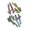
| Projections & slices | Image control
Images are generated by Spider. | ||||||||||||||||||||||||||||||||||||||||||||||||||||||||||||||||||||
| Voxel size | X=Y=Z: 1.08 Å | ||||||||||||||||||||||||||||||||||||||||||||||||||||||||||||||||||||
| Density |
| ||||||||||||||||||||||||||||||||||||||||||||||||||||||||||||||||||||
| Symmetry | Space group: 1 | ||||||||||||||||||||||||||||||||||||||||||||||||||||||||||||||||||||
| Details | EMDB XML:
CCP4 map header:
| ||||||||||||||||||||||||||||||||||||||||||||||||||||||||||||||||||||
-Supplemental data
- Sample components
-Entire : Ternary complex of Scap with Fab fragment
| Entire | Name: Ternary complex of Scap with Fab fragment |
|---|---|
| Components |
|
-Supramolecule #1: Ternary complex of Scap with Fab fragment
| Supramolecule | Name: Ternary complex of Scap with Fab fragment / type: complex / ID: 1 / Parent: 0 / Macromolecule list: all Details: Fab fragment generated by proteolytic cleavage of IgG antibody |
|---|---|
| Molecular weight | Theoretical: 172 KDa |
-Supramolecule #2: Scap L1-L7 domain
| Supramolecule | Name: Scap L1-L7 domain / type: complex / ID: 2 / Parent: 1 / Macromolecule list: #1 |
|---|---|
| Source (natural) | Organism:  |
| Molecular weight | Theoretical: 50 KDa |
-Supramolecule #3: 4G10 Fab, 4G10 light chain
| Supramolecule | Name: 4G10 Fab, 4G10 light chain / type: complex / ID: 3 / Parent: 1 / Macromolecule list: #2-#3 Details: Fab fragment generated by proteolytic cleavage of IgG antibody |
|---|---|
| Source (natural) | Organism:  |
-Macromolecule #1: Sterol regulatory element-binding protein cleavage-activating protein
| Macromolecule | Name: Sterol regulatory element-binding protein cleavage-activating protein type: protein_or_peptide / ID: 1 / Number of copies: 1 / Enantiomer: LEVO |
|---|---|
| Source (natural) | Organism:  |
| Molecular weight | Theoretical: 148.212594 KDa |
| Recombinant expression | Organism:  Homo sapiens (human) Homo sapiens (human) |
| Sequence | String: MTLTEKLRER ISRAFYNHGL LCASYPIPII LFTGLCILAC CYPLLKLPLP GTGPVEFSTP VKDYFPPSPD VVSQQGDLSE RPDWYVGAP VAYIQQIFVK ATVSPWQKNF LAVDVFRSPL SRVFQLVEEI RNHALRDSSG VKSLEEVCLQ VTDLLPGLKK L RNLLPEHG ...String: MTLTEKLRER ISRAFYNHGL LCASYPIPII LFTGLCILAC CYPLLKLPLP GTGPVEFSTP VKDYFPPSPD VVSQQGDLSE RPDWYVGAP VAYIQQIFVK ATVSPWQKNF LAVDVFRSPL SRVFQLVEEI RNHALRDSSG VKSLEEVCLQ VTDLLPGLKK L RNLLPEHG CLLLSPGNFW QNDRERFNAD PDIIKTIHQH EPKALQTSAT LKDLLFGLPG KYSGVNLYNR KRVVSYTVTL GL QRYDSRF LSSLRSRLKL LHPSPNCTLR EDSIVHVHFK EEIGIAELIP LVTTYIILFA YIYFSTRKID MVKSKWGLAL AAV VTVLSS LLMSVGLCTL FGLTPTLNGG EIFPYLVVVI GLENVLVLTK SVVSTPVDLE VKLRIAQGLS NESWSIMKNM ATEL GIILI GYFTLVPAIQ EFCLFAVVGL VSDFFLQMLF FTTVLSIDIR RMELADLNKR LPAEACLPPA KPASRSQRYE RQPAV RPAT PHTITLQPSS FRNLRLPKRL RVIYFFARTR LAQRLIMAGT VIWIGILVYT DPAGLRTYLT SQVTEQSPLG EAGLPP MPV PGGVLPAGDP KIDLSVFPSD PIQLSENQTQ QREQQAGLEP LGRLETNQHS WAQGPEGRGN GQTELGTEAE VTWGAED EE IWRKLSFRHW PSLFSYYNIT LAKRYISILP AIPVTLYLNP QEALEVRHPQ EANRYHPFLS SSGGKLNAEA QPDQTSSR L QGHRDVTLYK VAALGLASGI LLVLLLFCLY RLLCPKNYGQ NGLSHSRRRR GDLPCDDYGY SPPETEIVPL VLRGHLMDI ECLASDGMLL VSCCLVGQIR VWDAQTGDCL TVIPKPRLRR DSSGIFDYQE SWDHSPDGKT GLDDSFESSH QLKRMLSPPQ PPLFCDQPD LTSLIDTNFS EQVKVAESEP RLRAVGGRQK EAGYDFSSLV GKVYEEHSTS NCMNFGGLSA PHGQAGFCVG G STARSLGC GSEEGGCGGR RRSLGDESLS GFDKSSPLPS WGGDFESSVW SLDLQGNLIV AGRSNGKLEV WDAIEGTLRS SN DESQSGI TALVFLNNRI VAARLNGSLD FFSLETHTSL NHLQFRGAPS RSSIPSSPLF SSSDVIVCQL THTVSCAHQK PIT ALKAAA GRLVTGSQDH TLRVFRLEDS CCLFTLQGHS GAITAVYIDQ TMVLASGGQD GAICLWDVLT GSKVSHMYAH RGDV TSLTC TTSCVISSGL DDVISIWDRS SGIKLYSIQQ EMGCGSSLGV ISDNLLVTGG QGCVSFWDIG YGDLLQTVYL GKSNE SQPA RQILVLENAA IVCNFGSELS LVYVPSVLEK LDDYKDDDDK GSDYKDDDDK GSDYKDDDDK UniProtKB: Sterol regulatory element-binding protein cleavage-activating protein |
-Macromolecule #2: 4G10 heavy chain
| Macromolecule | Name: 4G10 heavy chain / type: protein_or_peptide / ID: 2 / Number of copies: 1 / Enantiomer: LEVO |
|---|---|
| Source (natural) | Organism:  |
| Molecular weight | Theoretical: 24.94266 KDa |
| Sequence | String: TGVHSEVQLQ QSGAELVRPG ASVKLSCTAS GFKIKDDYIH WVKQRPEQGL EWIGRIDPAN GHTRYAPKFQ DKATITADTS SNTAYLQLS SLTSEDTAVY YCTRYNDYDA FYFDYWGQGT TLTVSSASTK GPSVFPLAPS SKSTSGGTAA LGCLVKDYFP E PVTVSWNS ...String: TGVHSEVQLQ QSGAELVRPG ASVKLSCTAS GFKIKDDYIH WVKQRPEQGL EWIGRIDPAN GHTRYAPKFQ DKATITADTS SNTAYLQLS SLTSEDTAVY YCTRYNDYDA FYFDYWGQGT TLTVSSASTK GPSVFPLAPS SKSTSGGTAA LGCLVKDYFP E PVTVSWNS GALTSGVHTF PAVLQSSGLY SLSSVVTVPS SSLGTQTYIC NVNHKPSNTK VDKRVEPKSC DKT |
-Macromolecule #3: 4G10 light chain
| Macromolecule | Name: 4G10 light chain / type: protein_or_peptide / ID: 3 / Number of copies: 1 / Enantiomer: LEVO |
|---|---|
| Source (natural) | Organism:  |
| Molecular weight | Theoretical: 23.667178 KDa |
| Sequence | String: DIQMTQTTSS LSASLGDRVT ISCRASQDIR NYLNWYQQKP DGTVKLLIYY TSRLHSGVPS RFSGSGSGTD YSLTISNLEQ EDIATYFCQ QTNTLPWTFG GGTKVEIKRT VAAPSVFIFP PSDEQLKSGT ASVVCLLNNF YPREAKVQWK VDNALQSGNS Q ESVTEQDS ...String: DIQMTQTTSS LSASLGDRVT ISCRASQDIR NYLNWYQQKP DGTVKLLIYY TSRLHSGVPS RFSGSGSGTD YSLTISNLEQ EDIATYFCQ QTNTLPWTFG GGTKVEIKRT VAAPSVFIFP PSDEQLKSGT ASVVCLLNNF YPREAKVQWK VDNALQSGNS Q ESVTEQDS KDSTYSLSST LTLSKADYEK HKVYACEVTH QGLSSPVTKS FNRGEC |
-Experimental details
-Structure determination
| Method | cryo EM |
|---|---|
 Processing Processing | single particle reconstruction |
| Aggregation state | particle |
- Sample preparation
| Concentration | 5 mg/mL | ||||||||||||||||||
|---|---|---|---|---|---|---|---|---|---|---|---|---|---|---|---|---|---|---|---|
| Buffer | pH: 7.5 Component:
| ||||||||||||||||||
| Vitrification | Cryogen name: ETHANE / Instrument: FEI VITROBOT MARK IV |
- Electron microscopy
| Microscope | FEI TITAN KRIOS |
|---|---|
| Image recording | Film or detector model: GATAN K3 BIOQUANTUM (6k x 4k) / Average electron dose: 1.6 e/Å2 |
| Electron beam | Acceleration voltage: 300 kV / Electron source:  FIELD EMISSION GUN FIELD EMISSION GUN |
| Electron optics | Illumination mode: FLOOD BEAM / Imaging mode: BRIGHT FIELD |
| Experimental equipment |  Model: Titan Krios / Image courtesy: FEI Company |
 Movie
Movie Controller






 Z (Sec.)
 Y (Row.)
 X (Col.)


