 Movie
Movie Controller


+ Open data

- Basic information
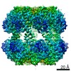
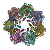
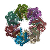
| Entry | Database: EMDB / ID: EMD-23000 | ||||||||||||
|---|---|---|---|---|---|---|---|---|---|---|---|---|---|
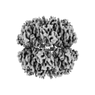
| Title | ClpP from Neisseria meningitidis - Compressed conformation | ||||||||||||
 Map data Map data | NmClpP compressed conformation | ||||||||||||
 Sample Sample |
| ||||||||||||
 Keywords Keywords | protease / ClpP / HYDROLASE | ||||||||||||
| Function / homology |  Function and homology information Function and homology information | ||||||||||||
| Biological species |  Neisseria meningitidis (bacteria) Neisseria meningitidis (bacteria) | ||||||||||||
| Method | single particle reconstruction / cryo EM / Resolution: 3.2 Å | ||||||||||||
 Authors Authors | Ripstein ZA / Vahidi S | ||||||||||||
| Funding support |  Canada, 3 items Canada, 3 items
| ||||||||||||
 Citation Citation | Journal: To Be Published Title: A pH-Dependent Conformational Switch Controls N. meningitidis ClpP Protease Function Authors: Ripstein ZR / Vahidi S / Rubinstein JL / Kay LE | ||||||||||||
| History |
|
- Structure visualization
| Movie |
Movie viewer |
|---|---|
| Structure viewer | EM map: SurfViewMolmilJmol/JSmol |
| Supplemental images |
- Downloads & links
-EMDB archive
| Map data | emd_23000.map.gz | 3.2 MB | EMDB map data format | |
|---|---|---|---|---|
| Header (meta data) | emd-23000-v30.xmlemd-23000.xml | 14.4 KB 14.4 KB | Display Display | EMDB header |
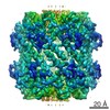
| Images |  emd_23000.png emd_23000.png | 94.8 KB | ||
| Filedesc metadata | emd-23000.cif.gz | 5.7 KB | ||
| Archive directory |  http://ftp.pdbj.org/pub/emdb/structures/EMD-23000ftp://ftp.pdbj.org/pub/emdb/structures/EMD-23000 http://ftp.pdbj.org/pub/emdb/structures/EMD-23000ftp://ftp.pdbj.org/pub/emdb/structures/EMD-23000 | HTTPS FTP |
-Validation report
| Summary document | emd_23000_validation.pdf.gz | 381.1 KB | Display | EMDB validaton report |
|---|---|---|---|---|
| Full document | emd_23000_full_validation.pdf.gz | 380.6 KB | Display | |
| Data in XML | emd_23000_validation.xml.gz | 6.5 KB | Display | |
| Data in CIF | emd_23000_validation.cif.gz | 7.4 KB | Display | |
| Arichive directory | https://ftp.pdbj.org/pub/emdb/validation_reports/EMD-23000ftp://ftp.pdbj.org/pub/emdb/validation_reports/EMD-23000 | HTTPS FTP |
-Related structure data
| Related structure data | 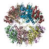 7kr2MC M: atomic model generated by this map C: citing same article ( |
|---|---|
| Similar structure data |

-Links
| EMDB pages | EMDB (EBI/PDBe) / EMDataResource |
|---|
-Map
| File | Download / File: emd_23000.map.gz / Format: CCP4 / Size: 64 MB / Type: IMAGE STORED AS FLOATING POINT NUMBER (4 BYTES) | ||||||||||||||||||||||||||||||||||||||||||||||||||||||||||||||||||||
|---|---|---|---|---|---|---|---|---|---|---|---|---|---|---|---|---|---|---|---|---|---|---|---|---|---|---|---|---|---|---|---|---|---|---|---|---|---|---|---|---|---|---|---|---|---|---|---|---|---|---|---|---|---|---|---|---|---|---|---|---|---|---|---|---|---|---|---|---|---|
| Annotation | NmClpP compressed conformation | ||||||||||||||||||||||||||||||||||||||||||||||||||||||||||||||||||||
| Voxel size | X=Y=Z: 1.06 Å | ||||||||||||||||||||||||||||||||||||||||||||||||||||||||||||||||||||
| Density |
| ||||||||||||||||||||||||||||||||||||||||||||||||||||||||||||||||||||
| Symmetry | Space group: 1 | ||||||||||||||||||||||||||||||||||||||||||||||||||||||||||||||||||||
| Details | EMDB XML:
CCP4 map header:
| ||||||||||||||||||||||||||||||||||||||||||||||||||||||||||||||||||||
-Supplemental data
- Sample components
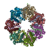
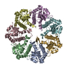
-Entire : ATP-dependent Clp protease proteolytic subunit
| Entire | Name: ATP-dependent Clp protease proteolytic subunit |
|---|---|
| Components |
|
-Supramolecule #1: ATP-dependent Clp protease proteolytic subunit
| Supramolecule | Name: ATP-dependent Clp protease proteolytic subunit / type: complex / ID: 1 / Parent: 0 / Macromolecule list: all |
|---|---|
| Source (natural) | Organism: Neisseria meningitidis (bacteria) |
| Molecular weight | Theoretical: 860 KDa |
-Macromolecule #1: ATP-dependent Clp protease proteolytic subunit
| Macromolecule | Name: ATP-dependent Clp protease proteolytic subunit / type: protein_or_peptide / ID: 1 / Number of copies: 14 / Enantiomer: LEVO / EC number: endopeptidase Clp |
|---|---|
| Source (natural) | Organism: Neisseria meningitidis (bacteria) |
| Molecular weight | Theoretical: 22.700902 KDa |
| Recombinant expression | Organism: |
| Sequence | String: MSFDNYLVPT VIEQSGRGER AFDIYSRLLK ERIVFLVGPV TDESANLVVA QLLFLESENP DKDIFFYINS PGGSVTAGMS IYDTMNFIK PDVSTLCLGQ AASMGAFLLS AGEKGKRFAL PNSRIMIHQP LISGGLGGQA SDIEIHAREL LKIKEKLNRL M AKHCDRDL ...String: MSFDNYLVPT VIEQSGRGER AFDIYSRLLK ERIVFLVGPV TDESANLVVA QLLFLESENP DKDIFFYINS PGGSVTAGMS IYDTMNFIK PDVSTLCLGQ AASMGAFLLS AGEKGKRFAL PNSRIMIHQP LISGGLGGQA SDIEIHAREL LKIKEKLNRL M AKHCDRDL ADLERDTDRD NFMSAEEAKE YGLIDQILEN RASLQL UniProtKB: ATP-dependent Clp protease proteolytic subunit |
-Experimental details
-Structure determination
| Method | cryo EM |
|---|---|
 Processing Processing | single particle reconstruction |
| Aggregation state | particle |
-Sample preparation
| Concentration | 2 mg/mL | ||||||||||
|---|---|---|---|---|---|---|---|---|---|---|---|
| Buffer | pH: 7 Component:
| ||||||||||
| Grid | Model: Homemade / Material: COPPER/RHODIUM / Mesh: 400 / Support film - Material: GOLD / Support film - topology: HOLEY / Support film - Film thickness: 30 / Pretreatment - Type: GLOW DISCHARGE / Pretreatment - Time: 15 sec. | ||||||||||
| Vitrification | Cryogen name: ETHANE-PROPANE / Chamber humidity: 100 % / Chamber temperature: 277 K / Instrument: FEI VITROBOT MARK III / Details: Blotted for 15 seconds at an offset of -5 mm. | ||||||||||
| Details | Mono-disperse complexes |
- Electron microscopy
| Microscope | TFS KRIOS |
|---|---|
| Temperature | Min: 70.0 K / Max: 77.0 K |
| Image recording | Film or detector model: FEI FALCON III (4k x 4k) / Detector mode: COUNTING / Digitization - Dimensions - Width: 4096 pixel / Digitization - Dimensions - Height: 4096 pixel / Number grids imaged: 1 / Number real images: 3594 / Average exposure time: 60.0 sec. / Average electron dose: 43.0 e/Å2 |
| Electron beam | Acceleration voltage: 300 kV / Electron source:  FIELD EMISSION GUN FIELD EMISSION GUN |
| Electron optics | C2 aperture diameter: 50.0 µm / Illumination mode: FLOOD BEAM / Imaging mode: BRIGHT FIELD / Cs: 2.7 mm / Nominal defocus max: 1.7 µm / Nominal defocus min: 0.9 µm / Nominal magnification: 75000 |
| Sample stage | Specimen holder model: FEI TITAN KRIOS AUTOGRID HOLDER / Cooling holder cryogen: NITROGEN |
| Experimental equipment |  Model: Titan Krios / Image courtesy: FEI Company |

