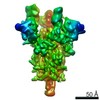
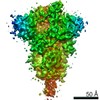
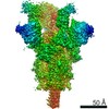
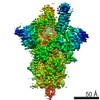
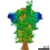
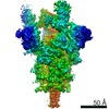
Journal: bioRxiv / Year: 2020 Title: Bi-paratopic and multivalent human VH domains neutralize SARS-CoV-2 by targeting distinct epitopes within the ACE2 binding interface of Spike. Authors: Colton J Bracken / Shion A Lim / Paige Solomon / Nicholas J Rettko / Duy P Nguyen / Beth Shoshana Zha / Kaitlin Schaefer / James R Byrnes / Jie Zhou / Irene Lui / Jia Liu / Katarina Pance / ...Authors: Colton J Bracken / Shion A Lim / Paige Solomon / Nicholas J Rettko / Duy P Nguyen / Beth Shoshana Zha / Kaitlin Schaefer / James R Byrnes / Jie Zhou / Irene Lui / Jia Liu / Katarina Pance / / Xin X Zhou / Kevin K Leung / James A Wells / Abstract: Neutralizing agents against SARS-CoV-2 are urgently needed for treatment and prophylaxis of COVID-19. Here, we present a strategy to rapidly identify and assemble synthetic human variable heavy (VH) ...Neutralizing agents against SARS-CoV-2 are urgently needed for treatment and prophylaxis of COVID-19. Here, we present a strategy to rapidly identify and assemble synthetic human variable heavy (VH) domain binders with high affinity toward neutralizing epitopes without the need for high-resolution structural information. We constructed a VH-phage library and targeted a known neutralizing site, the angiotensin-converting enzyme 2 (ACE2) binding interface of the trimeric SARS-CoV-2 Spike receptor-binding domain (Spike-RBD). Using a masked selection approach, we identified 85 unique VH binders to two non-overlapping epitopes within the ACE2 binding site on Spike-RBD. This enabled us to systematically link these VH domains into multivalent and bi-paratopic formats. These multivalent and bi-paratopic VH constructs showed a marked increase in affinity to Spike (up to 600-fold) and neutralization potency (up to 1400-fold) on pseudotyped SARS-CoV-2 virus when compared to the standalone VH domains. The most potent binder, a trivalent VH, neutralized authentic SARS-CoV-2 with half-minimal inhibitory concentration (IC ) of 4.0 nM (180 ng/mL). A cryo-EM structure of the trivalent VH bound to Spike shows each VH domain bound an RBD at the ACE2 binding site, explaining its increased neutralization potency and confirming our original design strategy. Our results demonstrate that targeted selection and engineering campaigns using a VH-phage library can enable rapid assembly of highly avid and potent molecules towards therapeutically important protein interfaces.
History
Deposition
Aug 25, 2020
-
Header (metadata) release
Oct 7, 2020
-
Map release
Oct 7, 2020
-
Update
Nov 13, 2024
-
Current status
Nov 13, 2024
Processing site: RCSB / Status: Released
-
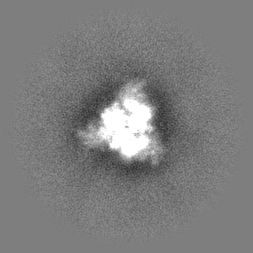
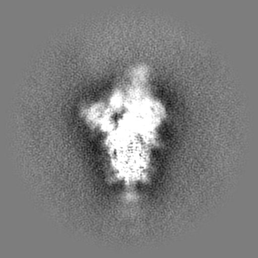
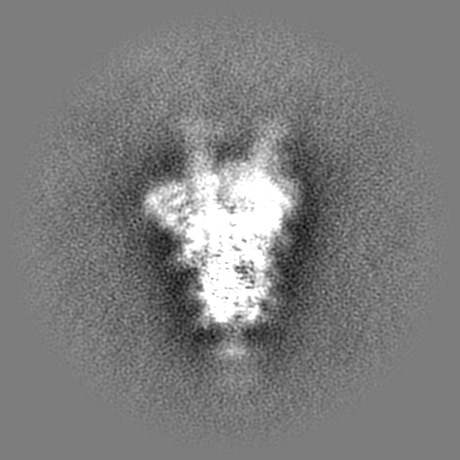
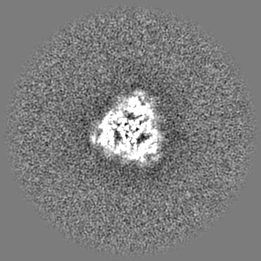
Structure visualization
Movie
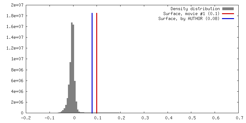
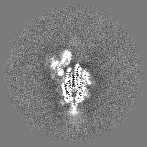
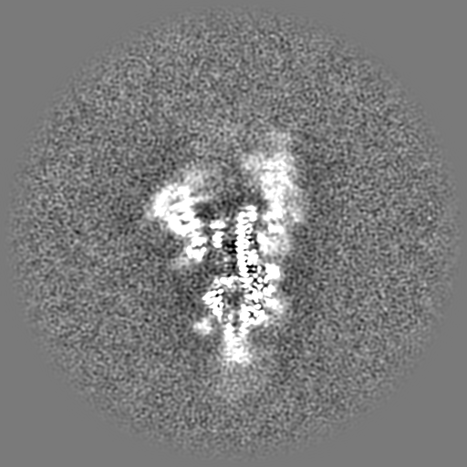
Surface view with section colored by density value
Model: UltrAuFoil R1.2/1.3 / Material: GOLD / Mesh: 200 / Support film - Material: CARBON / Support film - topology: HOLEY / Pretreatment - Type: GLOW DISCHARGE / Pretreatment - Time: 30 sec. / Pretreatment - Atmosphere: AIR
Vitrification
Cryogen name: ETHANE / Chamber humidity: 100 % / Chamber temperature: 277 K / Instrument: FEI VITROBOT MARK IV
-
Electron microscopy
Microscope
FEI TITAN KRIOS
Image recording
Film or detector model: GATAN K3 BIOQUANTUM (6k x 4k) / Average electron dose: 78.0 e/Å2
Electron beam
Acceleration voltage: 300 kV / Electron source: FIELD EMISSION GUN
Electron optics
Illumination mode: FLOOD BEAM / Imaging mode: BRIGHT FIELD
Experimental equipment
Model: Titan Krios / Image courtesy: FEI Company
+
Image processing #1
Image processing ID
1
Startup model
Type of model: NONE
Final reconstruction
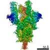
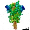
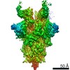
Resolution.type: BY AUTHOR / Resolution: 6.0 Å / Resolution method: OTHER Details: This reconstruction is a Gaussian low pass filtered map of a 3.2A gold standard FSC map, to better account for the VH density. The model was fit and relaxed into this map. Number images used: 21000
Initial angle assignment
Type: RANDOM ASSIGNMENT
Final angle assignment
Type: MAXIMUM LIKELIHOOD
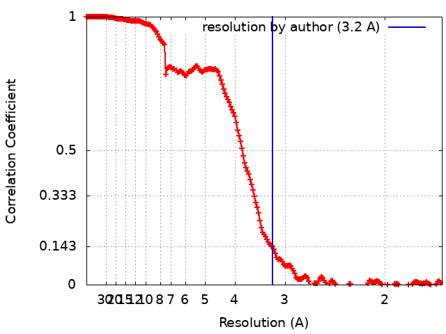
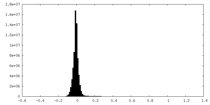
FSC plot (resolution estimation)
+
Image processing #2
Image processing ID
2
Startup model
Type of model: NONE
Final reconstruction
Resolution.type: BY AUTHOR / Resolution: 3.2 Å / Resolution method: FSC 0.143 CUT-OFF Details: Same reconstruction as above but filtered based on the gold standard FSC to 3.2A. Shows high resolution features but density for the low occupancy VH is poorly resolved. Number images used: 21000
In the structure databanks used in Yorodumi, some data are registered as the other names, "COVID-19 virus" and "2019-nCoV". Here are the details of the virus and the list of structure data.
Jan 31, 2019. EMDB accession codes are about to change! (news from PDBe EMDB page)
EMDB accession codes are about to change! (news from PDBe EMDB page)
The allocation of 4 digits for EMDB accession codes will soon come to an end. Whilst these codes will remain in use, new EMDB accession codes will include an additional digit and will expand incrementally as the available range of codes is exhausted. The current 4-digit format prefixed with “EMD-” (i.e. EMD-XXXX) will advance to a 5-digit format (i.e. EMD-XXXXX), and so on. It is currently estimated that the 4-digit codes will be depleted around Spring 2019, at which point the 5-digit format will come into force.
The EM Navigator/Yorodumi systems omit the EMD- prefix.
Related info.:Q: What is EMD? / ID/Accession-code notation in Yorodumi/EM Navigator
Yorodumi is a browser for structure data from EMDB, PDB, SASBDB, etc.
This page is also the successor to EM Navigator detail page, and also detail information page/front-end page for Omokage search.
The word "yorodu" (or yorozu) is an old Japanese word meaning "ten thousand". "mi" (miru) is to see.
Related info.:EMDB / PDB / SASBDB / Comparison of 3 databanks / Yorodumi Search / Aug 31, 2016. New EM Navigator & Yorodumi / Yorodumi Papers / Jmol/JSmol / Function and homology information / Changes in new EM Navigator and Yorodumi
 Movie
Movie Controller

 Yorodumi
 Open data
 Basic information
 Map data
 Sample
 Keywords
 Function and homology information
 Homo sapiens (human) /
 Severe acute respiratory syndrome coronavirus 2
 Authors
 United States, 4 items
  Citation
 Structure visualization
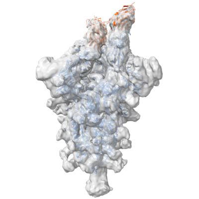
 Downloads & links
 emd_22514.png
 http://ftp.pdbj.org/pub/emdb/structures/EMD-22514
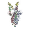


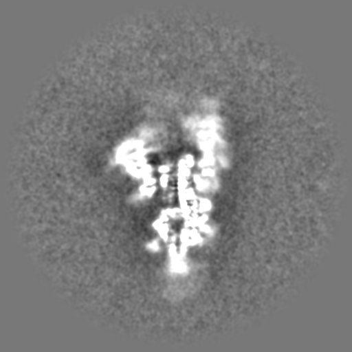
 Z (Sec.)
 Y (Row.)
 X (Col.)


 Sample components

 Cricetulus griseus (Chinese hamster)
 Processing
 Electron microscopy
 FIELD EMISSION GUN