 Movie
Movie Controller

[English] 日本語
 Yorodumi
- EMDB-2228: The Structure of Lactococcal Phage TP901-1 by electron microscopy... -
+ Open data

- Basic information
| Entry | Database: EMDB / ID: EMD-2228 | |||||||||
|---|---|---|---|---|---|---|---|---|---|---|
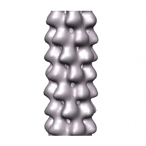
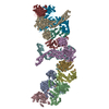
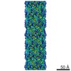
| Title | The Structure of Lactococcal Phage TP901-1 by electron microscopy: the helical tail | |||||||||
 Map data Map data | Helical Reconstruction of a tail fragment of TP901-1. | |||||||||
 Sample Sample |
| |||||||||
 Keywords Keywords | tail / tp901 / lactococcal phage / helical processing | |||||||||
| Biological species |  Lactococcus phage TP901-1 (virus) Lactococcus phage TP901-1 (virus) | |||||||||
| Method | single particle reconstruction / cryo EM / negative staining / Resolution: 20.0 Å | |||||||||
 Authors Authors | Bebeacua C / Lai L / Skovgaard Vegge C / Brondsted L / van Heel M / Veesler D / Cambillau C | |||||||||
 Citation Citation | Journal: J Virol / Year: 2013 Title: Visualizing a complete Siphoviridae member by single-particle electron microscopy: the structure of lactococcal phage TP901-1. Authors: Cecilia Bebeacua / Livia Lai / Christina Skovgaard Vegge / Lone Brøndsted / Marin van Heel / David Veesler / Christian Cambillau /  Abstract: Tailed phages are genome delivery machines exhibiting unequaled efficiency acquired over more than 3 billion years of evolution. Siphophages from the P335 and 936 families infect the Gram-positive ...Tailed phages are genome delivery machines exhibiting unequaled efficiency acquired over more than 3 billion years of evolution. Siphophages from the P335 and 936 families infect the Gram-positive bacterium Lactococcus lactis using receptor-binding proteins anchored to the host adsorption apparatus (baseplate). Crystallographic and electron microscopy (EM) studies have shed light on the distinct adsorption strategies used by phages of these two families, suggesting that they might also rely on different infection mechanisms. Here, we report electron microscopy reconstructions of the whole phage TP901-1 (P335 species) and propose a composite EM model of this gigantic molecular machine. Our results suggest conservation of structural proteins among tailed phages and add to the growing body of evidence pointing to a common evolutionary origin for these virions. Finally, we propose that host adsorption apparatus architectures have evolved in correlation with the nature of the receptors used during infection. | |||||||||
| History |
|
- Structure visualization
| Movie |
 Movie viewer Movie viewer |
|---|---|
| Structure viewer | EM map: SurfViewMolmilJmol/JSmol |
| Supplemental images |
- Downloads & links
-EMDB archive
| Map data | emd_2228.map.gz | 166.8 KB | EMDB map data format | |
|---|---|---|---|---|
| Header (meta data) | emd-2228-v30.xmlemd-2228.xml | 10.3 KB 10.3 KB | Display Display | EMDB header |
| Images | emd_2228.tif | 732.8 KB | ||
| Archive directory |  http://ftp.pdbj.org/pub/emdb/structures/EMD-2228ftp://ftp.pdbj.org/pub/emdb/structures/EMD-2228 http://ftp.pdbj.org/pub/emdb/structures/EMD-2228ftp://ftp.pdbj.org/pub/emdb/structures/EMD-2228 | HTTPS FTP |
-Validation report
| Summary document | emd_2228_validation.pdf.gz | 207 KB | Display | EMDB validaton report |
|---|---|---|---|---|
| Full document | emd_2228_full_validation.pdf.gz | 206.1 KB | Display | |
| Data in XML | emd_2228_validation.xml.gz | 5 KB | Display | |
| Arichive directory | https://ftp.pdbj.org/pub/emdb/validation_reports/EMD-2228ftp://ftp.pdbj.org/pub/emdb/validation_reports/EMD-2228 | HTTPS FTP |
-Related structure data


-Links
| EMDB pages | EMDB (EBI/PDBe) / EMDataResource |
|---|
-Map
| File | Download / File: emd_2228.map.gz / Format: CCP4 / Size: 1.4 MB / Type: IMAGE STORED AS FLOATING POINT NUMBER (4 BYTES) | ||||||||||||||||||||||||||||||||||||||||||||||||||||||||||||||||||||
|---|---|---|---|---|---|---|---|---|---|---|---|---|---|---|---|---|---|---|---|---|---|---|---|---|---|---|---|---|---|---|---|---|---|---|---|---|---|---|---|---|---|---|---|---|---|---|---|---|---|---|---|---|---|---|---|---|---|---|---|---|---|---|---|---|---|---|---|---|---|
| Annotation | Helical Reconstruction of a tail fragment of TP901-1. | ||||||||||||||||||||||||||||||||||||||||||||||||||||||||||||||||||||
| Projections & slices | Image control
Images are generated by Spider. | ||||||||||||||||||||||||||||||||||||||||||||||||||||||||||||||||||||
| Voxel size | X=Y=Z: 4.64 Å | ||||||||||||||||||||||||||||||||||||||||||||||||||||||||||||||||||||
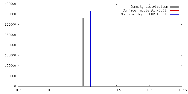
| Density |
| ||||||||||||||||||||||||||||||||||||||||||||||||||||||||||||||||||||
| Symmetry | Space group: 1 | ||||||||||||||||||||||||||||||||||||||||||||||||||||||||||||||||||||
| Details | EMDB XML:
CCP4 map header:
| ||||||||||||||||||||||||||||||||||||||||||||||||||||||||||||||||||||
 Z (Sec.)
 Y (Row.)
 X (Col.)


-Supplemental data
- Sample components
-Entire : Helical tail of the lactococcal phage TP901-1 (from a sample cont...
| Entire | Name: Helical tail of the lactococcal phage TP901-1 (from a sample containing the full phage) |
|---|---|
| Components |
|
-Supramolecule #1000: Helical tail of the lactococcal phage TP901-1 (from a sample cont...
| Supramolecule | Name: Helical tail of the lactococcal phage TP901-1 (from a sample containing the full phage) type: sample / ID: 1000 / Oligomeric state: Hexameric (C6)-Helical symmetry / Number unique components: 1 |
|---|
-Supramolecule #1: Lactococcus phage TP901-1
| Supramolecule | Name: Lactococcus phage TP901-1 / type: virus / ID: 1 Details: The sample contained the full phages with tail and baseplate. NCBI-ID: 35345 / Sci species name: Lactococcus phage TP901-1 / Database: NCBI / Virus type: VIRION / Virus isolate: STRAIN / Virus enveloped: No / Virus empty: No |
|---|---|
| Host (natural) | Organism:  Lactococcus lactis (lactic acid bacteria) / synonym: BACTERIA(EUBACTERIA) Lactococcus lactis (lactic acid bacteria) / synonym: BACTERIA(EUBACTERIA) |
| Virus shell | Shell ID: 1 / Diameter: 600 Å / T number (triangulation number): 7 |
-Experimental details
-Structure determination
| Method | negative staining, cryo EM |
|---|---|
 Processing Processing | single particle reconstruction |
| Aggregation state | particle |
-Sample preparation
| Buffer | pH: 7.5 Details: 100 mM sodium chloride, 10 mM magnesium sulfate, 50 mM Tris [pH 7.5], and 0.01% [wt/vol] gelatin |
|---|---|
| Staining | Type: NEGATIVE Details: The sample was incubated for one minute on glow-discharged grids and stained with 2% uranyl acetate for 10 seconds. |
| Grid | Details: 200 mesh carbon grids were glow discharged for 20 seconds. |
| Vitrification | Cryogen name: NITROGEN / Instrument: OTHER |
- Electron microscopy
| Microscope | FEI/PHILIPS CM200FEG |
|---|---|
| Alignment procedure | Legacy - Astigmatism: Objective lens astigmatism was corrected at 130,000 times magnification. |
| Specialist optics | Energy filter - Name: FEI |
| Date | Jun 1, 2008 |
| Image recording | Category: CCD / Film or detector model: GENERIC TVIPS (4k x 4k) / Number real images: 200 / Average electron dose: 10 e/Å2 |
| Electron beam | Acceleration voltage: 200 kV / Electron source:  FIELD EMISSION GUN FIELD EMISSION GUN |
| Electron optics | Illumination mode: SPOT SCAN / Imaging mode: BRIGHT FIELD / Cs: 2.2 mm / Nominal defocus max: 1.5 µm / Nominal defocus min: 0.5 µm / Nominal magnification: 50000 |
| Sample stage | Specimen holder: Nitrogen cooled / Specimen holder model: SIDE ENTRY, EUCENTRIC |
-Image processing
| CTF correction | Details: Images |
|---|---|
| Final reconstruction | Algorithm: OTHER / Resolution.type: BY AUTHOR / Resolution: 20.0 Å / Resolution method: OTHER / Software - Name: IMAGIC Details: The fragments were initially part of full-phage particles. After an initial refinement of the full-phage, the full phage was cut into fragments that were combined into one dataset and ...Details: The fragments were initially part of full-phage particles. After an initial refinement of the full-phage, the full phage was cut into fragments that were combined into one dataset and submitted to helical processing. Number images used: 4000 |
| Final angle assignment | Details: C6-Helical symmetry imposed |
