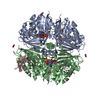
Movie
Movie Controller

[English] 日本語
 Yorodumi
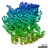
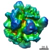
- EMDB-21156: Cryo-EM Structure of Escherichia coli 2-oxoglutarate dehydrogenas... -
+ Open data

- Basic information
| Entry | Database: EMDB / ID: EMD-21156 | |||||||||
|---|---|---|---|---|---|---|---|---|---|---|
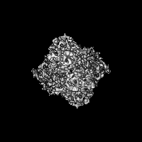
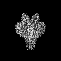
| Title | Cryo-EM Structure of Escherichia coli 2-oxoglutarate dehydrogenase E1 component sucA | |||||||||
 Map data Map data | Escherichia coli 2-oxoglutarate dehydrogenase E1 component sucA | |||||||||
 Sample Sample |
| |||||||||
 Keywords Keywords | TCA cycle / 2-oxoglutarate dehydrogenase complex (OGDH) / E1 component / sucA / AMP / Oxaloacetate (OAA) / Dimer / OXIDOREDUCTASE | |||||||||
| Function / homology |  Function and homology information Function and homology informationoxoglutarate dehydrogenase (succinyl-transferring) / oxoglutarate dehydrogenase (succinyl-transferring) activity / oxoglutarate dehydrogenase complex / thiamine pyrophosphate binding / tricarboxylic acid cycle / nucleotide binding / magnesium ion binding / identical protein binding / cytoplasm / cytosol Similarity search - Function | |||||||||
| Biological species |  | |||||||||
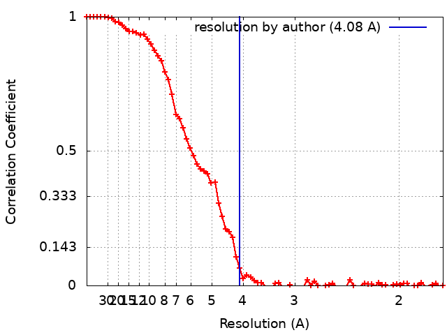
| Method | single particle reconstruction / cryo EM / Resolution: 4.08 Å | |||||||||
 Authors Authors | Gao H | |||||||||
 Citation Citation | Journal: To Be Published Title: Cryo-EM Structure of Escherichia coli 2-oxoglutarate dehydrogenase E1 component sucA Authors: Gao H | |||||||||
| History |
|
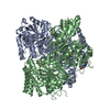
- Structure visualization
| Movie |
Movie viewer |
|---|---|
| Structure viewer | EM map: SurfViewMolmilJmol/JSmol |
| Supplemental images |

- Downloads & links
-EMDB archive
| Map data | emd_21156.map.gz | 28.5 MB | EMDB map data format | |
|---|---|---|---|---|
| Header (meta data) | emd-21156-v30.xmlemd-21156.xml | 11.6 KB 11.6 KB | Display Display | EMDB header |
| FSC (resolution estimation) | emd_21156_fsc.xml | 9.3 KB | Display | FSC data file |
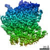
| Images | 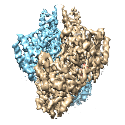 emd_21156.png emd_21156.png | 158.5 KB | ||
| Filedesc metadata | emd-21156.cif.gz | 5.5 KB | ||
| Archive directory |  http://ftp.pdbj.org/pub/emdb/structures/EMD-21156ftp://ftp.pdbj.org/pub/emdb/structures/EMD-21156 http://ftp.pdbj.org/pub/emdb/structures/EMD-21156ftp://ftp.pdbj.org/pub/emdb/structures/EMD-21156 | HTTPS FTP |
-Related structure data
| Related structure data |  6vefMC M: atomic model generated by this map C: citing same article ( |
|---|---|
| Similar structure data |

-Links
| EMDB pages | EMDB (EBI/PDBe) / EMDataResource |
|---|---|
| Related items in Molecule of the Month |


-Map
| File | Download / File: emd_21156.map.gz / Format: CCP4 / Size: 30.5 MB / Type: IMAGE STORED AS FLOATING POINT NUMBER (4 BYTES) | ||||||||||||||||||||||||||||||||||||||||||||||||||||||||||||
|---|---|---|---|---|---|---|---|---|---|---|---|---|---|---|---|---|---|---|---|---|---|---|---|---|---|---|---|---|---|---|---|---|---|---|---|---|---|---|---|---|---|---|---|---|---|---|---|---|---|---|---|---|---|---|---|---|---|---|---|---|---|
| Annotation | Escherichia coli 2-oxoglutarate dehydrogenase E1 component sucA | ||||||||||||||||||||||||||||||||||||||||||||||||||||||||||||
| Projections & slices | Image control
Images are generated by Spider. | ||||||||||||||||||||||||||||||||||||||||||||||||||||||||||||
| Voxel size | X=Y=Z: 0.877 Å | ||||||||||||||||||||||||||||||||||||||||||||||||||||||||||||
| Density |
| ||||||||||||||||||||||||||||||||||||||||||||||||||||||||||||
| Symmetry | Space group: 1 | ||||||||||||||||||||||||||||||||||||||||||||||||||||||||||||
| Details | EMDB XML:
CCP4 map header:
| ||||||||||||||||||||||||||||||||||||||||||||||||||||||||||||
 Z (Sec.)
 Y (Row.)
 X (Col.)


-Supplemental data
- Sample components
-Entire : Escherichia coli 2-oxoglutarate dehydrogenase E1 component sucA
| Entire | Name: Escherichia coli 2-oxoglutarate dehydrogenase E1 component sucA |
|---|---|
| Components |
|
-Supramolecule #1: Escherichia coli 2-oxoglutarate dehydrogenase E1 component sucA
| Supramolecule | Name: Escherichia coli 2-oxoglutarate dehydrogenase E1 component sucA type: complex / ID: 1 / Parent: 0 / Macromolecule list: #1 |
|---|---|
| Source (natural) | Organism: |
-Macromolecule #1: 2-oxoglutarate dehydrogenase E1 component
| Macromolecule | Name: 2-oxoglutarate dehydrogenase E1 component / type: protein_or_peptide / ID: 1 / Number of copies: 2 / Enantiomer: LEVO EC number: oxoglutarate dehydrogenase (succinyl-transferring) |
|---|---|
| Source (natural) | Organism: |
| Molecular weight | Theoretical: 94.942508 KDa |
| Sequence | String: DTNVKQVKVL QLINAYRFRG HQHANLDPLG LYQQDKVADL DPSFHDLTEA DFQETFNVGS FASGKETMKL GELLEALKQT YCGPIGAEY MHITSTEEKR WIQQRIESGR ATFNSEEKKR FLSELTAAEG LERYLGAKFP GAKRFSLEGD ALIPMLKEMI R HAGNSGTR ...String: DTNVKQVKVL QLINAYRFRG HQHANLDPLG LYQQDKVADL DPSFHDLTEA DFQETFNVGS FASGKETMKL GELLEALKQT YCGPIGAEY MHITSTEEKR WIQQRIESGR ATFNSEEKKR FLSELTAAEG LERYLGAKFP GAKRFSLEGD ALIPMLKEMI R HAGNSGTR EVVLGMAHRG RLNVLVNVLG KKPQDLFDEF AGKHKEHLGT GDVKYHMGFS SDFQTDGGLV HLALAFNPSH LE IVSPVVI GSVRARLDRL DEPSSNKVLP ITIHGDAAVT GQGVVQETLN MSKARGYEVG GTVRIVINNQ VGFTTSNPLD ARS TPYMTD IGKMVQAPIF HVNADDPEAV AFVTRLALDF RNTFKRDVFI DLVCYRRHGH NEADEPSATQ KIKKHPTPRK IYAD KLEQE KVATLEDATE MVNLYRDALD AGDCVVAEWR PMNMHSFTWS PYLNHEWDEE YPNKVEMKRL QELAKRISTV PEAVE MQSR VAKIYGDRQA MAAGEKLFDW GGAENLAYAT LVDEGIPVRL SGEDSGRGTF FHRHAVIHNQ SNGSTYTPLQ HIHNGQ GAF RVWDSVLSEE AVLAFEYGYA TAEPRTLTIW EAQFGDFANG AQVVIDQFIS SGEQKWGRMC GLVMLLPHGY EGQGPEH SS ARLERYLQLC AEQNMQVCVP STPAQVYHML RRQALRGMRR PLVVMSPKSL LRHPLAVSSL EELANGTFLP AIGEIDEL D PKGVKRVVMC SGKVYYDLLE QRRKNNQHDV AIVRIEQLYP FPHKAMQEVL QQFAHVKDFV WCQEEPLNQG AWYCSQHHF REVIPFGASL RYAGRPASAS PAVGHMSVHQ KQQQDLVNDA LNVE UniProtKB: Oxoglutarate dehydrogenase (succinyl-transferring) |
-Macromolecule #2: ADENOSINE MONOPHOSPHATE
| Macromolecule | Name: ADENOSINE MONOPHOSPHATE / type: ligand / ID: 2 / Number of copies: 2 / Formula: AMP |
|---|---|
| Molecular weight | Theoretical: 347.221 Da |
| Chemical component information |  ChemComp-AMP: |
-Macromolecule #3: OXALOACETATE ION
| Macromolecule | Name: OXALOACETATE ION / type: ligand / ID: 3 / Number of copies: 2 / Formula: OAA |
|---|---|
| Molecular weight | Theoretical: 131.064 Da |
| Chemical component information |  ChemComp-OAA: |
-Experimental details
-Structure determination
| Method | cryo EM |
|---|---|
 Processing Processing | single particle reconstruction |
| Aggregation state | particle |
-Sample preparation
| Buffer | pH: 7.5 |
|---|---|
| Grid | Model: Quantifoil R1.2/1.3 / Material: GOLD / Mesh: 300 / Pretreatment - Type: GLOW DISCHARGE / Pretreatment - Time: 80 sec. |
| Vitrification | Cryogen name: ETHANE / Chamber humidity: 100 % / Chamber temperature: 277 K / Instrument: FEI VITROBOT MARK IV |
- Electron microscopy
| Microscope | FEI TECNAI ARCTICA |
|---|---|
| Image recording | Film or detector model: GATAN K3 (6k x 4k) / Number grids imaged: 1 / Number real images: 800 / Average exposure time: 2.0 sec. / Average electron dose: 80.0 e/Å2 |
| Electron beam | Acceleration voltage: 200 kV / Electron source:  FIELD EMISSION GUN FIELD EMISSION GUN |
| Electron optics | C2 aperture diameter: 50.0 µm / Illumination mode: FLOOD BEAM / Imaging mode: BRIGHT FIELD / Cs: 2.7 mm |
| Experimental equipment |  Model: Talos Arctica / Image courtesy: FEI Company |