[English] 日本語
 Yorodumi
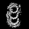
- EMDB-20681: Average of the proximal basal body from bovine respiratory cilia -
+ Open data
- Basic information
| Entry | Database: EMDB / ID: EMD-20681 | |||||||||
|---|---|---|---|---|---|---|---|---|---|---|
| Title | Average of the proximal basal body from bovine respiratory cilia | |||||||||
 Map data Map data | Average of the proximal basal body from bovine respiratory cilia | |||||||||
 Sample Sample |
| |||||||||
| Biological species |  | |||||||||
| Method | subtomogram averaging / cryo EM / Resolution: 33.2 Å | |||||||||
 Authors Authors | Greenan GA / Vale RD / Agard DA | |||||||||
| Funding support |  United States, 2 items United States, 2 items
| |||||||||
 Citation Citation |  Journal: J Cell Biol / Year: 2020 Journal: J Cell Biol / Year: 2020Title: Electron cryotomography of intact motile cilia defines the basal body to axoneme transition. Authors: Garrett A Greenan / Ronald D Vale / David A Agard /  Abstract: Cells use motile cilia to generate force in the extracellular space. The structure of a cilium can be classified into three subdomains: the intracellular basal body (BB) that templates cilium ...Cells use motile cilia to generate force in the extracellular space. The structure of a cilium can be classified into three subdomains: the intracellular basal body (BB) that templates cilium formation, the extracellular axoneme that generates force, and the transition zone (TZ) that bridges them. While the BB is composed of triplet microtubules (TMTs), the axoneme is composed of doublet microtubules (DMTs), meaning the cilium must convert between different microtubule geometries. Here, we performed electron cryotomography to define this conversion, and our reconstructions reveal identifying structural features of the BB, TZ, and axoneme. Each region is distinct in terms of microtubule number and geometry, microtubule inner proteins, and microtubule linkers. TMT to DMT conversion occurs within the BB, and microtubule geometry changes to axonemal by the end of the TZ, followed by the addition of axoneme-specific components essential for cilium motility. Our results provide the highest-resolution images of the motile cilium to date and reveal how BBs template axonemes. | |||||||||
| History |
|
- Structure visualization
| Movie |
 Movie viewer Movie viewer |
|---|---|
| Structure viewer | EM map:  SurfView SurfView Molmil Molmil Jmol/JSmol Jmol/JSmol |
| Supplemental images |
- Downloads & links
-EMDB archive
| Map data |  emd_20681.map.gz emd_20681.map.gz | 1.6 MB |  EMDB map data format EMDB map data format | |
|---|---|---|---|---|
| Header (meta data) |  emd-20681-v30.xml emd-20681-v30.xml emd-20681.xml emd-20681.xml | 7.9 KB 7.9 KB | Display Display |  EMDB header EMDB header |
| Images |  emd_20681.png emd_20681.png | 47.2 KB | ||
| Archive directory |  http://ftp.pdbj.org/pub/emdb/structures/EMD-20681 http://ftp.pdbj.org/pub/emdb/structures/EMD-20681 ftp://ftp.pdbj.org/pub/emdb/structures/EMD-20681 ftp://ftp.pdbj.org/pub/emdb/structures/EMD-20681 | HTTPS FTP |
-Validation report
| Summary document |  emd_20681_validation.pdf.gz emd_20681_validation.pdf.gz | 78.3 KB | Display |  EMDB validaton report EMDB validaton report |
|---|---|---|---|---|
| Full document |  emd_20681_full_validation.pdf.gz emd_20681_full_validation.pdf.gz | 77.4 KB | Display | |
| Data in XML |  emd_20681_validation.xml.gz emd_20681_validation.xml.gz | 495 B | Display | |
| Arichive directory |  https://ftp.pdbj.org/pub/emdb/validation_reports/EMD-20681 https://ftp.pdbj.org/pub/emdb/validation_reports/EMD-20681 ftp://ftp.pdbj.org/pub/emdb/validation_reports/EMD-20681 ftp://ftp.pdbj.org/pub/emdb/validation_reports/EMD-20681 | HTTPS FTP |
-Related structure data
| Related structure data | C: citing same article ( |
|---|---|
| Similar structure data |
- Links
Links
| EMDB pages |  EMDB (EBI/PDBe) / EMDB (EBI/PDBe) /  EMDataResource EMDataResource |
|---|
- Map
Map
| File |  Download / File: emd_20681.map.gz / Format: CCP4 / Size: 12.9 MB / Type: IMAGE STORED AS FLOATING POINT NUMBER (4 BYTES) Download / File: emd_20681.map.gz / Format: CCP4 / Size: 12.9 MB / Type: IMAGE STORED AS FLOATING POINT NUMBER (4 BYTES) | ||||||||||||||||||||||||||||||||||||||||||||||||||||||||||||||||||||
|---|---|---|---|---|---|---|---|---|---|---|---|---|---|---|---|---|---|---|---|---|---|---|---|---|---|---|---|---|---|---|---|---|---|---|---|---|---|---|---|---|---|---|---|---|---|---|---|---|---|---|---|---|---|---|---|---|---|---|---|---|---|---|---|---|---|---|---|---|---|
| Annotation | Average of the proximal basal body from bovine respiratory cilia | ||||||||||||||||||||||||||||||||||||||||||||||||||||||||||||||||||||
| Projections & slices | Image control
Images are generated by Spider. | ||||||||||||||||||||||||||||||||||||||||||||||||||||||||||||||||||||
| Voxel size | X=Y=Z: 8.18 Å | ||||||||||||||||||||||||||||||||||||||||||||||||||||||||||||||||||||
| Density |
| ||||||||||||||||||||||||||||||||||||||||||||||||||||||||||||||||||||
| Symmetry | Space group: 1 | ||||||||||||||||||||||||||||||||||||||||||||||||||||||||||||||||||||
| Details | EMDB XML:
CCP4 map header:
| ||||||||||||||||||||||||||||||||||||||||||||||||||||||||||||||||||||
-Supplemental data
- Sample components
-Entire : Nascent axoneme from bovine respiratory epithelial cells
| Entire | Name: Nascent axoneme from bovine respiratory epithelial cells |
|---|---|
| Components |
|
-Supramolecule #1: Nascent axoneme from bovine respiratory epithelial cells
| Supramolecule | Name: Nascent axoneme from bovine respiratory epithelial cells type: organelle_or_cellular_component / ID: 1 / Parent: 0 |
|---|---|
| Source (natural) | Organism:  |
-Experimental details
-Structure determination
| Method | cryo EM |
|---|---|
 Processing Processing | subtomogram averaging |
| Aggregation state | filament |
- Sample preparation
| Buffer | pH: 7.4 |
|---|---|
| Vitrification | Cryogen name: ETHANE / Chamber humidity: 100 % / Chamber temperature: 295 K / Instrument: FEI VITROBOT MARK IV / Details: 15 second blot. |
- Electron microscopy
| Microscope | FEI POLARA 300 |
|---|---|
| Image recording | Film or detector model: GATAN K2 SUMMIT (4k x 4k) / Detector mode: COUNTING / Average electron dose: 0.8 e/Å2 |
| Electron beam | Acceleration voltage: 300 kV / Electron source:  FIELD EMISSION GUN FIELD EMISSION GUN |
| Electron optics | C2 aperture diameter: 100.0 µm / Illumination mode: FLOOD BEAM / Imaging mode: BRIGHT FIELD |
| Experimental equipment |  Model: Tecnai Polara / Image courtesy: FEI Company |
- Image processing
| Final reconstruction | Number classes used: 1 / Applied symmetry - Point group: C1 (asymmetric) / Algorithm: BACK PROJECTION / Resolution.type: BY AUTHOR / Resolution: 33.2 Å / Resolution method: FSC 0.143 CUT-OFF / Number subtomograms used: 1350 |
|---|---|
| Extraction | Number tomograms: 21 / Number images used: 9450 |
| Final angle assignment | Type: MAXIMUM LIKELIHOOD |
 Movie
Movie Controller


 Z (Sec.)
 Y (Row.)
 X (Col.)


