 Movie
Movie Controller


+ Open data

- Basic information
| Entry | Database: EMDB / ID: EMD-2045 | |||||||||
|---|---|---|---|---|---|---|---|---|---|---|
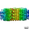
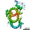
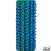
| Title | Subtomogram average of type VI secretion system (T6SS) sheath | |||||||||
 Map data Map data | electron cryotomography, sub-tomogram average | |||||||||
 Sample Sample |
| |||||||||
 Keywords Keywords | type VI secretion system / T6SS / bacterial secretion / sheath / VipA / VipB / VipAB | |||||||||
| Biological species |   Vibrio cholerae (bacteria) Vibrio cholerae (bacteria) | |||||||||
| Method | subtomogram averaging / cryo EM | |||||||||
 Authors Authors | Basler M / Pilhofer M / Henderson GP / Jensen GJ / Mekalanos JJ | |||||||||
 Citation Citation | Journal: Nature / Year: 2012 Title: Type VI secretion requires a dynamic contractile phage tail-like structure. Authors: M Basler / M Pilhofer / G P Henderson / G J Jensen / J J Mekalanos /  Abstract: Type VI secretion systems are bacterial virulence-associated nanomachines composed of proteins that are evolutionarily related to components of bacteriophage tails. Here we show that protein ...Type VI secretion systems are bacterial virulence-associated nanomachines composed of proteins that are evolutionarily related to components of bacteriophage tails. Here we show that protein secretion by the type VI secretion system of Vibrio cholerae requires the action of a dynamic intracellular tubular structure that is structurally and functionally homologous to contractile phage tail sheath. Time-lapse fluorescence light microscopy reveals that sheaths of the type VI secretion system cycle between assembly, quick contraction, disassembly and re-assembly. Whole-cell electron cryotomography further shows that the sheaths appear as long tubular structures in either extended or contracted conformations that are connected to the inner membrane by a distinct basal structure. These data support a model in which the contraction of the type VI secretion system sheath provides the energy needed to translocate proteins out of effector cells and into adjacent target cells. | |||||||||
| History |
|
- Structure visualization
| Movie |
 Movie viewer Movie viewer |
|---|---|
| Structure viewer | EM map: SurfViewMolmilJmol/JSmol |
| Supplemental images |

- Downloads & links
-EMDB archive
| Map data | emd_2045.map.gz | 1.6 MB | EMDB map data format | |
|---|---|---|---|---|
| Header (meta data) | emd-2045-v30.xmlemd-2045.xml | 7.7 KB 7.7 KB | Display Display | EMDB header |
| Images | EM-2045-T6SS.tif | 319.5 KB | ||
| Archive directory |  http://ftp.pdbj.org/pub/emdb/structures/EMD-2045ftp://ftp.pdbj.org/pub/emdb/structures/EMD-2045 http://ftp.pdbj.org/pub/emdb/structures/EMD-2045ftp://ftp.pdbj.org/pub/emdb/structures/EMD-2045 | HTTPS FTP |
-Related structure data
| Similar structure data |
|---|
-Links
| EMDB pages | EMDB (EBI/PDBe) / EMDataResource |
|---|
-Map
| File | Download / File: emd_2045.map.gz / Format: CCP4 / Size: 1.8 MB / Type: IMAGE STORED AS FLOATING POINT NUMBER (4 BYTES) | ||||||||||||||||||||||||||||||||||||||||||||||||||||||||||||||||||||
|---|---|---|---|---|---|---|---|---|---|---|---|---|---|---|---|---|---|---|---|---|---|---|---|---|---|---|---|---|---|---|---|---|---|---|---|---|---|---|---|---|---|---|---|---|---|---|---|---|---|---|---|---|---|---|---|---|---|---|---|---|---|---|---|---|---|---|---|---|---|
| Annotation | electron cryotomography, sub-tomogram average | ||||||||||||||||||||||||||||||||||||||||||||||||||||||||||||||||||||
| Projections & slices | Image control
Images are generated by Spider. generated in cubic-lattice coordinate | ||||||||||||||||||||||||||||||||||||||||||||||||||||||||||||||||||||
| Voxel size | X=Y=Z: 6.3 Å | ||||||||||||||||||||||||||||||||||||||||||||||||||||||||||||||||||||
| Density |
| ||||||||||||||||||||||||||||||||||||||||||||||||||||||||||||||||||||
| Symmetry | Space group: 1 | ||||||||||||||||||||||||||||||||||||||||||||||||||||||||||||||||||||
| Details | EMDB XML:
CCP4 map header:
| ||||||||||||||||||||||||||||||||||||||||||||||||||||||||||||||||||||
 Z (Sec.)
 Y (Row.)
 X (Col.)


-Supplemental data
- Sample components
-Entire : Type VI Secretion System sheath
| Entire | Name: Type VI Secretion System sheath |
|---|---|
| Components |
|
-Supramolecule #1000: Type VI Secretion System sheath
| Supramolecule | Name: Type VI Secretion System sheath / type: sample / ID: 1000 / Number unique components: 1 |
|---|
-Supramolecule #1: Type VI Secretion Sheath
| Supramolecule | Name: Type VI Secretion Sheath / type: organelle_or_cellular_component / ID: 1 / Recombinant expression: No / Database: NCBI |
|---|---|
| Source (natural) | Organism: Vibrio cholerae (bacteria) / Strain: 2740-80 |
-Experimental details
-Structure determination
| Method | cryo EM |
|---|---|
 Processing Processing | subtomogram averaging |
-Sample preparation
| Buffer | pH: 7.4 / Details: 50mM Tris, 150mM NaCl |
|---|---|
| Grid | Details: R 2/2 Quantifoil |
| Vitrification | Cryogen name: OTHER / Chamber humidity: 100 % / Instrument: FEI VITROBOT MARK II |
- Electron microscopy
| Microscope | FEI POLARA 300 |
|---|---|
| Specialist optics | Energy filter - Name: GIF Gatan |
| Date | May 30, 2011 |
| Image recording | Category: CCD / Film or detector model: GATAN ULTRASCAN 1000 (2k x 2k) |
| Electron beam | Acceleration voltage: 300 kV / Electron source:  FIELD EMISSION GUN FIELD EMISSION GUN |
| Electron optics | Illumination mode: FLOOD BEAM / Imaging mode: BRIGHT FIELD / Nominal magnification: 34000 |
| Sample stage | Specimen holder model: GATAN LIQUID NITROGEN / Tilt series - Axis1 - Min angle: -60 ° / Tilt series - Axis1 - Max angle: 60 ° |
| Experimental equipment |  Model: Tecnai Polara / Image courtesy: FEI Company |
-Image processing
| Details | Average number of tilts used in the 3D reconstructions: 120. Average tomographic tilt angle increment: 2. |
|---|---|
| Final reconstruction | Algorithm: OTHER / Software - Name: ETOMO,RAPTOR |
