 ムービー
ムービー コントローラー


+ データを開く

- 基本情報
基本情報
| 登録情報 | データベース: EMDB / ID: EMD-20622 | |||||||||
|---|---|---|---|---|---|---|---|---|---|---|
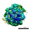
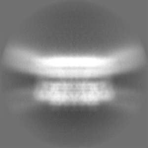
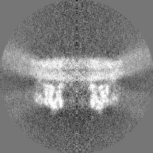
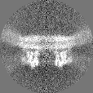
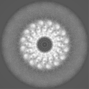
| タイトル | EM map of MPEG-1(w.t.) pre-pore complex bound to liposome | |||||||||
 マップデータ マップデータ | MPEG-1(w.t.) pre-pore complex bound to liposome | |||||||||
 試料 試料 |
| |||||||||
| 生物種 |  Homo sapiens (ヒト) Homo sapiens (ヒト) | |||||||||
| 手法 | 単粒子再構成法 / クライオ電子顕微鏡法 / 解像度: 8.94 Å | |||||||||
 データ登録者 データ登録者 | Pang SS / Bayly-Jones C | |||||||||
| 資金援助 |  オーストラリア, 1件 オーストラリア, 1件
| |||||||||
 引用 引用 | ジャーナル: Nat Commun / 年: 2019 タイトル: The cryo-EM structure of the acid activatable pore-forming immune effector Macrophage-expressed gene 1. 著者: Siew Siew Pang / Charles Bayly-Jones / Mazdak Radjainia / Bradley A Spicer / Ruby H P Law / Adrian W Hodel / Edward S Parsons / Susan M Ekkel / Paul J Conroy / Georg Ramm / Hariprasad ...著者: Siew Siew Pang / Charles Bayly-Jones / Mazdak Radjainia / Bradley A Spicer / Ruby H P Law / Adrian W Hodel / Edward S Parsons / Susan M Ekkel / Paul J Conroy / Georg Ramm / Hariprasad Venugopal / Phillip I Bird / Bart W Hoogenboom / Ilia Voskoboinik / Yann Gambin / Emma Sierecki / Michelle A Dunstone / James C Whisstock /   要旨: Macrophage-expressed gene 1 (MPEG1/Perforin-2) is a perforin-like protein that functions within the phagolysosome to damage engulfed microbes. MPEG1 is thought to form pores in target membranes, ...Macrophage-expressed gene 1 (MPEG1/Perforin-2) is a perforin-like protein that functions within the phagolysosome to damage engulfed microbes. MPEG1 is thought to form pores in target membranes, however, its mode of action remains unknown. We use cryo-Electron Microscopy (cryo-EM) to determine the 2.4 Å structure of a hexadecameric assembly of MPEG1 that displays the expected features of a soluble prepore complex. We further discover that MPEG1 prepore-like assemblies can be induced to perforate membranes through acidification, such as would occur within maturing phagolysosomes. We next solve the 3.6 Å cryo-EM structure of MPEG1 in complex with liposomes. These data reveal that a multi-vesicular body of 12 kDa (MVB12)-associated β-prism (MABP) domain binds membranes such that the pore-forming machinery of MPEG1 is oriented away from the bound membrane. This unexpected mechanism of membrane interaction suggests that MPEG1 remains bound to the phagolysosome membrane while simultaneously forming pores in engulfed bacterial targets. | |||||||||
| 履歴 |
|
- 構造の表示
構造の表示
| ムービー |
 ムービービューア ムービービューア |
|---|---|
| 構造ビューア | EMマップ: SurfViewMolmilJmol/JSmol |
| 添付画像 |

- ダウンロードとリンク
-EMDBアーカイブ
| マップデータ | emd_20622.map.gz | 20.8 MB | EMDBマップデータ形式 | |
|---|---|---|---|---|
| ヘッダ (付随情報) | emd-20622-v30.xmlemd-20622.xml | 17.8 KB 17.8 KB | 表示 表示 | EMDBヘッダ |
| FSC (解像度算出) | emd_20622_fsc.xml | 10.5 KB | 表示 | FSCデータファイル |
| 画像 | 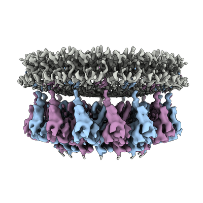 emd_20622.png emd_20622.png | 139.8 KB | ||
| マスクデータ | emd_20622_msk_1.map | 103 MB | マスクマップ | |
| その他 | emd_20622_additional.map.gzemd_20622_additional_1.map.gzemd_20622_half_map_1.map.gzemd_20622_half_map_2.map.gz | 74.9 MB 74.9 MB 75.9 MB 75.9 MB | ||
| アーカイブディレクトリ |  http://ftp.pdbj.org/pub/emdb/structures/EMD-20622ftp://ftp.pdbj.org/pub/emdb/structures/EMD-20622 http://ftp.pdbj.org/pub/emdb/structures/EMD-20622ftp://ftp.pdbj.org/pub/emdb/structures/EMD-20622 | HTTPS FTP |
-関連構造データ


-リンク
| EMDBのページ | EMDB (EBI/PDBe) / EMDataResource |
|---|
-マップ
| ファイル | ダウンロード / ファイル: emd_20622.map.gz / 形式: CCP4 / 大きさ: 103 MB / タイプ: IMAGE STORED AS FLOATING POINT NUMBER (4 BYTES) | ||||||||||||||||||||||||||||||||||||||||||||||||||||||||||||||||||||
|---|---|---|---|---|---|---|---|---|---|---|---|---|---|---|---|---|---|---|---|---|---|---|---|---|---|---|---|---|---|---|---|---|---|---|---|---|---|---|---|---|---|---|---|---|---|---|---|---|---|---|---|---|---|---|---|---|---|---|---|---|---|---|---|---|---|---|---|---|---|
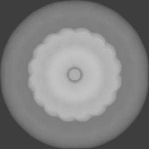
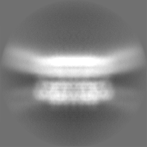
| 注釈 | MPEG-1(w.t.) pre-pore complex bound to liposome | ||||||||||||||||||||||||||||||||||||||||||||||||||||||||||||||||||||
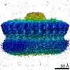
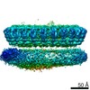
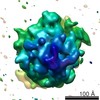
| 投影像・断面図 | 画像のコントロール
画像は Spider により作成 | ||||||||||||||||||||||||||||||||||||||||||||||||||||||||||||||||||||
| ボクセルのサイズ | X=Y=Z: 1.4 Å | ||||||||||||||||||||||||||||||||||||||||||||||||||||||||||||||||||||
| 密度 |
| ||||||||||||||||||||||||||||||||||||||||||||||||||||||||||||||||||||
| 対称性 | 空間群: 1 | ||||||||||||||||||||||||||||||||||||||||||||||||||||||||||||||||||||
| 詳細 | EMDB XML:
CCP4マップ ヘッダ情報:
| ||||||||||||||||||||||||||||||||||||||||||||||||||||||||||||||||||||
 Z (Sec.)
 Y (Row.)
 X (Col.)


-添付データ
-マスク #1
| ファイル | emd_20622_msk_1.map | ||||||||||||
|---|---|---|---|---|---|---|---|---|---|---|---|---|---|
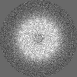
| 投影像・断面図 |
| ||||||||||||
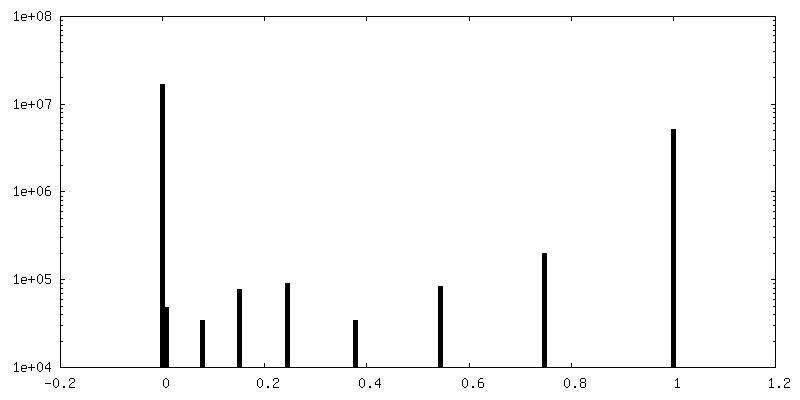
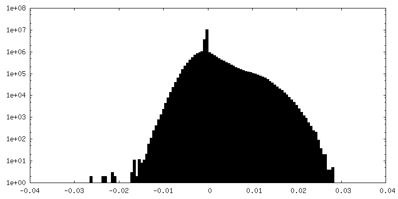
| 密度ヒストグラム |


-追加マップ: MPEG-1(w.t.) pre-pore complex bound to liposome, additional map
| ファイル | emd_20622_additional.map | ||||||||||||
|---|---|---|---|---|---|---|---|---|---|---|---|---|---|
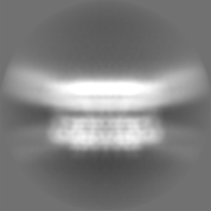
| 注釈 | MPEG-1(w.t.) pre-pore complex bound to liposome, additional map | ||||||||||||
| 投影像・断面図 |
| ||||||||||||
| 密度ヒストグラム |


-追加マップ: MPEG-1(w.t.) pre-pore complex bound to liposome, additional map
| ファイル | emd_20622_additional_1.map | ||||||||||||
|---|---|---|---|---|---|---|---|---|---|---|---|---|---|
| 注釈 | MPEG-1(w.t.) pre-pore complex bound to liposome, additional map | ||||||||||||
| 投影像・断面図 |
| ||||||||||||
| 密度ヒストグラム |


-ハーフマップ: MPEG-1(w.t.) pre-pore complex bound to liposome, half map 1
| ファイル | emd_20622_half_map_1.map | ||||||||||||
|---|---|---|---|---|---|---|---|---|---|---|---|---|---|
| 注釈 | MPEG-1(w.t.) pre-pore complex bound to liposome, half map 1 | ||||||||||||
| 投影像・断面図 |
| ||||||||||||
| 密度ヒストグラム |


-ハーフマップ: MPEG-1(w.t.) pre-pore complex bound to liposome, half map 2
| ファイル | emd_20622_half_map_2.map | ||||||||||||
|---|---|---|---|---|---|---|---|---|---|---|---|---|---|
| 注釈 | MPEG-1(w.t.) pre-pore complex bound to liposome, half map 2 | ||||||||||||
| 投影像・断面図 |
| ||||||||||||
| 密度ヒストグラム |


- 試料の構成要素
-全体 : MPEG-1 (w.t.) pre-pore complex bound to liposome
| 全体 | 名称: MPEG-1 (w.t.) pre-pore complex bound to liposome |
|---|---|
| 要素 |
|
-超分子 #1: MPEG-1 (w.t.) pre-pore complex bound to liposome
| 超分子 | 名称: MPEG-1 (w.t.) pre-pore complex bound to liposome / タイプ: complex / ID: 1 / 親要素: 0 / 含まれる分子: all |
|---|---|
| 由来(天然) | 生物種: Homo sapiens (ヒト) |
| 組換発現 | 生物種:   Spodoptera frugiperda (ツマジロクサヨトウ) Spodoptera frugiperda (ツマジロクサヨトウ) |
-分子 #1: Macrophage-Expressed Gene 1 protein
| 分子 | 名称: Macrophage-Expressed Gene 1 protein / タイプ: protein_or_peptide / ID: 1 / 光学異性体: LEVO |
|---|---|
| 由来(天然) | 生物種: Homo sapiens (ヒト) |
| 組換発現 | 生物種: Spodoptera frugiperda (ツマジロクサヨトウ) |
| 配列 | 文字列: KSGKPSGEMD EVGVQKCKNA LKLPVLEVLP GGGWDNLRNV DMGRVMELTY SNCRTTEDGQ YIIPDEIFTI PQKQSNLEMN SEILESWANY QSSTSYSINT ELSLFSKVNG KFSTEFQRMK TLQVKDQAIT TRVQVRNLVY TVKINPTLEL SSGFRKELLD ISDRLENNQT ...文字列: KSGKPSGEMD EVGVQKCKNA LKLPVLEVLP GGGWDNLRNV DMGRVMELTY SNCRTTEDGQ YIIPDEIFTI PQKQSNLEMN SEILESWANY QSSTSYSINT ELSLFSKVNG KFSTEFQRMK TLQVKDQAIT TRVQVRNLVY TVKINPTLEL SSGFRKELLD ISDRLENNQT RMATYLAELL VLNYGTHVTT SVDAGAALIQ EDHLRASFLQ DSQSSRSAVT ASAGLAFQNT VNFKFEENYT SQNVLTKSYL SNRTNSRVQS IGGVPFYPGI TLQAWQQGIT NHLVAIDRSG LPLHFFINPN MLPDLPGPLV KKVSKTVETA VKRYYTFNTY PGCTDLNSPN FNFQANTDDG SCEGKMTNFS FGGVYQECTQ LSGNRDVLLC QKLEQKNPLT GDFSCPSGYS PVHLLSQIHE EGYNHLECHR KCTLLVFCKT VCEDVFQVAK AEFRAFWCVA SSQVPENSGL LFGGLFSSKS INPMTNAQSC PAGYFPLRLF ENLKVCVSQD YELGSRFAVP FGGFFSCTVG NPLVDPAISR DLGAPSLKKC PGGFSQHPAL ISDGCQVSYC VKSGLFTGGS LPPARLPPFT RPPLMSQAAT NTVIVTNSEN ARSWIKDSQT HQWRLGEPIE LRRAMNVIHG DGGGLSHHHH HH |
-実験情報
-構造解析
| 手法 | クライオ電子顕微鏡法 |
|---|---|
 解析 解析 | 単粒子再構成法 |
| 試料の集合状態 | particle |
-試料調製
| 濃度 | 1.7 mg/mL |
|---|---|
| 緩衝液 | pH: 7.2 |
| グリッド | 詳細: unspecified |
| 凍結 | 凍結剤: ETHANE |
| 詳細 | MPEG-1 protein mixed with POPC:POPS liposomes |
- 電子顕微鏡法
| 顕微鏡 | FEI TITAN KRIOS |
|---|---|
| 撮影 | フィルム・検出器のモデル: GATAN K2 SUMMIT (4k x 4k) 平均電子線量: 53.6 e/Å2 |
| 電子線 | 加速電圧: 300 kV / 電子線源:  FIELD EMISSION GUN FIELD EMISSION GUN |
| 電子光学系 | 照射モード: FLOOD BEAM / 撮影モード: BRIGHT FIELD |
| 実験機器 |  モデル: Titan Krios / 画像提供: FEI Company |
-画像解析
| 最終 再構成 | 想定した対称性 - 点群: C16 (16回回転対称) / 解像度のタイプ: BY AUTHOR / 解像度: 8.94 Å / 解像度の算出法: FSC 0.143 CUT-OFF / 使用した粒子像数: 4217 |
|---|---|
| 初期 角度割当 | タイプ: RANDOM ASSIGNMENT |
| 最終 角度割当 | タイプ: MAXIMUM LIKELIHOOD |
| FSC曲線 (解像度の算出) |  |
