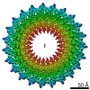
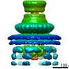
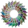
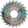
- EMDB-20830: Cryo-EM reconstruction of the inner membrane rings containing the... -
+
Open data
ID or keywords:
Loading...
-
Basic information
Entry
Database: EMDB / ID: EMD-20830
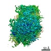
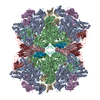
Title
Cryo-EM reconstruction of the inner membrane rings containing the core components of the export apparatus and associated membranes of the needle complex from Salmonella typhimurium type III secretion system.
Map data
Sample
Complex: PrgH and PrgK, the protein components of the inner rings of the Salmonella's needle complex, in complex with the core components of the export apparatus and associated membranes.
National Institutes of Health/National Institute Of Allergy and Infectious Diseases
Citation
Journal: Proc Natl Acad Sci U S A / Year: 2019 Title: High-resolution view of the type III secretion export apparatus in situ reveals membrane remodeling and a secretion pathway. Authors: Carmen Butan / Maria Lara-Tejero / Wenwei Li / Jun Liu / Jorge E Galán / Abstract: Type III protein secretion systems are essential virulence factors for many important pathogenic bacteria. The entire protein secretion machine is composed of several substructures that organize into ...Type III protein secretion systems are essential virulence factors for many important pathogenic bacteria. The entire protein secretion machine is composed of several substructures that organize into a holostructure or injectisome. The core component of the injectisome is the needle complex, which houses the export apparatus that serves as a gate for the passage of the secreted proteins through the bacterial inner membrane. Here, we describe a high-resolution structure of the export apparatus of the type III secretion system in association with the needle complex and the underlying bacterial membrane, both in isolation and in situ. We show the precise location of the core export apparatus components within the injectisome and bacterial envelope and demonstrate that their deployment results in major membrane remodeling and thinning, which may be central for the protein translocation process. We also show that InvA, a critical export apparatus component, forms a multiring cytoplasmic conduit that provides a pathway for the type III secretion substrates to reach the entrance of the export gate. Combined with structure-guided mutagenesis, our studies provide major insight into potential mechanisms of protein translocation and injectisome assembly.
History
Deposition
Oct 15, 2019
-
Header (metadata) release
Nov 13, 2019
-
Map release
Jul 1, 2020
-
Update
Jul 1, 2020
-
Current status
Jul 1, 2020
Processing site: RCSB / Status: Released
-
Structure visualization
Movie
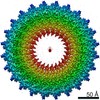
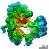
Surface view with section colored by density value
Entire : PrgH and PrgK, the protein components of the inner rings of the S...
Entire
Name: PrgH and PrgK, the protein components of the inner rings of the Salmonella's needle complex, in complex with the core components of the export apparatus and associated membranes.
Components
Complex: PrgH and PrgK, the protein components of the inner rings of the Salmonella's needle complex, in complex with the core components of the export apparatus and associated membranes.
-
Supramolecule #1: PrgH and PrgK, the protein components of the inner rings of the S...
Supramolecule
Name: PrgH and PrgK, the protein components of the inner rings of the Salmonella's needle complex, in complex with the core components of the export apparatus and associated membranes. type: complex / ID: 1 / Parent: 0
In the structure databanks used in Yorodumi, some data are registered as the other names, "COVID-19 virus" and "2019-nCoV". Here are the details of the virus and the list of structure data.
Jan 31, 2019. EMDB accession codes are about to change! (news from PDBe EMDB page)
EMDB accession codes are about to change! (news from PDBe EMDB page)
The allocation of 4 digits for EMDB accession codes will soon come to an end. Whilst these codes will remain in use, new EMDB accession codes will include an additional digit and will expand incrementally as the available range of codes is exhausted. The current 4-digit format prefixed with “EMD-” (i.e. EMD-XXXX) will advance to a 5-digit format (i.e. EMD-XXXXX), and so on. It is currently estimated that the 4-digit codes will be depleted around Spring 2019, at which point the 5-digit format will come into force.
The EM Navigator/Yorodumi systems omit the EMD- prefix.
Related info.:Q: What is EMD? / ID/Accession-code notation in Yorodumi/EM Navigator
Yorodumi is a browser for structure data from EMDB, PDB, SASBDB, etc.
This page is also the successor to EM Navigator detail page, and also detail information page/front-end page for Omokage search.
The word "yorodu" (or yorozu) is an old Japanese word meaning "ten thousand". "mi" (miru) is to see.
Related info.:EMDB / PDB / SASBDB / Comparison of 3 databanks / Yorodumi Search / Aug 31, 2016. New EM Navigator & Yorodumi / Yorodumi Papers / Jmol/JSmol / Function and homology information / Changes in new EM Navigator and Yorodumi
 Movie
Movie Controller

 Yorodumi
 Open data
 Basic information
 Map data
 Sample
 Salmonella enterica subsp. enterica serovar Typhimurium (bacteria)
 Authors
 Citation
 Structure visualization
 Movie viewer
 Downloads & links
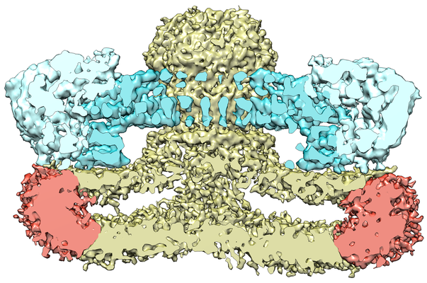 emd_20830.png
 http://ftp.pdbj.org/pub/emdb/structures/EMD-20830
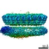

 Z (Sec.)
 Y (Row.)
 X (Col.)


 Sample components
 Processing
 Electron microscopy
 FIELD EMISSION GUN