- EMDB-1417: Cryo-EM study of the Spinach chloroplast ribosome reveals the str... -
+
データを開く
IDまたはキーワード:
読み込み中...
-
基本情報
登録情報
データベース: EMDB / ID: EMD-1417
タイトル
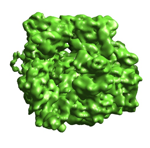
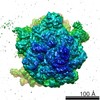
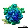
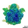
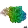
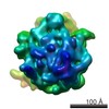
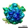
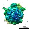
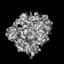
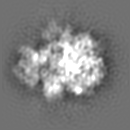
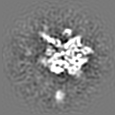
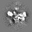
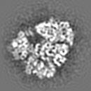
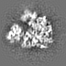
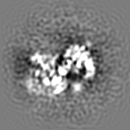
Cryo-EM study of the Spinach chloroplast ribosome reveals the structural and functional roles of plastid-specific ribosomal proteins
マップデータ
Spinach Chloroplast 70S ribosome
試料
試料: Spinacea oleracea chloroplast 70S ribosome
複合体: Spinach Chloroplast 70S Ribosome
機能・相同性
機能・相同性情報
plastid small ribosomal subunit / mitochondrial large ribosomal subunit / mitochondrial small ribosomal subunit / mitochondrial translation / chloroplast / DNA-templated transcription termination / large ribosomal subunit / ribosomal small subunit assembly / transferase activity / ribosome biogenesis ...plastid small ribosomal subunit / mitochondrial large ribosomal subunit / mitochondrial small ribosomal subunit / mitochondrial translation / chloroplast / DNA-templated transcription termination / large ribosomal subunit / ribosomal small subunit assembly / transferase activity / ribosome biogenesis / ribosomal small subunit biogenesis / small ribosomal subunit / ribosomal large subunit assembly / small ribosomal subunit rRNA binding / cytosolic small ribosomal subunit / large ribosomal subunit rRNA binding / cytosolic large ribosomal subunit / negative regulation of translation / rRNA binding / structural constituent of ribosome / ribosome / translation / ribonucleoprotein complex / response to antibiotic / mRNA binding / mitochondrion / RNA binding 類似検索 - 分子機能
Ribosomal protein L32p, plant/cyanobacteria type / Ribosomal protein L1, bacterial-type / Ribosomal protein L1, conserved site / Ribosomal protein L1 signature. / Ribosomal protein L1, 3-layer alpha/beta-sandwich / Ribosomal protein L1-like / Ribosomal protein L1/ribosomal biogenesis protein / Ribosomal protein L1p/L10e family / Ribosomal protein L11, bacterial-type / Ribosomal protein S14, bacterial/plastid ...Ribosomal protein L32p, plant/cyanobacteria type / Ribosomal protein L1, bacterial-type / Ribosomal protein L1, conserved site / Ribosomal protein L1 signature. / Ribosomal protein L1, 3-layer alpha/beta-sandwich / Ribosomal protein L1-like / Ribosomal protein L1/ribosomal biogenesis protein / Ribosomal protein L1p/L10e family / Ribosomal protein L11, bacterial-type / Ribosomal protein S14, bacterial/plastid / Ribosomal protein S16, conserved site / Ribosomal protein S16 signature. / Ribosomal protein L11, conserved site / Ribosomal protein L11 signature. / Ribosomal protein L16 signature 1. / Ribosomal protein L21, conserved site / Ribosomal protein L21 signature. / Ribosomal protein L16 signature 2. / Ribosomal protein L16, conserved site / Ribosomal protein L11, N-terminal / Ribosomal protein L11, N-terminal domain / Ribosomal protein L11/L12 / Ribosomal protein L11, C-terminal / Ribosomal protein L11, C-terminal domain superfamily / Ribosomal protein L11/L12, N-terminal domain superfamily / Ribosomal protein L11/L12 / Ribosomal protein L11, RNA binding domain / Ribosomal protein L33, conserved site / Ribosomal protein L33 signature. / Ribosomal protein L35, conserved site / Ribosomal protein L35 signature. / Ribosomal protein L35, non-mitochondrial / Ribosomal protein S3, bacterial-type / : / Ribosomal protein S19, bacterial-type / Ribosomal protein S7, bacterial/organellar-type / Ribosomal protein S11, bacterial-type / Ribosomal protein L5, bacterial-type / Ribosomal protein S4, bacterial-type / Ribosomal protein S5, bacterial-type / : / Ribosomal protein S6, plastid/chloroplast / Ribosomal protein L20 signature. / Ribosomal protein L14P, bacterial-type / Ribosomal protein S2, bacteria/mitochondria/plastid / Ribosomal protein L35 / Ribosomal protein L35 superfamily / Ribosomal protein L22, bacterial/chloroplast-type / Ribosomal protein L35 / Ribosomal protein L2, bacterial/organellar-type / Ribosomal protein L33 / Ribosomal protein S18, conserved site / Ribosomal protein S18 signature. / Ribosomal protein L33 / Ribosomal protein S9, bacterial/plastid / Ribosomal protein L33 superfamily / Ribosomal protein S16 / Ribosomal protein S16 domain superfamily / Ribosomal protein S16 / Ribosomal protein L16 / Ribosomal protein S15, bacterial-type / Ribosomal protein S6 / Ribosomal protein S6 / Ribosomal protein S6 superfamily / Ribosomal protein L20 / Ribosomal protein S12, bacterial-type / Ribosomal protein L20 / Ribosomal protein L20, C-terminal / Ribosomal protein L19 / Ribosomal protein L19 / Ribosomal protein L19 superfamily / : / Large ribosomal subunit protein uL24, C-terminal domain / Translation elongation factor EF1B/ribosomal protein S6 / Ribosomal protein L34 / Ribosomal protein L34 / Ribosomal protein L24 / Ribosomal protein S18 / Ribosomal protein S18 / Ribosomal protein L21 / Ribosomal protein L32p / Ribosomal protein S18 superfamily / Ribosomal protein L21-like / L21-like superfamily / Ribosomal prokaryotic L21 protein / 50S ribosomal protein uL4 / Ribosomal protein L13, bacterial-type / Ribosomal protein S2 signature 2. / Ribosomal protein S3, conserved site / Ribosomal protein S3 signature. / : / Ribosomal protein L5, conserved site / Ribosomal protein L5 signature. / Ribosomal protein S14, conserved site / Ribosomal protein S14 signature. / Ribosomal protein L2 signature. / Ribosomal protein S2 signature 1. / Type-2 KH domain profile. / K Homology domain, type 2 / Ribosomal protein S3, C-terminal 類似検索 - ドメイン・相同性
Large ribosomal subunit protein uL4c / Small ribosomal subunit protein uS11c / Small ribosomal subunit protein uS14c / Small ribosomal subunit protein uS19c / Large ribosomal subunit protein uL2cz/uL2cy / Small ribosomal subunit protein uS2c / Large ribosomal subunit protein uL22c / Small ribosomal subunit protein uS3c / Large ribosomal subunit protein uL14c / Small ribosomal subunit protein uS8c ...Large ribosomal subunit protein uL4c / Small ribosomal subunit protein uS11c / Small ribosomal subunit protein uS14c / Small ribosomal subunit protein uS19c / Large ribosomal subunit protein uL2cz/uL2cy / Small ribosomal subunit protein uS2c / Large ribosomal subunit protein uL22c / Small ribosomal subunit protein uS3c / Large ribosomal subunit protein uL14c / Small ribosomal subunit protein uS8c / Large ribosomal subunit protein uL13c / Small ribosomal subunit protein uS4c / Large ribosomal subunit protein uL16c / Large ribosomal subunit protein bL35c / Large ribosomal subunit protein bL21c / Large ribosomal subunit protein uL24c / Large ribosomal subunit protein bL20c / Large ribosomal subunit protein bL32c / Large ribosomal subunit protein bL33c / Small ribosomal subunit protein bS16c / Large ribosomal subunit protein uL11c / Small ribosomal subunit protein uS12cz/uS12cy / Small ribosomal subunit protein uS7cz/uS7cy / Small ribosomal subunit protein uS13c / Large ribosomal subunit protein uL5c / Large ribosomal subunit protein bL34c / Small ribosomal subunit protein uS9c / Small ribosomal subunit protein bS6c alpha / Large ribosomal subunit protein bL19c / Large ribosomal subunit protein uL1c / Large ribosomal subunit protein uL23c / Small ribosomal subunit protein uS15c / Small ribosomal subunit protein bS18c / Small ribosomal subunit protein uS5c 類似検索 - 構成要素
ジャーナル: Proc Natl Acad Sci U S A / 年: 2007 タイトル: Cryo-EM study of the spinach chloroplast ribosome reveals the structural and functional roles of plastid-specific ribosomal proteins. 著者: Manjuli R Sharma / Daniel N Wilson / Partha P Datta / Chandana Barat / Frank Schluenzen / Paola Fucini / Rajendra K Agrawal / 要旨: Protein synthesis in the chloroplast is carried out by chloroplast ribosomes (chloro-ribosome) and regulated in a light-dependent manner. Chloroplast or plastid ribosomal proteins (PRPs) generally ...Protein synthesis in the chloroplast is carried out by chloroplast ribosomes (chloro-ribosome) and regulated in a light-dependent manner. Chloroplast or plastid ribosomal proteins (PRPs) generally are larger than their bacterial counterparts, and chloro-ribosomes contain additional plastid-specific ribosomal proteins (PSRPs); however, it is unclear to what extent these proteins play structural or regulatory roles during translation. We have obtained a three-dimensional cryo-EM map of the spinach 70S chloro-ribosome, revealing the overall structural organization to be similar to bacterial ribosomes. Fitting of the conserved portions of the x-ray crystallographic structure of the bacterial 70S ribosome into our cryo-EM map of the chloro-ribosome reveals the positions of PRP extensions and the locations of the PSRPs. Surprisingly, PSRP1 binds in the decoding region of the small (30S) ribosomal subunit, in a manner that would preclude the binding of messenger and transfer RNAs to the ribosome, suggesting that PSRP1 is a translation factor rather than a ribosomal protein. PSRP2 and PSRP3 appear to structurally compensate for missing segments of the 16S rRNA within the 30S subunit, whereas PSRP4 occupies a position buried within the head of the 30S subunit. One of the two PSRPs in the large (50S) ribosomal subunit lies near the tRNA exit site. Furthermore, we find a mass of density corresponding to chloro-ribosome recycling factor; domain II of this factor appears to interact with the flexible C-terminal domain of PSRP1. Our study provides evolutionary insights into the structural and functional roles that the PSRPs play during protein synthesis in chloroplasts.
名称: Spinach Chloroplast 70S Ribosome / タイプ: complex / ID: 1 / Name.synonym: chloro-ribosome 詳細: The SSU 30S has PSRP1,2,3, and 4 identified. LSU 50S did not have PRPL25 and PRPL30 density present. pRRF (plastid ribosome recycling factor)is tightly bound to LSU 50S subunit. One of the ...詳細: The SSU 30S has PSRP1,2,3, and 4 identified. LSU 50S did not have PRPL25 and PRPL30 density present. pRRF (plastid ribosome recycling factor)is tightly bound to LSU 50S subunit. One of the two PSRPs on the LSU 50S subunit is identified. 組換発現: No / Ribosome-details: ribosome-eukaryote: ALL
凍結剤: ETHANE / チャンバー内湿度: 100 % / チャンバー内温度: 277 K / 装置: HOMEMADE PLUNGER / 詳細: Vitrification instrument: Cryo-plunger 手法: 5 microliters applied to the grid then blotted for 3 seconds with Whatman number 1 filter paper before plunging
-
電子顕微鏡法
顕微鏡
FEI TECNAI F20
温度
平均: 93 K
アライメント法
Legacy - 非点収差: objective lens astigmatism was corrected at 250K times magnification
 ムービー
ムービー コントローラー


 データを開く
 基本情報
基本情報 マップデータ
 試料
試料 機能・相同性情報
 Spinacia oleracea (ホウレンソウ)
 データ登録者
 引用
引用
 構造の表示
構造の表示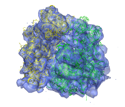
 ダウンロードとリンク
 1417-3BBN-3BBO.png
 http://ftp.pdbj.org/pub/emdb/structures/EMD-1417


 Z (Sec.)
 Y (Row.)
 X (Col.)


 試料の構成要素
 解析
解析 電子顕微鏡法
 FIELD EMISSION GUN

