 Movie
Movie Controller


+ Open data

- Basic information
| Entry |  | ||||||||||||||||||
|---|---|---|---|---|---|---|---|---|---|---|---|---|---|---|---|---|---|---|---|
| Title | Cryo-EM structure of the 1 hpf zebrafish embryo 80S ribosome | ||||||||||||||||||
 Map data Map data | |||||||||||||||||||
 Sample Sample |
| ||||||||||||||||||
| Function / homology |  Function and homology information Function and homology informationHypusine synthesis from eIF5A-lysine / Platelet degranulation / optokinetic behavior / L13a-mediated translational silencing of Ceruloplasmin expression / SRP-dependent cotranslational protein targeting to membrane / RMTs methylate histone arginines / TNFR1-induced NF-kappa-B signaling pathway / TNFR1-mediated ceramide production / : / : ...Hypusine synthesis from eIF5A-lysine / Platelet degranulation / optokinetic behavior / L13a-mediated translational silencing of Ceruloplasmin expression / SRP-dependent cotranslational protein targeting to membrane / RMTs methylate histone arginines / TNFR1-induced NF-kappa-B signaling pathway / TNFR1-mediated ceramide production / : / : / Formation of a pool of free 40S subunits / Formation of the ternary complex, and subsequently, the 43S complex / Ribosomal scanning and start codon recognition / : / Protein methylation / Nonsense Mediated Decay (NMD) independent of the Exon Junction Complex (EJC) / Nonsense Mediated Decay (NMD) enhanced by the Exon Junction Complex (EJC) / mTORC1-mediated signalling / brain segmentation / Regulation of TNFR1 signaling / convergent extension involved in gastrulation / : / Neutrophil degranulation / : / primitive hemopoiesis / embryonic retina morphogenesis in camera-type eye / anatomical structure development / death domain binding / positive regulation of translational termination / hemoglobin biosynthetic process / ribosome hibernation / mitochondrial cytochrome c oxidase assembly / positive regulation of translational elongation / chordate embryonic development / ribosomal subunit / embryonic brain development / exocrine pancreas development / positive regulation of gastrulation / positive regulation of translational fidelity / definitive hemopoiesis / mitochondrial large ribosomal subunit / pancreas development / regulation of establishment of cell polarity / cytoplasmic side of rough endoplasmic reticulum membrane / laminin receptor activity / translational elongation / negative regulation of Wnt signaling pathway / regulation of innate immune response / regulation of cell division / erythrocyte maturation / hemopoiesis / intrinsic apoptotic signaling pathway by p53 class mediator / endonucleolytic cleavage to generate mature 3'-end of SSU-rRNA from (SSU-rRNA, 5.8S rRNA, LSU-rRNA) / mitochondrial respiratory chain complex I assembly / hematopoietic stem cell differentiation / mRNA transport / protein-RNA complex assembly / positive regulation of translational initiation / vasculogenesis / erythrocyte development / translation regulator activity / translation elongation factor activity / nuclear pore / laminin binding / rough endoplasmic reticulum / endonucleolytic cleavage in ITS1 to separate SSU-rRNA from 5.8S rRNA and LSU-rRNA from tricistronic rRNA transcript (SSU-rRNA, 5.8S rRNA, LSU-rRNA) / translation repressor activity / translation initiation factor binding / translation initiation factor activity / cellular response to amino acid starvation / class I DNA-(apurinic or apyrimidinic site) endonuclease activity / DNA-(apurinic or apyrimidinic site) lyase / rescue of stalled ribosome / negative regulation of autophagy / maturation of LSU-rRNA from tricistronic rRNA transcript (SSU-rRNA, 5.8S rRNA, LSU-rRNA) / maturation of SSU-rRNA from tricistronic rRNA transcript (SSU-rRNA, 5.8S rRNA, LSU-rRNA) / erythrocyte differentiation / positive regulation of RNA splicing / maturation of LSU-rRNA / ribosomal large subunit biogenesis / maturation of SSU-rRNA / small-subunit processome / apoptotic signaling pathway / positive regulation of apoptotic signaling pathway / protein kinase C binding / regulation of erythrocyte differentiation / brain development / ribosomal large subunit assembly / mRNA 5'-UTR binding / modification-dependent protein catabolic process / spindle / rRNA processing / large ribosomal subunit / protein tag activity / regulation of protein localization / protein transport / cellular response to xenobiotic stimulus / ribosome binding / glucose homeostasis / regulation of translation Similarity search - Function | ||||||||||||||||||
| Biological species |  | ||||||||||||||||||
| Method | single particle reconstruction / cryo EM / Resolution: 3.2 Å | ||||||||||||||||||
 Authors Authors | Leesch F / Lorenzo-Orts L / Grishkovskaya I / Kandolf S / Belacic K / Meinhart A / Haselbach D / Pauli A | ||||||||||||||||||
| Funding support |  Austria, Austria,  Switzerland, 5 items Switzerland, 5 items
| ||||||||||||||||||
 Citation Citation | Journal: Nature / Year: 2023 Title: A molecular network of conserved factors keeps ribosomes dormant in the egg. Authors: Friederike Leesch / Laura Lorenzo-Orts / Carina Pribitzer / Irina Grishkovskaya / Josef Roehsner / Anastasia Chugunova / Manuel Matzinger / Elisabeth Roitinger / Katarina Belačić / Susanne ...Authors: Friederike Leesch / Laura Lorenzo-Orts / Carina Pribitzer / Irina Grishkovskaya / Josef Roehsner / Anastasia Chugunova / Manuel Matzinger / Elisabeth Roitinger / Katarina Belačić / Susanne Kandolf / Tzi-Yang Lin / Karl Mechtler / Anton Meinhart / David Haselbach / Andrea Pauli / Abstract: Ribosomes are produced in large quantities during oogenesis and are stored in the egg. However, the egg and early embryo are translationally repressed. Here, using mass spectrometry and cryo-electron ...Ribosomes are produced in large quantities during oogenesis and are stored in the egg. However, the egg and early embryo are translationally repressed. Here, using mass spectrometry and cryo-electron microscopy analyses of ribosomes isolated from zebrafish (Danio rerio) and Xenopus laevis eggs and embryos, we provide molecular evidence that ribosomes transition from a dormant state to an active state during the first hours of embryogenesis. Dormant ribosomes are associated with four conserved factors that form two modules, consisting of Habp4-eEF2 and death associated protein 1b (Dap1b) or Dap in complex with eIF5a. Both modules occupy functionally important sites and act together to stabilize ribosomes and repress translation. Dap1b (also known as Dapl1 in mammals) is a newly discovered translational inhibitor that stably inserts into the polypeptide exit tunnel. Addition of recombinant zebrafish Dap1b protein is sufficient to block translation and reconstitute the dormant egg ribosome state in a mammalian translation extract in vitro. Thus, a developmentally programmed, conserved ribosome state has a key role in ribosome storage and translational repression in the egg. | ||||||||||||||||||
| History |
|
- Structure visualization
| Supplemental images |
|---|

- Downloads & links
-EMDB archive
| Map data | emd_13111.map.gz | 364.2 MB | EMDB map data format | |
|---|---|---|---|---|
| Header (meta data) | emd-13111-v30.xmlemd-13111.xml | 96.9 KB 96.9 KB | Display Display | EMDB header |
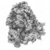
| Images | 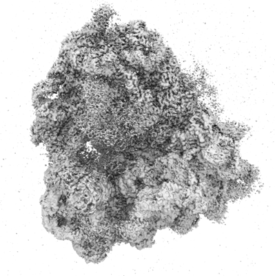 emd_13111.png emd_13111.png | 157 KB | ||
| Archive directory |  http://ftp.pdbj.org/pub/emdb/structures/EMD-13111ftp://ftp.pdbj.org/pub/emdb/structures/EMD-13111 http://ftp.pdbj.org/pub/emdb/structures/EMD-13111ftp://ftp.pdbj.org/pub/emdb/structures/EMD-13111 | HTTPS FTP |
-Validation report
| Summary document | emd_13111_validation.pdf.gz | 469.2 KB | Display | EMDB validaton report |
|---|---|---|---|---|
| Full document | emd_13111_full_validation.pdf.gz | 468.8 KB | Display | |
| Data in XML | emd_13111_validation.xml.gz | 7.9 KB | Display | |
| Data in CIF | emd_13111_validation.cif.gz | 9.1 KB | Display | |
| Arichive directory | https://ftp.pdbj.org/pub/emdb/validation_reports/EMD-13111ftp://ftp.pdbj.org/pub/emdb/validation_reports/EMD-13111 | HTTPS FTP |
-Related structure data
| Related structure data |  7oyaMC  7oybC  7oycC  7oydC M: atomic model generated by this map C: citing same article ( |
|---|---|
| Similar structure data |


-Links
| EMDB pages | EMDB (EBI/PDBe) / EMDataResource |
|---|---|
| Related items in Molecule of the Month |


-Map
| File | Download / File: emd_13111.map.gz / Format: CCP4 / Size: 421.9 MB / Type: IMAGE STORED AS FLOATING POINT NUMBER (4 BYTES) | ||||||||||||||||||||
|---|---|---|---|---|---|---|---|---|---|---|---|---|---|---|---|---|---|---|---|---|---|
| Voxel size | X=Y=Z: 1.06 Å | ||||||||||||||||||||
| Density |
| ||||||||||||||||||||
| Symmetry | Space group: 1 | ||||||||||||||||||||
| Details | EMDB XML:
|
-Supplemental data
- Sample components
+Entire : 80S ribosome from 1 hpf zebrafish embryos
+Supramolecule #1: 80S ribosome from 1 hpf zebrafish embryos
+Macromolecule #1: 18S rRNA
+Macromolecule #76: 28S rRNA
+Macromolecule #77: 5S rRNA
+Macromolecule #78: 5.8S rRNA
+Macromolecule #2: 40S ribosomal protein SA
+Macromolecule #3: 40S ribosomal protein S3a
+Macromolecule #4: 40S ribosomal protein S2
+Macromolecule #5: 40S ribosomal protein S4, X isoform
+Macromolecule #6: 40S ribosomal protein S6
+Macromolecule #7: 40S ribosomal protein S7
+Macromolecule #8: 40S ribosomal protein S8
+Macromolecule #9: 40S ribosomal protein S9
+Macromolecule #10: 40S ribosomal protein S11
+Macromolecule #11: 40S ribosomal protein S13
+Macromolecule #12: Ribosomal protein S14
+Macromolecule #13: 40S ribosomal protein S17
+Macromolecule #14: 40S ribosomal protein S21
+Macromolecule #15: 40S ribosomal protein S15a
+Macromolecule #16: 40S ribosomal protein S23
+Macromolecule #17: 40S ribosomal protein S24
+Macromolecule #18: 40S ribosomal protein S26
+Macromolecule #19: 40S ribosomal protein S27
+Macromolecule #20: 40S ribosomal protein S30
+Macromolecule #21: DNA-(apurinic or apyrimidinic site) lyase
+Macromolecule #22: Ribosomal protein S5
+Macromolecule #23: Ribosomal protein S10
+Macromolecule #24: 40S ribosomal protein S15
+Macromolecule #25: Ribosomal protein S16
+Macromolecule #26: 40S ribosomal protein S20
+Macromolecule #27: 40S ribosomal protein S28
+Macromolecule #28: 40S ribosomal protein S29
+Macromolecule #29: Guanine nucleotide-binding protein subunit beta-2-like 1
+Macromolecule #30: 40S ribosomal protein S25
+Macromolecule #31: 40S ribosomal protein S19
+Macromolecule #32: 40S ribosomal protein S18
+Macromolecule #33: Zgc:103482
+Macromolecule #34: 60S ribosomal protein L27
+Macromolecule #35: 60S ribosomal protein L29
+Macromolecule #36: 60S ribosomal protein L30
+Macromolecule #37: 60S ribosomal protein L31
+Macromolecule #38: Ribosomal protein L32
+Macromolecule #39: 60S ribosomal protein L35a
+Macromolecule #40: 60S ribosomal protein L34
+Macromolecule #41: 60S ribosomal protein L35
+Macromolecule #42: 60S ribosomal protein L36
+Macromolecule #43: Ribosomal protein L37
+Macromolecule #44: 60S ribosomal protein L38
+Macromolecule #45: 60S ribosomal protein L36a
+Macromolecule #46: Zgc:171772
+Macromolecule #47: 60S ribosomal protein L8
+Macromolecule #48: Ribosomal protein L3
+Macromolecule #49: Ribosomal protein L4
+Macromolecule #50: Ribosomal protein L5b
+Macromolecule #51: 60S ribosomal protein L6
+Macromolecule #52: Ribosomal protein L7
+Macromolecule #53: 60S ribosomal protein L7a
+Macromolecule #54: 60S ribosomal protein L9
+Macromolecule #55: 60S ribosomal protein L11
+Macromolecule #56: 60S ribosomal protein L13
+Macromolecule #57: 60S ribosomal protein L14
+Macromolecule #58: Ribosomal protein L15
+Macromolecule #59: 60S ribosomal protein L13a
+Macromolecule #60: 60S ribosomal protein L17
+Macromolecule #61: Ribosomal protein L18
+Macromolecule #62: 60S ribosomal protein L19
+Macromolecule #63: 60S ribosomal protein L21
+Macromolecule #64: Ribosomal protein L22
+Macromolecule #65: 60S ribosomal protein L23
+Macromolecule #66: 60S ribosomal protein L24
+Macromolecule #67: Ribosomal protein L23a
+Macromolecule #68: ATPase H+ transporting V0 subunit e1
+Macromolecule #69: Ribosomal protein L39
+Macromolecule #70: 60S ribosomal protein L41
+Macromolecule #71: 60S ribosomal protein L28
+Macromolecule #72: 60S ribosomal protein L10
+Macromolecule #73: 60S ribosomal protein L18a
+Macromolecule #74: 60S ribosomal protein L40
+Macromolecule #75: 60S ribosomal protein L27a
+Macromolecule #79: Novel protein similar to human proliferation-associated 2G4 prote...
+Macromolecule #80: Eukaryotic translation elongation factor 2
+Macromolecule #81: Eukaryotic translation initiation factor 5A
+Macromolecule #82: Dap1b
+Macromolecule #83: ZINC ION
+Macromolecule #84: MAGNESIUM ION
-Experimental details
-Structure determination
| Method | cryo EM |
|---|---|
 Processing Processing | single particle reconstruction |
| Aggregation state | particle |
-Sample preparation
| Buffer | pH: 7.6 Component:
| ||||||||||||||||||
|---|---|---|---|---|---|---|---|---|---|---|---|---|---|---|---|---|---|---|---|
| Grid | Model: Quantifoil R3.5/1 / Material: COPPER / Mesh: 200 / Support film - Material: CARBON / Support film - topology: CONTINUOUS / Support film - Film thickness: 2.0 nm / Pretreatment - Type: GLOW DISCHARGE / Pretreatment - Time: 60 sec. / Pretreatment - Atmosphere: AIR | ||||||||||||||||||
| Vitrification | Cryogen name: ETHANE / Chamber humidity: 70 % / Instrument: LEICA EM GP |
- Electron microscopy
| Microscope | FEI TITAN KRIOS |
|---|---|
| Image recording | Film or detector model: FEI FALCON III (4k x 4k) / Detector mode: INTEGRATING / Average electron dose: 43.0 e/Å2 |
| Electron beam | Acceleration voltage: 300 kV / Electron source:  FIELD EMISSION GUN FIELD EMISSION GUN |
| Electron optics | Illumination mode: FLOOD BEAM / Imaging mode: BRIGHT FIELD / Cs: 2.7 mm |
| Sample stage | Specimen holder model: FEI TITAN KRIOS AUTOGRID HOLDER |
| Experimental equipment |  Model: Titan Krios / Image courtesy: FEI Company |
+Image processing
-Atomic model buiding 1
| Refinement | Protocol: OTHER |
|---|---|
| Output model | PDB-7oya: |
