[English] 日本語
Yorodumi
- EMDB-12239: Bacterial 30S ribosomal subunit assembly complex state M (body domain) -

+
Open data


ID or keywords:

Loading...

-
Basic information

Entry
Database: EMDB / ID: EMD-12239
TitleBacterial 30S ribosomal subunit assembly complex state M (body domain)
Map data
Sample
  • Complex: Bacterial 30S ribosomal subunit assembly complex state M (body domain)
    • RNA: x 1 types
    • Protein or peptide: x 12 types
  • Ligand: x 1 types
KeywordsCryo-EM / 30S biogenesis / ribosome assembly / RbfA / RsgA / YjeQ / RimP / KsgA / RsmA / RIBOSOME
Function / homology
Function and homology information


ornithine decarboxylase inhibitor activity / misfolded RNA binding / Group I intron splicing / RNA folding / four-way junction DNA binding / negative regulation of translational initiation / regulation of mRNA stability / mRNA regulatory element binding translation repressor activity / positive regulation of RNA splicing / DNA endonuclease activity ...ornithine decarboxylase inhibitor activity / misfolded RNA binding / Group I intron splicing / RNA folding / four-way junction DNA binding / negative regulation of translational initiation / regulation of mRNA stability / mRNA regulatory element binding translation repressor activity / positive regulation of RNA splicing / DNA endonuclease activity / transcription antitermination / DNA-templated transcription termination / maintenance of translational fidelity / mRNA 5'-UTR binding / ribosomal small subunit biogenesis / small ribosomal subunit rRNA binding / regulation of translation / ribosomal small subunit assembly / small ribosomal subunit / cytosolic small ribosomal subunit / cytoplasmic translation / tRNA binding / molecular adaptor activity / rRNA binding / ribosome / structural constituent of ribosome / translation / response to antibiotic / zinc ion binding / cytosol / cytoplasm
Similarity search - Function
Ribosomal protein S21, conserved site / Ribosomal protein S21 signature. / Ribosomal protein S21 superfamily / Ribosomal protein S21 / Ribosomal protein S16, conserved site / Ribosomal protein S16 signature. / Ribosomal protein S21 / Ribosomal protein S6, conserved site / Ribosomal protein S6 signature. / Ribosomal protein S11, bacterial-type ...Ribosomal protein S21, conserved site / Ribosomal protein S21 signature. / Ribosomal protein S21 superfamily / Ribosomal protein S21 / Ribosomal protein S16, conserved site / Ribosomal protein S16 signature. / Ribosomal protein S21 / Ribosomal protein S6, conserved site / Ribosomal protein S6 signature. / Ribosomal protein S11, bacterial-type / Ribosomal protein S20 / Ribosomal protein S20 superfamily / Ribosomal protein S20 / Ribosomal protein S4, bacterial-type / 30S ribosomal protein S17 / Ribosomal protein S5, bacterial-type / Ribosomal protein S6, plastid/chloroplast / Ribosomal protein S18, conserved site / Ribosomal protein S18 signature. / Ribosomal protein S16 / Ribosomal protein S16 domain superfamily / Ribosomal protein S16 / Ribosomal protein S15, bacterial-type / Ribosomal protein S6 / Ribosomal protein S6 / Ribosomal protein S6 superfamily / Ribosomal protein S12, bacterial-type / Translation elongation factor EF1B/ribosomal protein S6 / Ribosomal protein S18 / Ribosomal protein S18 / Ribosomal protein S18 superfamily / : / Ribosomal protein S5, N-terminal, conserved site / Ribosomal protein S5 signature. / Ribosomal protein S5 / Ribosomal protein S17, conserved site / Ribosomal protein S17 signature. / S5 double stranded RNA-binding domain profile. / Ribosomal protein S5, N-terminal / Ribosomal protein S5, N-terminal domain / Ribosomal protein S5, C-terminal / Ribosomal protein S5, C-terminal domain / Ribosomal protein S8 signature. / Ribosomal protein S4/S9 N-terminal domain / Ribosomal protein S4, conserved site / Ribosomal protein S4 signature. / Ribosomal protein S4/S9 N-terminal domain / Ribosomal protein S4/S9, N-terminal / Ribosomal protein S15 signature. / Ribosomal protein S4/S9 / Ribosomal protein S8 / Ribosomal protein S8 superfamily / Ribosomal protein S8 / S4 RNA-binding domain profile. / Ribosomal S11, conserved site / Ribosomal protein S11 signature. / Ribosomal protein S11 / S4 RNA-binding domain / S4 domain / Ribosomal protein S11 / RNA-binding S4 domain / RNA-binding S4 domain superfamily / Ribosomal protein S12 signature. / Ribosomal protein S12/S23 / Ribosomal protein S12/S23 / Ribosomal protein S17/S11 / Ribosomal protein S17 / Ribosomal protein S15 / Ribosomal_S15 / Ribosomal protein S15 / Ribosomal protein S11 superfamily / S15/NS1, RNA-binding / Ribosomal protein S5 domain 2-type fold, subgroup / Ribosomal protein S5 domain 2-type fold / Nucleic acid-binding, OB-fold
Similarity search - Domain/homology
Small ribosomal subunit protein bS6 / Small ribosomal subunit protein uS11 / Small ribosomal subunit protein uS12 / Small ribosomal subunit protein bS16 / Small ribosomal subunit protein bS18 / Small ribosomal subunit protein bS20 / Small ribosomal subunit protein uS4 / Small ribosomal subunit protein uS5 / Small ribosomal subunit protein uS8 / Small ribosomal subunit protein uS15 ...Small ribosomal subunit protein bS6 / Small ribosomal subunit protein uS11 / Small ribosomal subunit protein uS12 / Small ribosomal subunit protein bS16 / Small ribosomal subunit protein bS18 / Small ribosomal subunit protein bS20 / Small ribosomal subunit protein uS4 / Small ribosomal subunit protein uS5 / Small ribosomal subunit protein uS8 / Small ribosomal subunit protein uS15 / Small ribosomal subunit protein uS17 / Small ribosomal subunit protein bS21
Similarity search - Component
Biological speciesEscherichia coli (strain K12) (bacteria)
Methodsingle particle reconstruction / cryo EM / Resolution: 2.88 Å
AuthorsSchedlbauer A / Iturrioz I
Funding support Spain, 2 items
OrganizationGrant numberCountry
Ministry of Economy and Competitiveness (MINECO)CTQ2017-82222-R Spain
European CommissionPCIG14-GA-2013-632072 Spain
CitationJournal: Sci Adv / Year: 2021
Title: A conserved rRNA switch is central to decoding site maturation on the small ribosomal subunit.
Authors: Andreas Schedlbauer / Idoia Iturrioz / Borja Ochoa-Lizarralde / Tammo Diercks / Jorge Pedro López-Alonso / José Luis Lavin / Tatsuya Kaminishi / Retina Çapuni / Neha Dhimole / Elisa de ...Authors: Andreas Schedlbauer / Idoia Iturrioz / Borja Ochoa-Lizarralde / Tammo Diercks / Jorge Pedro López-Alonso / José Luis Lavin / Tatsuya Kaminishi / Retina Çapuni / Neha Dhimole / Elisa de Astigarraga / David Gil-Carton / Paola Fucini / Sean R Connell /
Abstract: While a structural description of the molecular mechanisms guiding ribosome assembly in eukaryotic systems is emerging, bacteria use an unrelated core set of assembly factors for which high- ...While a structural description of the molecular mechanisms guiding ribosome assembly in eukaryotic systems is emerging, bacteria use an unrelated core set of assembly factors for which high-resolution structural information is still missing. To address this, we used single-particle cryo-electron microscopy to visualize the effects of bacterial ribosome assembly factors RimP, RbfA, RsmA, and RsgA on the conformational landscape of the 30 ribosomal subunit and obtained eight snapshots representing late steps in the folding of the decoding center. Analysis of these structures identifies a conserved secondary structure switch in the 16 ribosomal RNA central to decoding site maturation and suggests both a sequential order of action and molecular mechanisms for the assembly factors in coordinating and controlling this switch. Structural and mechanistic parallels between bacterial and eukaryotic systems indicate common folding features inherent to all ribosomes.
History
DepositionJan 25, 2021-
Header (metadata) releaseJul 7, 2021-
Map releaseJul 7, 2021-
UpdateApr 24, 2024-
Current statusApr 24, 2024Processing site: PDBe / Status: Released

-
Structure visualization

Movie
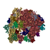
  • Surface view with section colored by density value
  • Surface level: 0.016
  • Imaged by UCSF Chimera
  • Download
  • Surface view colored by height
  • Surface level: 0.016
  • Imaged by UCSF Chimera
  • Download
  • Surface view with fitted model
  • Atomic models: PDB-7bod
  • Surface level: 0.018
  • Imaged by UCSF Chimera
  • Download
Movie viewer
Structure viewerEM map:
SurfViewMolmilJmol/JSmol
Supplemental images

Downloads & links

-
Map

FileDownload / File: emd_12239.map.gz / Format: CCP4 / Size: 244.1 MB / Type: IMAGE STORED AS FLOATING POINT NUMBER (4 BYTES)
Projections & slices

Image control

Size
Brightness
Contrast
Others
AxesZ (Sec.)Y (Row.)X (Col.)
1.05 Å/pix.
x 400 pix.
= 420. Å
1.05 Å/pix.
x 400 pix.
= 420. Å
1.05 Å/pix.
x 400 pix.
= 420. Å

Surface

Projections

Slices (1/3)

Slices (1/2)

Slices (2/3)

Images are generated by Spider.

Voxel sizeX=Y=Z: 1.05 Å
Density
Contour LevelBy AUTHOR: 0.016 / Movie #1: 0.016
Minimum - Maximum-0.028794914 - 0.08695554
Average (Standard dev.)-0.00011659608 (±0.0029321678)
SymmetrySpace group: 1
Details

EMDB XML:

Map geometry
Axis orderXYZ
Origin000
Dimensions400400400
Spacing400400400
CellA=B=C: 419.99997 Å
α=β=γ: 90.0 °

CCP4 map header:

modeImage stored as Reals
Å/pix. X/Y/Z1.051.051.05
M x/y/z400400400
origin x/y/z0.0000.0000.000
length x/y/z420.000420.000420.000
α/β/γ90.00090.00090.000
start NX/NY/NZ000
NX/NY/NZ450450450
MAP C/R/S123
start NC/NR/NS000
NC/NR/NS400400400
D min/max/mean-0.0290.087-0.000

-
Supplemental data

-
Mask #1

Fileemd_12239_msk_1.map
Projections & Slices
AxesZYX

Projections

Slices (1/2)
Density Histograms

-
Half map: #1

Fileemd_12239_half_map_1.map
Projections & Slices
AxesZYX

Projections

Slices (1/2)
Density Histograms

-
Half map: #2

Fileemd_12239_half_map_2.map
Projections & Slices
AxesZYX

Projections

Slices (1/2)
Density Histograms

-
Sample components

+
Entire : Bacterial 30S ribosomal subunit assembly complex state M (body domain)

EntireName: Bacterial 30S ribosomal subunit assembly complex state M (body domain)
Components
  • Complex: Bacterial 30S ribosomal subunit assembly complex state M (body domain)
    • RNA: 16S rRNA (body domain of 30S subunit)
    • Protein or peptide: 30S ribosomal protein S4
    • Protein or peptide: 30S ribosomal protein S5
    • Protein or peptide: 30S ribosomal protein S6
    • Protein or peptide: 30S ribosomal protein S8
    • Protein or peptide: 30S ribosomal protein S11
    • Protein or peptide: 30S ribosomal protein S12
    • Protein or peptide: 30S ribosomal protein S15
    • Protein or peptide: 30S ribosomal protein S16
    • Protein or peptide: 30S ribosomal protein S17
    • Protein or peptide: 30S ribosomal protein S18
    • Protein or peptide: 30S ribosomal protein S20
    • Protein or peptide: 30S ribosomal protein S21
  • Ligand: MAGNESIUM ION

+
Supramolecule #1: Bacterial 30S ribosomal subunit assembly complex state M (body domain)

SupramoleculeName: Bacterial 30S ribosomal subunit assembly complex state M (body domain)
type: complex / ID: 1 / Parent: 0 / Macromolecule list: #1-#13 / Details: 30S body from multibody refinement
Source (natural)Organism: Escherichia coli (strain K12) (bacteria)

+
Macromolecule #1: 16S rRNA (body domain of 30S subunit)

MacromoleculeName: 16S rRNA (body domain of 30S subunit) / type: rna / ID: 1 / Number of copies: 1
Source (natural)Organism: Escherichia coli (strain K12) (bacteria) / Strain: K12
Molecular weightTheoretical: 499.873406 KDa
SequenceString: AAAUUGAAGA GUUUGAUCAU GGCUCAGAUU GAACGCUGGC GGCAGGCCUA ACACAUGCAA GUCGAACGGU AACAGGAAGA AGCUUGCUU CUUUGCUGAC GAGUGGCGGA CGGGUGAGUA AUGUCUGGGA AACUGCCUGA UGGAGGGGGA UAACUACUGG A AACGGUAG ...String:
AAAUUGAAGA GUUUGAUCAU GGCUCAGAUU GAACGCUGGC GGCAGGCCUA ACACAUGCAA GUCGAACGGU AACAGGAAGA AGCUUGCUU CUUUGCUGAC GAGUGGCGGA CGGGUGAGUA AUGUCUGGGA AACUGCCUGA UGGAGGGGGA UAACUACUGG A AACGGUAG CUAAUACCGC AUAACGUCGC AAGACCAAAG AGGGGGACCU UCGGGCCUCU UGCCAUCGGA UGUGCCCAGA UG GGAUUAG CUAGUAGGUG GGGUAACGGC UCACCUAGGC GACGAUCCCU AGCUGGUCUG AGAGGAUGAC CAGCCACACU GGA ACUGAG ACACGGUCCA GACUCCUACG GGAGGCAGCA GUGGGGAAUA UUGCACAAUG GGCGCAAGCC UGAUGCAGCC AUGC CGCGU GUAUGAAGAA GGCCUUCGGG UUGUAAAGUA CUUUCAGCGG GGAGGAAGGG AGUAAAGUUA AUACCUUUGC UCAUU GACG UUACCCGCAG AAGAAGCACC GGCUAACUCC G(PSU)GCCAGCAG CC(G7M)CGGUAAU ACGGAGGGUG CAAGCGUU A AUCGGAAUUA CUGGGCGUAA AGCGCACGCA GGCGGUUUGU UAAGUCAGAU GUGAAAUCCC CGGGCUCAAC CUGGGAACU GCAUCUGAUA CUGGCAAGCU UGAGUCUCGU AGAGGGGGGU AGAAUUCCAG GUGUAGCGGU GAAAUGCGUA GAGAUCUGGA GGAAUACCG GUGGCGAAGG CGGCCCCCUG GACGAAGACU GACGCUCAGG UGCGAAAGCG UGGGGAGCAA ACAGGAUUAG A UACCCUGG UAGUCCACGC CGUAAACGAU GUCGACUUGG AGGUUGUGCC CUUGAGGCGU GGCUUCCGGA GCUAACGCGU UA AGUCGAC CGCCUGGGGA GUACGGCCGC AAGGUUAAAA CUCAAAUGAA UUGACGGGGG CCCGCACAAG CGGUGGAGCA UGU GGUUUA AUUCGAU(2MG)(5MC)A ACGCGAAGAA CCUUACCUGG UCUUGACAUC CACGGAAGUU UUCAGAGAUG AGAAUG UGC CUUCGGGAAC CGUGAGACAG GUGCUGCAUG GCUGUCGUCA GCUCGUGUUG UGAAAUGUUG GGUUAAGUCC CGCAACG AG CGCAACCCUU AUCCUUUGUU GCCAGCGGUC CGGCCGGGAA CUCAAAGGAG ACUGCCAGUG AUAAACUGGA GGAAGGUG G GGAUGACGUC AAGUCAUCAU G(2MG)CCCUUACG ACCAGGGCUA CACACGUGCU ACAAUGGCGC AUACAAAGAG AAGCG ACCU CGCGAGAGCA AGCGGACCUC AUAAAGUGCG UCGUAGUCCG GAUUGGAGUC UGCAACUCGA CUCCAUGAAG UCGGAA UCG CUAGUAAUCG UGGAUCAGAA UGCCACGGUG AAUACGUUCC CGGGCCUUGU ACACACCG(4OC)C CGU(5MC)ACACC AUGGGAGUGG GUUGCAAAAG AAGUAGGUAG CUUAACCUUC GGGAGGGCGC UUACCACUUU GUGAUUCAUG ACUGGGGUGA AGUCG(UR3)AAC AAGGUAACCG UAGG(2MG)G(MA6)(MA6)CC UGCGGUUGGA UCACCUCCUU A

+
Macromolecule #2: 30S ribosomal protein S4

MacromoleculeName: 30S ribosomal protein S4 / type: protein_or_peptide / ID: 2 / Number of copies: 1 / Enantiomer: LEVO
Source (natural)Organism: Escherichia coli (strain K12) (bacteria) / Strain: K12
Molecular weightTheoretical: 23.514199 KDa
SequenceString: MARYLGPKLK LSRREGTDLF LKSGVRAIDT KCKIEQAPGQ HGARKPRLSD YGVQLREKQK VRRIYGVLER QFRNYYKEAA RLKGNTGEN LLALLEGRLD NVVYRMGFGA TRAEARQLVS HKAIMVNGRV VNIASYQVSP NDVVSIREKA KKQSRVKAAL E LAEQREKP ...String:
MARYLGPKLK LSRREGTDLF LKSGVRAIDT KCKIEQAPGQ HGARKPRLSD YGVQLREKQK VRRIYGVLER QFRNYYKEAA RLKGNTGEN LLALLEGRLD NVVYRMGFGA TRAEARQLVS HKAIMVNGRV VNIASYQVSP NDVVSIREKA KKQSRVKAAL E LAEQREKP TWLEVDAGKM EGTFKRKPER SDLSADINEH LIVELYSK

UniProtKB: Small ribosomal subunit protein uS4

+
Macromolecule #3: 30S ribosomal protein S5

MacromoleculeName: 30S ribosomal protein S5 / type: protein_or_peptide / ID: 3 / Number of copies: 1 / Enantiomer: LEVO
Source (natural)Organism: Escherichia coli (strain K12) (bacteria) / Strain: K12
Molecular weightTheoretical: 17.629398 KDa
SequenceString:
MAHIEKQAGE LQEKLIAVNR VSKTVKGGRI FSFTALTVVG DGNGRVGFGY GKAREVPAAI QKAMEKARRN MINVALNNGT LQHPVKGVH TGSRVFMQPA SEGTGIIAGG AMRAVLEVAG VHNVLAKAYG STNPINVVRA TIDGLENMNS PEMVAAKRGK S VEEILGK

UniProtKB: Small ribosomal subunit protein uS5

+
Macromolecule #4: 30S ribosomal protein S6

MacromoleculeName: 30S ribosomal protein S6 / type: protein_or_peptide / ID: 4 / Number of copies: 1 / Enantiomer: LEVO
Source (natural)Organism: Escherichia coli (strain K12) (bacteria) / Strain: K12
Molecular weightTheoretical: 15.727512 KDa
SequenceString:
MRHYEIVFMV HPDQSEQVPG MIERYTAAIT GAEGKIHRLE DWGRRQLAYP INKLHKAHYV LMNVEAPQEV IDELETTFRF NDAVIRSMV MRTKHAVTEA SPMVKAKDER RERRDDFANE TADDAEAGDS EEEEEE

UniProtKB: Small ribosomal subunit protein bS6

+
Macromolecule #5: 30S ribosomal protein S8

MacromoleculeName: 30S ribosomal protein S8 / type: protein_or_peptide / ID: 5 / Number of copies: 1 / Enantiomer: LEVO
Source (natural)Organism: Escherichia coli (strain K12) (bacteria) / Strain: K12
Molecular weightTheoretical: 14.146557 KDa
SequenceString:
MSMQDPIADM LTRIRNGQAA NKAAVTMPSS KLKVAIANVL KEEGFIEDFK VEGDTKPELE LTLKYFQGKA VVESIQRVSR PGLRIYKRK DELPKVMAGL GIAVVSTSKG VMTDRAARQA GLGGEIICYV A

UniProtKB: Small ribosomal subunit protein uS8

+
Macromolecule #6: 30S ribosomal protein S11

MacromoleculeName: 30S ribosomal protein S11 / type: protein_or_peptide / ID: 6 / Number of copies: 1 / Enantiomer: LEVO
Source (natural)Organism: Escherichia coli (strain K12) (bacteria) / Strain: K12
Molecular weightTheoretical: 13.870975 KDa
SequenceString:
MAKAPIRARK RVRKQVSDGV AHIHASFNNT IVTITDRQGN ALGWATAGGS GFRGSRKSTP FAAQVAAERC ADAVKEYGIK NLEVMVKGP GPGRESTIRA LNAAGFRITN ITDVTPIPHN GCRPPKKRRV

UniProtKB: Small ribosomal subunit protein uS11

+
Macromolecule #7: 30S ribosomal protein S12

MacromoleculeName: 30S ribosomal protein S12 / type: protein_or_peptide / ID: 7 / Number of copies: 1 / Enantiomer: LEVO
Source (natural)Organism: Escherichia coli (strain K12) (bacteria) / Strain: K12
Molecular weightTheoretical: 13.814249 KDa
SequenceString:
MATVNQLVRK PRARKVAKSN VPALEACPQK RGVCTRVYTT TPKKPNSALR KVCRVRLTNG FEVTSYIGGE GHNLQEHSVI LIRGGRVK(D2T) LPGVRYHTVR GALDCSGVKD RKQARSKYGV KRPKA

UniProtKB: Small ribosomal subunit protein uS12

+
Macromolecule #8: 30S ribosomal protein S15

MacromoleculeName: 30S ribosomal protein S15 / type: protein_or_peptide / ID: 8 / Number of copies: 1 / Enantiomer: LEVO
Source (natural)Organism: Escherichia coli (strain K12) (bacteria) / Strain: K12
Molecular weightTheoretical: 10.290816 KDa
SequenceString:
MSLSTEATAK IVSEFGRDAN DTGSTEVQVA LLTAQINHLQ GHFAEHKKDH HSRRGLLRMV SQRRKLLDYL KRKDVARYTQ LIERLGLRR

UniProtKB: Small ribosomal subunit protein uS15

+
Macromolecule #9: 30S ribosomal protein S16

MacromoleculeName: 30S ribosomal protein S16 / type: protein_or_peptide / ID: 9 / Number of copies: 1 / Enantiomer: LEVO
Source (natural)Organism: Escherichia coli (strain K12) (bacteria) / Strain: K12
Molecular weightTheoretical: 9.207572 KDa
SequenceString:
MVTIRLARHG AKKRPFYQVV VADSRNARNG RFIERVGFFN PIASEKEEGT RLDLDRIAHW VGQGATISDR VAALIKEVNK AA

UniProtKB: Small ribosomal subunit protein bS16

+
Macromolecule #10: 30S ribosomal protein S17

MacromoleculeName: 30S ribosomal protein S17 / type: protein_or_peptide / ID: 10 / Number of copies: 1 / Enantiomer: LEVO
Source (natural)Organism: Escherichia coli (strain K12) (bacteria) / Strain: K12
Molecular weightTheoretical: 9.724491 KDa
SequenceString:
MTDKIRTLQG RVVSDKMEKS IVVAIERFVK HPIYGKFIKR TTKLHVHDEN NECGIGDVVE IRECRPLSKT KSWTLVRVVE KAVL

UniProtKB: Small ribosomal subunit protein uS17

+
Macromolecule #11: 30S ribosomal protein S18

MacromoleculeName: 30S ribosomal protein S18 / type: protein_or_peptide / ID: 11 / Number of copies: 1 / Enantiomer: LEVO
Source (natural)Organism: Escherichia coli (strain K12) (bacteria) / Strain: K12
Molecular weightTheoretical: 9.005472 KDa
SequenceString:
MARYFRRRKF CRFTAEGVQE IDYKDIATLK NYITESGKIV PSRITGTRAK YQRQLARAIK RARYLSLLPY TDRHQ

UniProtKB: Small ribosomal subunit protein bS18

+
Macromolecule #12: 30S ribosomal protein S20

MacromoleculeName: 30S ribosomal protein S20 / type: protein_or_peptide / ID: 12 / Number of copies: 1 / Enantiomer: LEVO
Source (natural)Organism: Escherichia coli (strain K12) (bacteria) / Strain: K12
Molecular weightTheoretical: 9.708464 KDa
SequenceString:
MANIKSAKKR AIQSEKARKH NASRRSMMRT FIKKVYAAIE AGDKAAAQKA FNEMQPIVDR QAAKGLIHKN KAARHKANLT AQINKLA

UniProtKB: Small ribosomal subunit protein bS20

+
Macromolecule #13: 30S ribosomal protein S21

MacromoleculeName: 30S ribosomal protein S21 / type: protein_or_peptide / ID: 13 / Number of copies: 1 / Enantiomer: LEVO
Source (natural)Organism: Escherichia coli (strain K12) (bacteria) / Strain: K12
Molecular weightTheoretical: 8.524039 KDa
SequenceString:
MPVIKVRENE PFDVALRRFK RSCEKAGVLA EVRRREFYEK PTTERKRAKA SAVKRHAKKL ARENARRTRL Y

UniProtKB: Small ribosomal subunit protein bS21

+
Macromolecule #14: MAGNESIUM ION

MacromoleculeName: MAGNESIUM ION / type: ligand / ID: 14 / Number of copies: 101 / Formula: MG
Molecular weightTheoretical: 24.305 Da

-
Experimental details

-
Structure determination

Methodcryo EM
Processingsingle particle reconstruction
Aggregation stateparticle

-
Sample preparation

BufferpH: 7.8
GridModel: Quantifoil R2/2 / Support film - Material: CARBON / Support film - topology: CONTINUOUS
VitrificationCryogen name: ETHANE / Chamber humidity: 100 % / Chamber temperature: 277 K / Instrument: FEI VITROBOT MARK III

-
Electron microscopy

MicroscopeFEI TITAN KRIOS
Image recordingFilm or detector model: GATAN K2 SUMMIT (4k x 4k) / Detector mode: COUNTING / Average electron dose: 38.8 e/Å2
Electron beamAcceleration voltage: 300 kV / Electron source: FIELD EMISSION GUN
Electron opticsIllumination mode: SPOT SCAN / Imaging mode: BRIGHT FIELD
Sample stageSpecimen holder model: FEI TITAN KRIOS AUTOGRID HOLDER / Cooling holder cryogen: NITROGEN
Experimental equipment
Model: Titan Krios / Image courtesy: FEI Company

+
Image processing

Particle selectionNumber selected: 231521
Startup modelType of model: OTHER / Details: Relion initial model
Final reconstructionResolution.type: BY AUTHOR / Resolution: 2.88 Å / Resolution method: FSC 0.143 CUT-OFF / Software - Name: RELION (ver. 3.0) / Number images used: 25682
Initial angle assignmentType: MAXIMUM LIKELIHOOD / Software - Name: RELION (ver. 3.0)
Final angle assignmentType: MAXIMUM LIKELIHOOD / Software - Name: RELION (ver. 3.0)
Final 3D classificationSoftware - Name: RELION (ver. 3.0)
FSC plot (resolution estimation)

-
Atomic model buiding 1

Initial modelPDB ID:

Chain - Source name: PDB / Chain - Initial model type: experimental model
RefinementSpace: REAL / Protocol: BACKBONE TRACE
Output model

PDB-7bod:
Bacterial 30S ribosomal subunit assembly complex state M (body domain)

+
About Yorodumi

-
News

-
Feb 9, 2022. New format data for meta-information of EMDB entries

New format data for meta-information of EMDB entries

  • Version 3 of the EMDB header file is now the official format.
  • The previous official version 1.9 will be removed from the archive.

Related info.:EMDB header

External links:wwPDB to switch to version 3 of the EMDB data model

-
Aug 12, 2020. Covid-19 info

Covid-19 info

URL: https://pdbj.org/emnavi/covid19.php

New page: Covid-19 featured information page in EM Navigator.

Related info.:Covid-19 info / Mar 5, 2020. Novel coronavirus structure data

+
Mar 5, 2020. Novel coronavirus structure data

Novel coronavirus structure data

Related info.:Yorodumi Speices / Aug 12, 2020. Covid-19 info

External links:COVID-19 featured content - PDBj / Molecule of the Month (242):Coronavirus Proteases

+
Jan 31, 2019. EMDB accession codes are about to change! (news from PDBe EMDB page)

EMDB accession codes are about to change! (news from PDBe EMDB page)

  • The allocation of 4 digits for EMDB accession codes will soon come to an end. Whilst these codes will remain in use, new EMDB accession codes will include an additional digit and will expand incrementally as the available range of codes is exhausted. The current 4-digit format prefixed with “EMD-” (i.e. EMD-XXXX) will advance to a 5-digit format (i.e. EMD-XXXXX), and so on. It is currently estimated that the 4-digit codes will be depleted around Spring 2019, at which point the 5-digit format will come into force.
  • The EM Navigator/Yorodumi systems omit the EMD- prefix.

Related info.:Q: What is EMD? / ID/Accession-code notation in Yorodumi/EM Navigator

External links:EMDB Accession Codes are Changing Soon! / Contact to PDBj

+
Jul 12, 2017. Major update of PDB

Major update of PDB

  • wwPDB released updated PDB data conforming to the new PDBx/mmCIF dictionary.
  • This is a major update changing the version number from 4 to 5, and with Remediation, in which all the entries are updated.
  • In this update, many items about electron microscopy experimental information are reorganized (e.g. em_software).
  • Now, EM Navigator and Yorodumi are based on the updated data.

External links:wwPDB Remediation / Enriched Model Files Conforming to OneDep Data Standards Now Available in the PDB FTP Archive

-
Yorodumi

Thousand views of thousand structures

  • Yorodumi is a browser for structure data from EMDB, PDB, SASBDB, etc.
  • This page is also the successor to EM Navigator detail page, and also detail information page/front-end page for Omokage search.
  • The word "yorodu" (or yorozu) is an old Japanese word meaning "ten thousand". "mi" (miru) is to see.

Related info.:EMDB / PDB / SASBDB / Comparison of 3 databanks / Yorodumi Search / Aug 31, 2016. New EM Navigator & Yorodumi / Yorodumi Papers / Jmol/JSmol / Function and homology information / Changes in new EM Navigator and Yorodumi

Read more