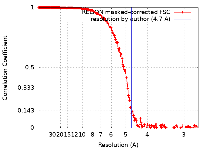
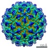
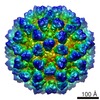
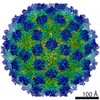
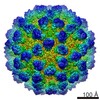
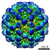
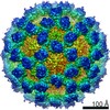
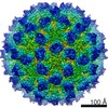
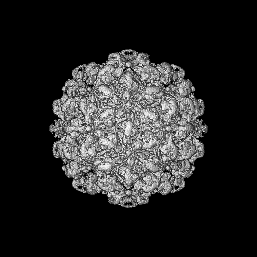
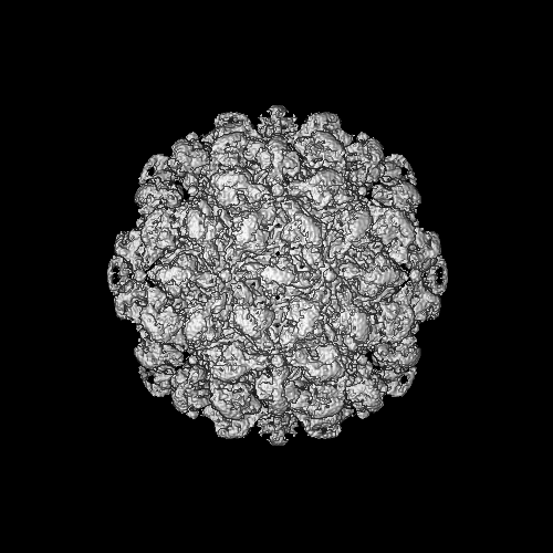
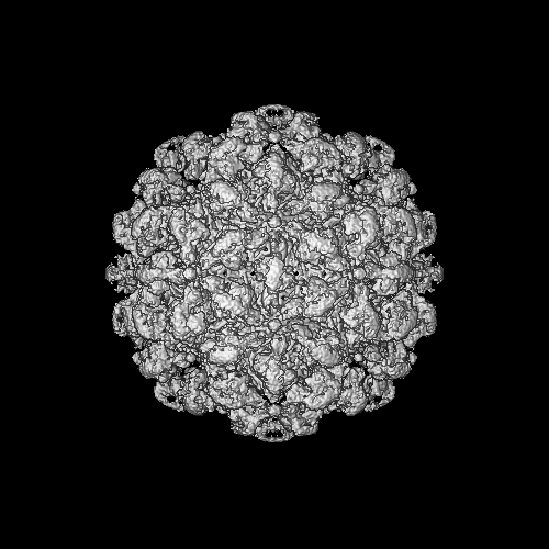
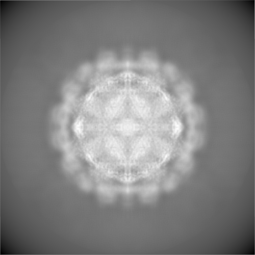
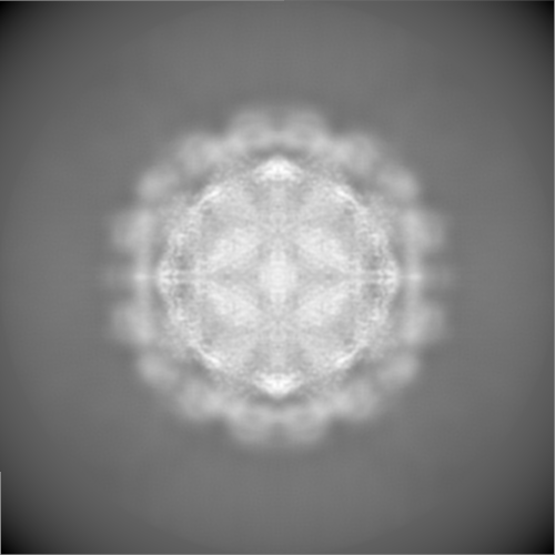
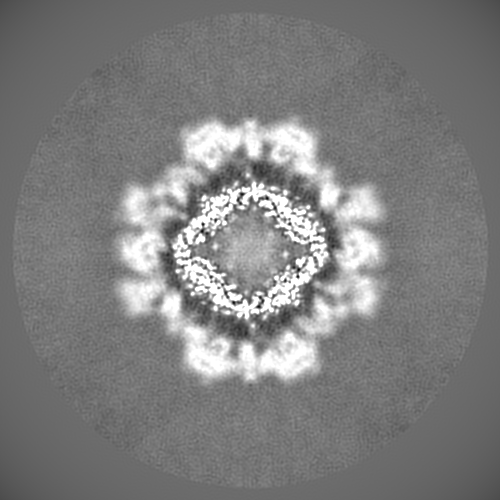
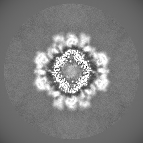
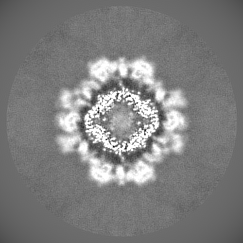
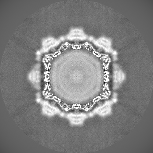
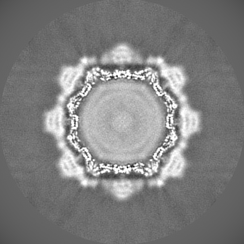
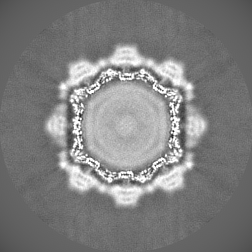
Journal: J Virol / Year: 2020 Title: Nanobody-Mediated Neutralization Reveals an Achilles Heel for Norovirus. Authors: Anna D Koromyslova / Jessica M Devant / Turgay Kilic / Charles D Sabin / Virginie Malak / Grant S Hansman / Abstract: Human norovirus frequently causes outbreaks of acute gastroenteritis. Although discovered more than five decades ago, antiviral development has, until recently, been hampered by the lack of a ...Human norovirus frequently causes outbreaks of acute gastroenteritis. Although discovered more than five decades ago, antiviral development has, until recently, been hampered by the lack of a reliable human norovirus cell culture system. Nevertheless, a lot of pathogenesis studies were accomplished using murine norovirus (MNV), which can be grown routinely in cell culture. In this study, we analyzed a sizeable library of nanobodies that were raised against the murine norovirus virion with the main purpose of developing nanobody-based inhibitors. We discovered two types of neutralizing nanobodies and analyzed the inhibition mechanisms using X-ray crystallography, cryo-electron microscopy (cryo-EM), and cell culture techniques. The first type bound on the top region of the protruding (P) domain. Interestingly, this nanobody binding region closely overlapped the MNV receptor-binding site and collectively shared numerous P domain-binding residues. In addition, we showed that these nanobodies competed with the soluble receptor, and this action blocked virion attachment to cultured cells. The second type bound at a dimeric interface on the lower side of the P dimer. We discovered that these nanobodies disrupted a structural change in the capsid associated with binding cofactors (i.e., metal cations/bile acid). Indeed, we found that capsids underwent major conformational changes following addition of Mg or Ca Ultimately, these nanobodies directly obstructed a structural modification reserved for a postreceptor attachment stage. Altogether, our new data show that nanobody-based inhibition could occur by blocking functional and structural capsid properties. This research discovered and analyzed two different types of MNV-neutralizing nanobodies. The top-binding nanobodies sterically inhibited the receptor-binding site, whereas the dimeric-binding nanobodies interfered with a structural modification associated with cofactor binding. Moreover, we found that the capsid contained a number of vulnerable regions that were essential for viral replication. In fact, the capsid appeared to be organized in a state of flux, which could be important for cofactor/receptor-binding functions. Blocking these capsid-binding events with nanobodies directly inhibited essential capsid functions. Moreover, a number of MNV-specific nanobody binding epitopes were comparable to human norovirus-specific nanobody inhibitors. Therefore, this additional structural and inhibition information could be further exploited in the development of human norovirus antivirals.
History
Deposition
Jan 10, 2020
-
Header (metadata) release
Apr 22, 2020
-
Map release
Apr 22, 2020
-
Update
Jul 22, 2020
-
Current status
Jul 22, 2020
Processing site: PDBe / Status: Released
-
Structure visualization
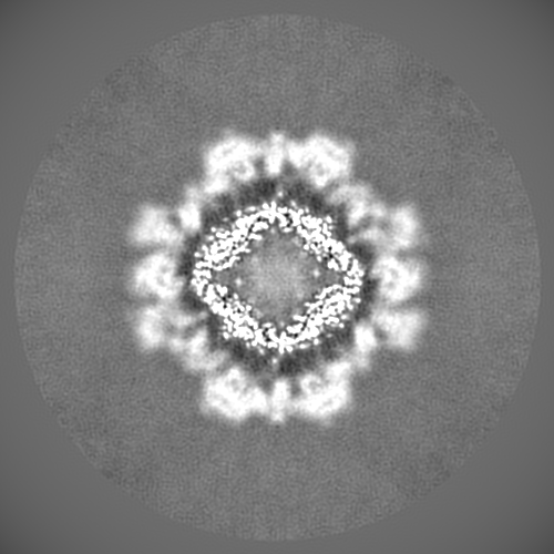
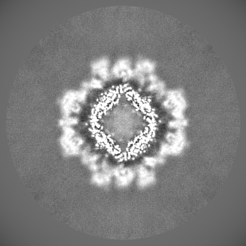
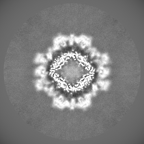
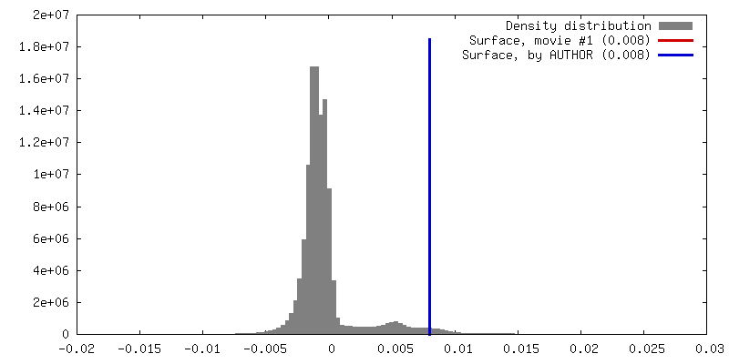
Movie
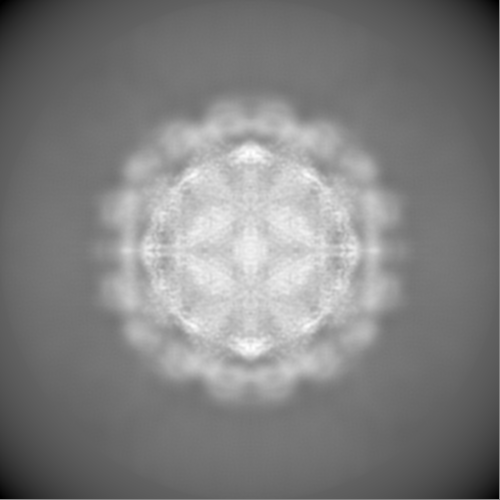
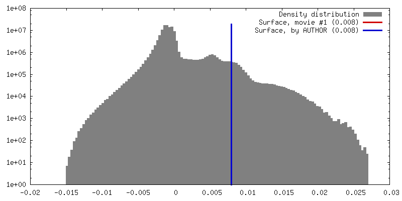
Surface view with section colored by density value
In the structure databanks used in Yorodumi, some data are registered as the other names, "COVID-19 virus" and "2019-nCoV". Here are the details of the virus and the list of structure data.
Jan 31, 2019. EMDB accession codes are about to change! (news from PDBe EMDB page)
EMDB accession codes are about to change! (news from PDBe EMDB page)
The allocation of 4 digits for EMDB accession codes will soon come to an end. Whilst these codes will remain in use, new EMDB accession codes will include an additional digit and will expand incrementally as the available range of codes is exhausted. The current 4-digit format prefixed with “EMD-” (i.e. EMD-XXXX) will advance to a 5-digit format (i.e. EMD-XXXXX), and so on. It is currently estimated that the 4-digit codes will be depleted around Spring 2019, at which point the 5-digit format will come into force.
The EM Navigator/Yorodumi systems omit the EMD- prefix.
Related info.:Q: What is EMD? / ID/Accession-code notation in Yorodumi/EM Navigator
Yorodumi is a browser for structure data from EMDB, PDB, SASBDB, etc.
This page is also the successor to EM Navigator detail page, and also detail information page/front-end page for Omokage search.
The word "yorodu" (or yorozu) is an old Japanese word meaning "ten thousand". "mi" (miru) is to see.
Related info.:EMDB / PDB / SASBDB / Comparison of 3 databanks / Yorodumi Search / Aug 31, 2016. New EM Navigator & Yorodumi / Yorodumi Papers / Jmol/JSmol / Function and homology information / Changes in new EM Navigator and Yorodumi
 Movie
Movie Controller

 Yorodumi
 Open data
 Basic information
 Map data
 Sample
 Murine norovirus
 Authors
 Citation
 Structure visualization
 Movie viewer
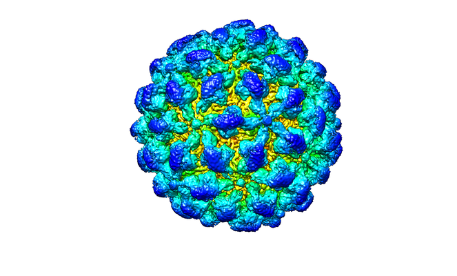
 Downloads & links
 emd_10597.png
 http://ftp.pdbj.org/pub/emdb/structures/EMD-10597


 Z (Sec.)
 Y (Row.)
 X (Col.)


 Sample components
 Processing
 Electron microscopy
 FIELD EMISSION GUN