Movie
Movie Controller


+ Open data

- Basic information
| Entry | Database: EMDB / ID: EMD-10091 | |||||||||
|---|---|---|---|---|---|---|---|---|---|---|
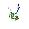
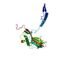
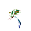
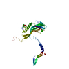
| Title | Granulovirus occlusion bodies by serial electron diffraction | |||||||||
 Map data Map data | ||||||||||
 Sample Sample |
| |||||||||
 Keywords Keywords | granulovirus / occlusion body / serial crystallography / viral protein | |||||||||
| Function / homology | Polyhedrin / Polyhedrin / viral occlusion body / structural molecule activity / Granulin Function and homology information Function and homology information | |||||||||
| Biological species |  Cydia pomonella granulosis virus (isolate Mexico/1963) / Cydia pomonella granulosis virus (isolate Mexican) Cydia pomonella granulosis virus (isolate Mexico/1963) / Cydia pomonella granulosis virus (isolate Mexican) | |||||||||
| Method | electron crystallography / cryo EM / Resolution: 1.6 Å | |||||||||
 Authors Authors | Buecker R / Mehrabi P | |||||||||
 Citation Citation | Journal: Nat Commun / Year: 2020 Title: Serial protein crystallography in an electron microscope. Authors: Robert Bücker / Pascal Hogan-Lamarre / Pedram Mehrabi / Eike C Schulz / Lindsey A Bultema / Yaroslav Gevorkov / Wolfgang Brehm / Oleksandr Yefanov / Dominik Oberthür / Günther H Kassier / ...Authors: Robert Bücker / Pascal Hogan-Lamarre / Pedram Mehrabi / Eike C Schulz / Lindsey A Bultema / Yaroslav Gevorkov / Wolfgang Brehm / Oleksandr Yefanov / Dominik Oberthür / Günther H Kassier / R J Dwayne Miller /   Abstract: Serial X-ray crystallography at free-electron lasers allows to solve biomolecular structures from sub-micron-sized crystals. However, beam time at these facilities is scarce, and involved sample ...Serial X-ray crystallography at free-electron lasers allows to solve biomolecular structures from sub-micron-sized crystals. However, beam time at these facilities is scarce, and involved sample delivery techniques are required. On the other hand, rotation electron diffraction (MicroED) has shown great potential as an alternative means for protein nano-crystallography. Here, we present a method for serial electron diffraction of protein nanocrystals combining the benefits of both approaches. In a scanning transmission electron microscope, crystals randomly dispersed on a sample grid are automatically mapped, and a diffraction pattern at fixed orientation is recorded from each at a high acquisition rate. Dose fractionation ensures minimal radiation damage effects. We demonstrate the method by solving the structure of granulovirus occlusion bodies and lysozyme to resolutions of 1.55 Å and 1.80 Å, respectively. Our method promises to provide rapid structure determination for many classes of materials with minimal sample consumption, using readily available instrumentation. | |||||||||
| History |
|
- Structure visualization
| Movie |
Movie viewer |
|---|---|
| Structure viewer | EM map: SurfViewMolmilJmol/JSmol |
| Supplemental images |

- Downloads & links
-EMDB archive
| Map data | emd_10091.map.gz | 2.2 MB | EMDB map data format | |
|---|---|---|---|---|
| Header (meta data) | emd-10091-v30.xmlemd-10091.xml | 12.8 KB 12.8 KB | Display Display | EMDB header |
| Images |  emd_10091.png emd_10091.png | 104.9 KB | ||
| Filedesc metadata | emd-10091.cif.gz | 5.5 KB | ||
| Filedesc structureFactors | emd_10091_sf.cif.gz | 687.9 KB | ||
| Archive directory |  http://ftp.pdbj.org/pub/emdb/structures/EMD-10091ftp://ftp.pdbj.org/pub/emdb/structures/EMD-10091 http://ftp.pdbj.org/pub/emdb/structures/EMD-10091ftp://ftp.pdbj.org/pub/emdb/structures/EMD-10091 | HTTPS FTP |
-Related structure data
| Related structure data |  6s2oMC  6s2nC M: atomic model generated by this map C: citing same article ( |
|---|---|
| Similar structure data |


-Links
| EMDB pages | EMDB (EBI/PDBe) / EMDataResource |
|---|
-Map
| File | Download / File: emd_10091.map.gz / Format: CCP4 / Size: 41.4 MB / Type: IMAGE STORED AS FLOATING POINT NUMBER (4 BYTES) | ||||||||||||||||||||||||||||||||||||||||||||||||||||||||||||
|---|---|---|---|---|---|---|---|---|---|---|---|---|---|---|---|---|---|---|---|---|---|---|---|---|---|---|---|---|---|---|---|---|---|---|---|---|---|---|---|---|---|---|---|---|---|---|---|---|---|---|---|---|---|---|---|---|---|---|---|---|---|
| Projections & slices | Image control
Images are generated by Spider. generated in cubic-lattice coordinate | ||||||||||||||||||||||||||||||||||||||||||||||||||||||||||||
| Voxel size | X=Y=Z: 0.3822 Å | ||||||||||||||||||||||||||||||||||||||||||||||||||||||||||||
| Density |
| ||||||||||||||||||||||||||||||||||||||||||||||||||||||||||||
| Symmetry | Space group: 1 | ||||||||||||||||||||||||||||||||||||||||||||||||||||||||||||
| Details | EMDB XML:
CCP4 map header:
| ||||||||||||||||||||||||||||||||||||||||||||||||||||||||||||
 Z (Sec.)
 Y (Row.)
 X (Col.)


-Supplemental data
- Sample components
-Entire : Cydia pomonella granulosis virus (isolate Mexican)
| Entire | Name: Cydia pomonella granulosis virus (isolate Mexican) |
|---|---|
| Components |
|
-Supramolecule #1: Cydia pomonella granulosis virus (isolate Mexican)
| Supramolecule | Name: Cydia pomonella granulosis virus (isolate Mexican) / type: virus / ID: 1 / Parent: 0 / Macromolecule list: #1 / NCBI-ID: 654905 Sci species name: Cydia pomonella granulosis virus (isolate Mexican) Virus type: VIRION / Virus isolate: OTHER / Virus enveloped: No / Virus empty: No |
|---|
-Macromolecule #1: Granulin
| Macromolecule | Name: Granulin / type: protein_or_peptide / ID: 1 / Number of copies: 1 / Enantiomer: LEVO |
|---|---|
| Source (natural) | Organism: Cydia pomonella granulosis virus (isolate Mexico/1963) Strain: isolate Mexico/1963 |
| Molecular weight | Theoretical: 29.378559 KDa |
| Recombinant expression | Organism: Cydia pomonella granulosis virus (isolate Mexican) |
| Sequence | String: MGYNKSLRYS RHDGTSCVID NHHLKSLGAV LNDVRRKKDR IREAEYEPII DIADQYMVTE DPFRGPGKNV RITLFKEIRR VHPDTMKLV CNWSGKEFLR ETWTRFISEE FPITTDQEIM DLWFELQLRP MHPNRCYKFT MQYALGAHPD YVAHDVIRQQ D PYYVGPNN ...String: MGYNKSLRYS RHDGTSCVID NHHLKSLGAV LNDVRRKKDR IREAEYEPII DIADQYMVTE DPFRGPGKNV RITLFKEIRR VHPDTMKLV CNWSGKEFLR ETWTRFISEE FPITTDQEIM DLWFELQLRP MHPNRCYKFT MQYALGAHPD YVAHDVIRQQ D PYYVGPNN IERINLSKKG FAFPLTCLQS VYNDNFERFF DDVLWPYFYR PLVYVGTTSA EIEEIMIEVS LLFKIKEFAP DV PLFTGPA Y UniProtKB: Granulin |
-Macromolecule #2: water
| Macromolecule | Name: water / type: ligand / ID: 2 / Number of copies: 90 / Formula: HOH |
|---|---|
| Molecular weight | Theoretical: 18.015 Da |
| Chemical component information |  ChemComp-HOH: |
-Experimental details
-Structure determination
| Method | cryo EM |
|---|---|
 Processing Processing | electron crystallography |
| Aggregation state | 3D array |
-Sample preparation
| Buffer | pH: 7 |
|---|---|
| Vitrification | Cryogen name: ETHANE |
- Electron microscopy
| Microscope | FEI TECNAI F20 |
|---|---|
| Temperature | Min: 93.0 K / Max: 100.0 K |
| Image recording | Film or detector model: OTHER / Digitization - Dimensions - Width: 1536 pixel / Digitization - Dimensions - Height: 512 pixel / Number grids imaged: 1 / Number diffraction images: 32000 / Average exposure time: 0.01 sec. / Average electron dose: 4.7 e/Å2 |
| Electron beam | Acceleration voltage: 200 kV / Electron source:  FIELD EMISSION GUN FIELD EMISSION GUN |
| Electron optics | C2 aperture diameter: 5.0 µm / Illumination mode: OTHER / Imaging mode: DIFFRACTION / Camera length: 2580 mm |
| Sample stage | Specimen holder model: GATAN 626 SINGLE TILT LIQUID NITROGEN CRYO TRANSFER HOLDER Cooling holder cryogen: NITROGEN / Tilt angle: 0 |
| Experimental equipment |  Model: Tecnai F20 / Image courtesy: FEI Company |
-Image processing
| Final reconstruction | Resolution.type: BY AUTHOR / Resolution: 1.6 Å / Resolution method: DIFFRACTION PATTERN/LAYERLINES / Software - Name: Coot |
|---|---|
| Molecular replacement | Software - Name: PHENIX (ver. 1.17) |
| Merging software list | Software - details: CrystFEL |
| Crystallography statistics | Number intensities measured: 9650574 / Number structure factors: 23422 / Fourier space coverage: 100 / R sym: 0.093 / R merge: 0.093 / Overall phase error: 13.64 / Overall phase residual: 1 / Phase error rejection criteria: 0 / High resolution: 1.6 Å / Shell - Shell ID: 1 / Shell - High resolution: 10.0 Å / Shell - Low resolution: 1.55 Å / Shell - Number structure factors: 1 / Shell - Phase residual: 1 / Shell - Fourier space coverage: 1 / Shell - Multiplicity: 1 |