 Movie
Movie Controller


+ Open data

- Basic information
| Entry | Database: EMDB / ID: EMD-0201 | |||||||||
|---|---|---|---|---|---|---|---|---|---|---|
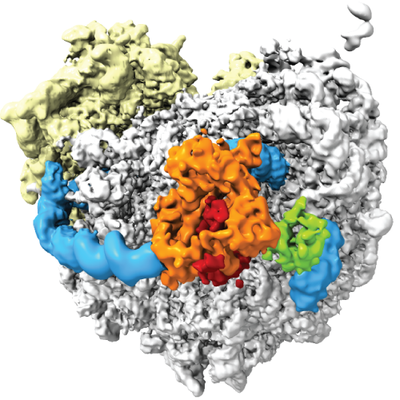
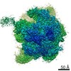
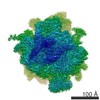
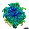
| Title | Cryo-EM structure of the ribosome-NatA complex | |||||||||
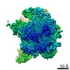
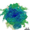
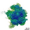
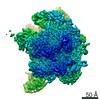
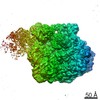
 Map data Map data | Ribosome-NatA complex refined on NatA. Postprocessed map was low pass filtered to 6 Angstrom. | |||||||||
 Sample Sample |
| |||||||||
| Function / homology |  Function and homology information Function and homology information N-terminal protein amino acid propionylation / peptide-glutamate-alpha-N-acetyltransferase activity / N-terminal amino-acid Nalpha-acetyltransferase NatA / N-terminal methionine Nalpha-acetyltransferase NatE / peptide-serine-alpha-N-acetyltransferase activity / NatA complex / N-terminal protein amino acid acetylation / peptide alpha-N-acetyltransferase activity / mitotic sister chromatid cohesion / ribosome binding ...N-terminal protein amino acid propionylation / peptide-glutamate-alpha-N-acetyltransferase activity / N-terminal amino-acid Nalpha-acetyltransferase NatA / N-terminal methionine Nalpha-acetyltransferase NatE / peptide-serine-alpha-N-acetyltransferase activity / NatA complex / N-terminal protein amino acid acetylation / peptide alpha-N-acetyltransferase activity / mitotic sister chromatid cohesion / ribosome binding / mitochondrion / identical protein binding / cytoplasm N-terminal protein amino acid propionylation / peptide-glutamate-alpha-N-acetyltransferase activity / N-terminal amino-acid Nalpha-acetyltransferase NatA / N-terminal methionine Nalpha-acetyltransferase NatE / peptide-serine-alpha-N-acetyltransferase activity / NatA complex / N-terminal protein amino acid acetylation / peptide alpha-N-acetyltransferase activity / mitotic sister chromatid cohesion / ribosome binding ...N-terminal protein amino acid propionylation / peptide-glutamate-alpha-N-acetyltransferase activity / N-terminal amino-acid Nalpha-acetyltransferase NatA / N-terminal methionine Nalpha-acetyltransferase NatE / peptide-serine-alpha-N-acetyltransferase activity / NatA complex / N-terminal protein amino acid acetylation / peptide alpha-N-acetyltransferase activity / mitotic sister chromatid cohesion / ribosome binding / mitochondrion / identical protein binding / cytoplasmSimilarity search - Function | |||||||||
| Biological species |  Saccharomyces cerevisiae S288C (yeast) / Saccharomyces cerevisiae (strain ATCC 204508 / S288c) (yeast) Saccharomyces cerevisiae S288C (yeast) / Saccharomyces cerevisiae (strain ATCC 204508 / S288c) (yeast) | |||||||||
| Method | single particle reconstruction / cryo EM / Resolution: 4.8 Å | |||||||||
 Authors Authors | Knorr AG / Becker T / Berninghausen O / Beckmann R | |||||||||
 Citation Citation | Journal: Nat Struct Mol Biol / Year: 2019 Title: Ribosome-NatA architecture reveals that rRNA expansion segments coordinate N-terminal acetylation. Authors: Alexandra G Knorr / Christian Schmidt / Petr Tesina / Otto Berninghausen / Thomas Becker / Birgitta Beatrix / Roland Beckmann /  Abstract: The majority of eukaryotic proteins are N-terminally α-acetylated by N-terminal acetyltransferases (NATs). Acetylation usually occurs co-translationally and defects have severe consequences. ...The majority of eukaryotic proteins are N-terminally α-acetylated by N-terminal acetyltransferases (NATs). Acetylation usually occurs co-translationally and defects have severe consequences. Nevertheless, it is unclear how these enzymes act in concert with the translating ribosome. Here, we report the structure of a native ribosome-NatA complex from Saccharomyces cerevisiae. NatA (comprising Naa10, Naa15 and Naa50) displays a unique mode of ribosome interaction by contacting eukaryotic-specific ribosomal RNA expansion segments in three out of four binding patches. Thereby, NatA is dynamically positioned directly underneath the ribosomal exit tunnel to facilitate modification of the emerging nascent peptide chain. Methionine amino peptidases, but not chaperones or signal recognition particle, would be able to bind concomitantly. This work assigns a function to the hitherto enigmatic ribosomal RNA expansion segments and provides mechanistic insights into co-translational protein maturation by N-terminal acetylation. | |||||||||
| History |
|
- Structure visualization
| Movie |
Movie viewer |
|---|---|
| Structure viewer | EM map: SurfViewMolmilJmol/JSmol |
| Supplemental images |
- Downloads & links
-EMDB archive
| Map data | emd_0201.map.gz | 264.3 MB | EMDB map data format | |
|---|---|---|---|---|
| Header (meta data) | emd-0201-v30.xmlemd-0201.xml | 12.6 KB 12.6 KB | Display Display | EMDB header |
| Images |  emd_0201.png emd_0201.png | 224.2 KB | ||
| Archive directory |  http://ftp.pdbj.org/pub/emdb/structures/EMD-0201ftp://ftp.pdbj.org/pub/emdb/structures/EMD-0201 http://ftp.pdbj.org/pub/emdb/structures/EMD-0201ftp://ftp.pdbj.org/pub/emdb/structures/EMD-0201 | HTTPS FTP |
-Related structure data
| Related structure data |  6hd5MC  0202C  0203C 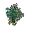 6hd7C M: atomic model generated by this map C: citing same article ( |
|---|---|
| Similar structure data |

-Links
| EMDB pages | EMDB (EBI/PDBe) / EMDataResource |
|---|---|
| Related items in Molecule of the Month |


-Map
| File | Download / File: emd_0201.map.gz / Format: CCP4 / Size: 282.6 MB / Type: IMAGE STORED AS FLOATING POINT NUMBER (4 BYTES) | ||||||||||||||||||||||||||||||||||||||||||||||||||||||||||||
|---|---|---|---|---|---|---|---|---|---|---|---|---|---|---|---|---|---|---|---|---|---|---|---|---|---|---|---|---|---|---|---|---|---|---|---|---|---|---|---|---|---|---|---|---|---|---|---|---|---|---|---|---|---|---|---|---|---|---|---|---|---|
| Annotation | Ribosome-NatA complex refined on NatA. Postprocessed map was low pass filtered to 6 Angstrom. | ||||||||||||||||||||||||||||||||||||||||||||||||||||||||||||
| Voxel size | X=Y=Z: 1.084 Å | ||||||||||||||||||||||||||||||||||||||||||||||||||||||||||||
| Density |
| ||||||||||||||||||||||||||||||||||||||||||||||||||||||||||||
| Symmetry | Space group: 1 | ||||||||||||||||||||||||||||||||||||||||||||||||||||||||||||
| Details | EMDB XML:
CCP4 map header:
| ||||||||||||||||||||||||||||||||||||||||||||||||||||||||||||
-Supplemental data
- Sample components
-Entire : Ribosome-NatA complex
| Entire | Name: Ribosome-NatA complex |
|---|---|
| Components |
|
-Supramolecule #1: Ribosome-NatA complex
| Supramolecule | Name: Ribosome-NatA complex / type: complex / ID: 1 / Parent: 0 / Macromolecule list: all Details: Map was refined on NatA. Coordinates are deposited for NatA only. |
|---|---|
| Source (natural) | Organism: Saccharomyces cerevisiae S288C (yeast) |
-Macromolecule #1: N-terminal acetyltransferase A complex subunit NAT1
| Macromolecule | Name: N-terminal acetyltransferase A complex subunit NAT1 / type: protein_or_peptide / ID: 1 / Details: ribosome binding subunit / Number of copies: 1 / Enantiomer: LEVO |
|---|---|
| Source (natural) | Organism: Saccharomyces cerevisiae (strain ATCC 204508 / S288c) (yeast) |
| Molecular weight | Theoretical: 99.050133 KDa |
| Sequence | String: MSRKRSTKPK PAAKIALKKE NDQFLEALKL YEGKQYKKSL KLLDAILKKD GSHVDSLALK GLDLYSVGEK DDAASYVANA IRKIEGASA SPICCHVLGI YMRNTKEYKE SIKWFTAALN NGSTNKQIYR DLATLQSQIG DFKNALVSRK KYWEAFLGYR A NWTSLAVA ...String: MSRKRSTKPK PAAKIALKKE NDQFLEALKL YEGKQYKKSL KLLDAILKKD GSHVDSLALK GLDLYSVGEK DDAASYVANA IRKIEGASA SPICCHVLGI YMRNTKEYKE SIKWFTAALN NGSTNKQIYR DLATLQSQIG DFKNALVSRK KYWEAFLGYR A NWTSLAVA QDVNGERQQA INTLSQFEKL AEGKISDSEK YEHSECLMYK NDIMYKAASD NQDKLQNVLK HLNDIEPCVF DK FGLLERK ATIYMKLGQL KDASIVYRTL IKRNPDNFKY YKLLEVSLGI QGDNKLKKAL YGKLEQFYPR CEPPKFIPLT FLQ DKEELS KKLREYVLPQ LERGVPATFS NVKPLYQRRK SKVSPLLEKI VLDYLSGLDP TQDPIPFIWT NYYLSQHFLF LKDF PKAQE YIDAALDHTP TLVEFYILKA RILKHLGLMD TAAGILEEGR QLDLQDRFIN CKTVKYFLRA NNIDKAVEVA SLFTK NDDS VNGIKDLHLV EASWFIVEQA EAYYRLYLDR KKKLDDLASL KKEVESDKSE QIANDIKENQ WLVRKYKGLA LKRFNA IPK FYKQFEDDQL DFHSYCMRKG TPRAYLEMLE WGKALYTKPM YVRAMKEASK LYFQMHDDRL KRKSDSLDEN SDEIQNN GQ NSSSQKKKAK KEAAAMNKRK ETEAKSVAAY PSDQDNDVFG EKLIETSTPM EDFATEFYNN YSMQVREDER DYILDFEF N YRIGKLALCF ASLNKFAKRF GTTSGLFGSM AIVLLHATRN DTPFDPILKK VVTKSLEKEY SENFPLNEIS NNSFDWLNF YQEKFGKNDI NGLLFLYRYR DDVPIGSSNL KEMIISSLSP LEPHSQNEIL QYYL |
-Macromolecule #2: N-terminal acetyltransferase A complex catalytic subunit ARD1
| Macromolecule | Name: N-terminal acetyltransferase A complex catalytic subunit ARD1 type: protein_or_peptide / ID: 2 / Details: catalytic subunit / Number of copies: 1 / Enantiomer: LEVO EC number: N-terminal amino-acid Nalpha-acetyltransferase NatA |
|---|---|
| Source (natural) | Organism: Saccharomyces cerevisiae (strain ATCC 204508 / S288c) (yeast) |
| Molecular weight | Theoretical: 27.635168 KDa |
| Sequence | String: MPINIRRATI NDIICMQNAN LHNLPENYMM KYYMYHILSW PEASFVATTT TLDCEDSDEQ DENDKLELTL DGTNDGRTIK LDPTYLAPG EKLVGYVLVK MNDDPDQQNE PPNGHITSLS VMRTYRRMGI AENLMRQALF ALREVHQAEY VSLHVRQSNR A ALHLYRDT ...String: MPINIRRATI NDIICMQNAN LHNLPENYMM KYYMYHILSW PEASFVATTT TLDCEDSDEQ DENDKLELTL DGTNDGRTIK LDPTYLAPG EKLVGYVLVK MNDDPDQQNE PPNGHITSLS VMRTYRRMGI AENLMRQALF ALREVHQAEY VSLHVRQSNR A ALHLYRDT LAFEVLSIEK SYYQDGEDAY AMKKVLKLEE LQISNFTHRR LKENEEKLED DLESDLLEDI IKQGVNDIIV |
-Macromolecule #3: N-alpha-acetyltransferase NAT5
| Macromolecule | Name: N-alpha-acetyltransferase NAT5 / type: protein_or_peptide / ID: 3 / Details: ribosome binding subunit / Number of copies: 1 / Enantiomer: LEVO EC number: N-terminal methionine Nalpha-acetyltransferase NatE |
|---|---|
| Source (natural) | Organism: Saccharomyces cerevisiae (strain ATCC 204508 / S288c) (yeast) |
| Molecular weight | Theoretical: 19.753727 KDa |
| Sequence | String: MGRDICTLDN VYANNLGMLT KLAHVTVPNL YQDAFFSALF AEDSLVAKNK KPSSKKDVHF TQMAYYSEIP VGGLVAKLVP KKQNELSLK GIQIEFLGVL PNYRHKSIGS KLLKFAEDKC SECHQHNVFV YLPAVDDLTK QWFIAHGFEQ VGETVNNFIK G VNGDEQDA ILLKKHIS |
-Experimental details
-Structure determination
| Method | cryo EM |
|---|---|
 Processing Processing | single particle reconstruction |
| Aggregation state | particle |
-Sample preparation
| Buffer | pH: 7.5 |
|---|---|
| Vitrification | Cryogen name: ETHANE |
- Electron microscopy
| Microscope | FEI TITAN KRIOS |
|---|---|
| Electron beam | Acceleration voltage: 300 kV / Electron source: FIELD EMISSION GUN |
| Electron optics | Illumination mode: FLOOD BEAM / Imaging mode: BRIGHT FIELDBright-field microscopy |
| Image recording | Film or detector model: FEI FALCON II (4k x 4k) / Average electron dose: 2.5 e/Å2 |
| Experimental equipment |  Model: Titan Krios / Image courtesy: FEI Company |
-Image processing
| CTF correction | Software - Name: Gctf |
|---|---|
| Initial angle assignment | Type: PROJECTION MATCHING / Software - Name: RELION |
| Final angle assignment | Type: PROJECTION MATCHING / Software - Name: RELION |
| Final reconstruction | Resolution.type: BY AUTHOR / Resolution: 4.8 Å / Resolution method: FSC 0.143 CUT-OFF / Number images used: 262507 |
-Atomic model buiding 1
| Refinement | Protocol: RIGID BODY FIT |
|---|---|
| Output model | PDB-6hd5: |
