Loading
PDBj
MenuPDBj@FacebookPDBj@TwitterPDBj@YouTubewwPDB FoundationwwPDB
RCSB PDBPDBeBMRBAdv. SearchSearch help

7A2R
DownloadVisualize
BU of 7a2r by Molmil
Crystal structure of the Fyn SH3 domain L112V-S114N-S115T-E121L-R123H mutant at pH 6.0
Descriptor: Tyrosine-protein kinase Fyn
Authors:Camara-Artigas, A, Plaza-Garrido, M, Salinas-Garcia, M.C.
Deposit date:2020-08-18
Release date:2021-08-25
Last modified:2024-01-31
Method:X-RAY DIFFRACTION (1.05 Å)
Cite:Crystal structure of the Fyn SH3 domain L112V-S114N-S115T-E121L-R123H mutant at pH 6.0
To be published
7A31
DownloadVisualize
BU of 7a31 by Molmil
Crystal structure of the c-Src SH3 domain mutant S94A-T98D-V111L-N113S-T114S at pH 4.5
Descriptor: Proto-oncogene tyrosine-protein kinase Src
Authors:Camara-Artigas, A, Plaza-Garrido, M, Salinas-Garcia, M.C.
Deposit date:2020-08-18
Release date:2021-08-25
Last modified:2024-01-31
Method:X-RAY DIFFRACTION (0.94 Å)
Cite:Crystal structure of the c-Src SH3 domain mutant S94A-T98D-V111L-N113S-T114S at pH 4.5
To be published
7A3B
DownloadVisualize
BU of 7a3b by Molmil
Intertwined dimer of the c-Src SH3 domain mutant E106D
Descriptor: GLYCEROL, Proto-oncogene tyrosine-protein kinase Src, TRIETHYLENE GLYCOL
Authors:Camara-Artigas, A, Plaza-Garrido, M, Salinas-Garcia, M.C.
Deposit date:2020-08-18
Release date:2021-08-25
Last modified:2024-01-31
Method:X-RAY DIFFRACTION (1.91 Å)
Cite:Intertwined dimer of the c-Src SH3 domain mutant E106D
To be published
7A2L
DownloadVisualize
BU of 7a2l by Molmil
Crystal structure of the Fyn SH3 domain mutant E129Q in space group C21 at pH 4.0
Descriptor: FORMIC ACID, SODIUM ION, Tyrosine-protein kinase Fyn
Authors:Camara-Artigas, A, Plaza-Garrido, M, Salinas-Garcia, M.C.
Deposit date:2020-08-18
Release date:2021-08-25
Last modified:2024-01-31
Method:X-RAY DIFFRACTION (1.9 Å)
Cite:Crystal structure of the Fyn SH3 domain mutant E129Q in space group C21 at pH 4.0
To be published
7A2V
DownloadVisualize
BU of 7a2v by Molmil
Crystal structure of the Fyn SH3 domain L112V-S114N-S115T-E129Q at pH 4.0
Descriptor: Tyrosine-protein kinase Fyn
Authors:Camara-Artigas, A, Plaza-Garrido, M, Salinas-Garcia, M.C.
Deposit date:2020-08-18
Release date:2021-08-25
Last modified:2024-01-31
Method:X-RAY DIFFRACTION (1.81 Å)
Cite:Crystal structure of the Fyn SH3 domain L112V-S114N-S115T-E129Q at pH 4.0
To be published
7A33
DownloadVisualize
BU of 7a33 by Molmil
Crystal structure of the c-Src SH3 domain mutant S94A-T98D-V111L-N113S-T114S at pH 3.0
Descriptor: FORMIC ACID, Proto-oncogene tyrosine-protein kinase Src, SODIUM ION
Authors:Camara-Artigas, A, Plaza-Garrido, M, Salinas-Garcia, M.C.
Deposit date:2020-08-18
Release date:2021-08-25
Last modified:2024-01-31
Method:X-RAY DIFFRACTION (0.96 Å)
Cite:Crystal structure of the c-Src SH3 domain mutant S94A-T98D-V111L-N113S-T114S at pH 3.0
To be published
7A32
DownloadVisualize
BU of 7a32 by Molmil
Crystal structure of the c-Src SH3 domain mutant S94A-T98D-V111L-N113S-T114S at pH 7.0
Descriptor: GLYCEROL, Proto-oncogene tyrosine-protein kinase Src, SULFATE ION
Authors:Camara-Artigas, A, Plaza-Garrido, M, Salinas-Garcia, M.C.
Deposit date:2020-08-18
Release date:2021-08-25
Last modified:2024-01-31
Method:X-RAY DIFFRACTION (1.15 Å)
Cite:Crystal structure of the c-Src SH3 domain mutant S94A-T98D-V111L-N113S-T114S at pH 7.0
To be published
7A2T
DownloadVisualize
BU of 7a2t by Molmil
Crystal structure of the Fyn SH3 domain L112V-S114N-S115T-E121L-R123H mutant at pH 4.0
Descriptor: ACETATE ION, Tyrosine-protein kinase Fyn
Authors:Camara-Artigas, A, Plaza-Garrido, M, Salinas-Garcia, M.C.
Deposit date:2020-08-18
Release date:2021-08-25
Last modified:2024-01-31
Method:X-RAY DIFFRACTION (1.22 Å)
Cite:Crystal structure of the Fyn SH3 domain L112V-S114N-S115T-E121L-R123H mutant at pH 4.0
To be published
7A35
DownloadVisualize
BU of 7a35 by Molmil
Crystal structure of the c-Src SH3 domain mutant V111L-N113S-T114S at pH 7.0
Descriptor: Proto-oncogene tyrosine-protein kinase Src, SULFATE ION
Authors:Camara-Artigas, A, Plaza-Garrido, M, Salinas-Garcia, M.C.
Deposit date:2020-08-18
Release date:2021-08-25
Last modified:2024-01-31
Method:X-RAY DIFFRACTION (1.314 Å)
Cite:Crystal structure of the c-Src SH3 domain mutant V111L-N113S-T114S at pH 7.0
To be published
7A2U
DownloadVisualize
BU of 7a2u by Molmil
Crystal structure of the Fyn SH3 domain L112V-S114N-S115T at pH 4.5
Descriptor: ACETATE ION, Tyrosine-protein kinase Fyn
Authors:Camara-Artigas, A, Plaza-Garrido, M, Salinas-Garcia, M.C.
Deposit date:2020-08-18
Release date:2021-08-25
Last modified:2024-01-31
Method:X-RAY DIFFRACTION (1.7 Å)
Cite:Crystal structure of the Fyn SH3 domain L112V-S114N-S115T at pH 4.5
To be published
7A34
DownloadVisualize
BU of 7a34 by Molmil
Intertwined dimer of the c-Src SH3 domain mutant V111L-N113S-T114S
Descriptor: DI(HYDROXYETHYL)ETHER, Proto-oncogene tyrosine-protein kinase Src, TRIETHYLENE GLYCOL
Authors:Camara-Artigas, A, Plaza-Garrido, M, Salinas-Garcia, M.C.
Deposit date:2020-08-18
Release date:2021-08-25
Last modified:2024-01-31
Method:X-RAY DIFFRACTION (1.85 Å)
Cite:Intertwined dimer of the c-Src SH3 domain mutant V111L-N113S-T114S
To be published
7A2M
DownloadVisualize
BU of 7a2m by Molmil
Crystal structure of the Fyn SH3 domain A95S-D99T mutant in C21 space group
Descriptor: FORMIC ACID, SODIUM ION, SULFATE ION, ...
Authors:Camara-Artigas, A, Plaza-Garrido, M, Salinas-Garcia, M.C.
Deposit date:2020-08-18
Release date:2021-08-25
Last modified:2024-01-31
Method:X-RAY DIFFRACTION (1.5 Å)
Cite:Crystal structure of the Fyn SH3 domain A95S-D99T mutant in C21 space group
To be published
7A37
DownloadVisualize
BU of 7a37 by Molmil
Crystal structure of the c-Src SH3 domain mutant V111L-N113S-T114S in 3 M urea
Descriptor: Proto-oncogene tyrosine-protein kinase Src, UREA
Authors:Camara-Artigas, A, Plaza-Garrido, M, Salinas-Garcia, M.C.
Deposit date:2020-08-18
Release date:2021-08-25
Last modified:2024-01-31
Method:X-RAY DIFFRACTION (1.52 Å)
Cite:Crystal structure of the c-Src SH3 domain mutant V111L-N113S-T114S in 3 M urea
To be published
7A2S
DownloadVisualize
BU of 7a2s by Molmil
Crystal structure of the Fyn SH3 domain L112V-S114N-S115T-E121L-R123H mutant at pH 5.0
Descriptor: Tyrosine-protein kinase Fyn
Authors:Camara-Artigas, A, Plaza-Garrido, M, Salinas-Garcia, M.C.
Deposit date:2020-08-18
Release date:2021-08-25
Last modified:2024-01-31
Method:X-RAY DIFFRACTION (1.02 Å)
Cite:Crystal structure of the Fyn SH3 domain L112V-S114N-S115T-E121L-R123H mutant at pH 5.0
To Be Published
7A2K
DownloadVisualize
BU of 7a2k by Molmil
Crystal structure of the Fyn SH3 domain in space group P1 at pH 7.5
Descriptor: SODIUM ION, Tyrosine-protein kinase Fyn
Authors:Camara-Artigas, A, Plaza-Garrido, M, Salinas-Garcia, M.C.
Deposit date:2020-08-18
Release date:2021-08-25
Last modified:2024-01-31
Method:X-RAY DIFFRACTION (1.5 Å)
Cite:Crystal structure of the Fyn SH3 domain in space group P1 at pH 7.5
To be published
7A3A
DownloadVisualize
BU of 7a3a by Molmil
Intertwined dimer of the c-Src SH3 domain mutant V111L-N113S-T114S-Q128E at pH 6.0
Descriptor: 1,2-ETHANEDIOL, DI(HYDROXYETHYL)ETHER, Proto-oncogene tyrosine-protein kinase Src, ...
Authors:Camara-Artigas, A, Plaza-Garrido, M, Salinas-Garcia, M.C.
Deposit date:2020-08-18
Release date:2021-08-25
Last modified:2024-01-31
Method:X-RAY DIFFRACTION (1.8 Å)
Cite:Intertwined dimer of the c-Src SH3 domain mutant V111L-N113S-T114S-Q128E at pH 6.0
To be published
7A36
DownloadVisualize
BU of 7a36 by Molmil
Crystal structure of the c-Src SH3 domain mutant V111L-N113S-T114S in 7 M urea
Descriptor: Proto-oncogene tyrosine-protein kinase Src, SODIUM ION, UREA
Authors:Camara-Artigas, A, Plaza-Garrido, M, Salinas-Garcia, M.C.
Deposit date:2020-08-18
Release date:2021-08-25
Last modified:2024-01-31
Method:X-RAY DIFFRACTION (1.5 Å)
Cite:Crystal structure of the c-Src SH3 domain mutant V111L-N113S-T114S in 7 M urea
To be published
7A2N
DownloadVisualize
BU of 7a2n by Molmil
Crystal structure of the Fyn SH3 domain A95S-D99T mutant in space group P21
Descriptor: FORMIC ACID, SODIUM ION, Tyrosine-protein kinase Fyn
Authors:Camara-Artigas, A, Plaza-Garrido, M, Salinas-Garcia, M.C.
Deposit date:2020-08-18
Release date:2021-08-25
Last modified:2024-01-31
Method:X-RAY DIFFRACTION (1.4 Å)
Cite:Crystal structure of the Fyn SH3 domain A95S-D99T mutant in space group P21
To be published
7A3E
DownloadVisualize
BU of 7a3e by Molmil
Intertwined dimer of the c-Src SH3 domain mutant T126S
Descriptor: DI(HYDROXYETHYL)ETHER, Proto-oncogene tyrosine-protein kinase Src, SULFATE ION, ...
Authors:Camara-Artigas, A, Plaza-Garrido, M, Salinas-Garcia, M.C.
Deposit date:2020-08-18
Release date:2021-08-25
Last modified:2024-01-31
Method:X-RAY DIFFRACTION (1.52 Å)
Cite:Intertwined dimer of the c-Src SH3 domain mutant T126S
To be published
7A2W
DownloadVisualize
BU of 7a2w by Molmil
Crystal structure of the Fyn SH3 domain L112V-S114N-S115T-E121L-R123H mutant in complex with VSL12 at pH 3.0
Descriptor: CITRIC ACID, Tyrosine-protein kinase Fyn, VSL12
Authors:Camara-Artigas, A, Plaza-Garrido, M, Salinas-Garcia, M.C.
Deposit date:2020-08-18
Release date:2021-08-25
Last modified:2024-01-31
Method:X-RAY DIFFRACTION (0.99 Å)
Cite:Crystal structure of the Fyn SH3 domain L112V-S114N-S115T-E121L-R123H mutant in complex with VSL12 at pH 3.0
To be published
7A38
DownloadVisualize
BU of 7a38 by Molmil
Crystal structure of the c-Src SH3 domain mutant V111L-N113S-T114S-Q128E at pH 6.0
Descriptor: 2-(N-MORPHOLINO)-ETHANESULFONIC ACID, Proto-oncogene tyrosine-protein kinase Src, SULFATE ION
Authors:Camara-Artigas, A, Plaza-Garrido, M, Salinas-Garcia, M.C.
Deposit date:2020-08-18
Release date:2021-08-25
Last modified:2024-01-31
Method:X-RAY DIFFRACTION (1.62 Å)
Cite:Crystal structure of the c-Src SH3 domain mutant V111L-N113S-T114S-Q128E at pH 6.0
To be published
1E6H
DownloadVisualize
BU of 1e6h by Molmil
A-SPECTRIN SH3 DOMAIN A11V, M25I, V44I, V58L MUTANTS
Descriptor: SPECTRIN ALPHA CHAIN
Authors:Vega, M.C, Serrano, L.
Deposit date:2000-08-17
Release date:2002-05-23
Last modified:2023-12-13
Method:X-RAY DIFFRACTION (2.01 Å)
Cite:Conformational Strain in the Hydrophobic Core and its Implications for Protein Folding and Design
Nat.Struct.Biol., 9, 2002
1E6G
DownloadVisualize
BU of 1e6g by Molmil
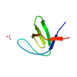
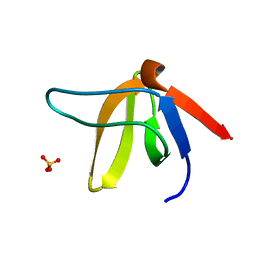
A-SPECTRIN SH3 DOMAIN A11V, V23L, M25I, V53I, V58L MUTANT
Descriptor: SPECTRIN ALPHA CHAIN, SULFATE ION
Authors:Vega, M.C, Serrano, L.
Deposit date:2000-08-15
Release date:2002-05-23
Last modified:2023-12-13
Method:X-RAY DIFFRACTION (2.3 Å)
Cite:Conformational Strain in the Hydrophobic Core and its Implications for Protein Folding and Design
Nat.Struct.Biol., 9, 2002
1GCQ
DownloadVisualize
BU of 1gcq by Molmil
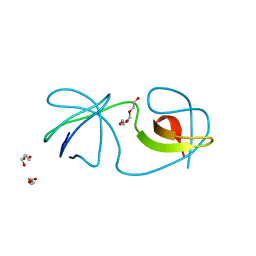
CRYSTAL STRUCTURE OF VAV AND GRB2 SH3 DOMAINS
Descriptor: (4R)-2-METHYLPENTANE-2,4-DIOL, GROWTH FACTOR RECEPTOR-BOUND PROTEIN 2, VAV PROTO-ONCOGENE
Authors:Nishida, M, Nagata, K, Hachimori, Y, Ogura, K, Inagaki, F.
Deposit date:2000-08-08
Release date:2001-08-08
Last modified:2023-12-27
Method:X-RAY DIFFRACTION (1.68 Å)
Cite:Novel recognition mode between Vav and Grb2 SH3 domains.
EMBO J., 20, 2001
1GFD
DownloadVisualize
BU of 1gfd by Molmil
SOLUTION STRUCTURE AND LIGAND-BINDING SITE OF THE C-TERMINAL SH3 DOMAIN OF GRB2
Descriptor: GROWTH FACTOR RECEPTOR-BOUND PROTEIN 2
Authors:Kohda, D, Terasawa, H, Hatanaka, H, Inagaki, F.
Deposit date:1994-06-13
Release date:1994-08-31
Last modified:2024-05-01
Method:SOLUTION NMR
Cite:Solution structure and ligand-binding site of the carboxy-terminal SH3 domain of GRB2.
Structure, 2, 1994

220113

數據於2024-05-22公開中

PDB statisticsPDBj update infoContact PDBjnumon