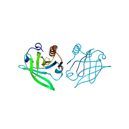
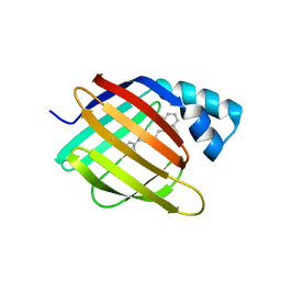
Loading
PDBj
MenuPDBj@FacebookPDBj@TwitterPDBj@YouTubewwPDB FoundationwwPDB
RCSB PDBPDBeBMRBAdv. SearchSearch help

1A57
DownloadVisualize
BU of 1a57 by Molmil
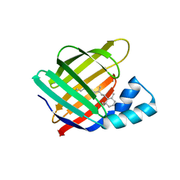
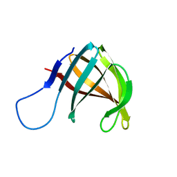
THE THREE-DIMENSIONAL STRUCTURE OF A HELIX-LESS VARIANT OF INTESTINAL FATTY ACID BINDING PROTEIN, NMR, 20 STRUCTURES
Descriptor: INTESTINAL FATTY ACID-BINDING PROTEIN
Authors:Steele, R.A, Emmert, D.A, Kao, J, Hodsdon, M.E, Frieden, C, Cistola, D.P.
Deposit date:1998-02-20
Release date:1998-05-27
Last modified:2021-11-03
Method:SOLUTION NMR
Cite:The three-dimensional structure of a helix-less variant of intestinal fatty acid-binding protein.
Protein Sci., 7, 1998
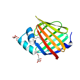
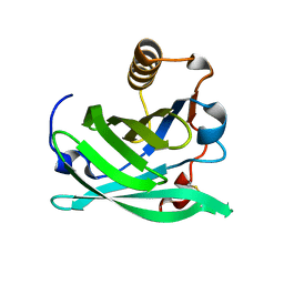
1AB0
DownloadVisualize
BU of 1ab0 by Molmil
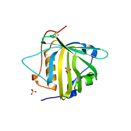
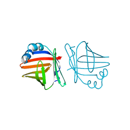
C1G/V32D/F57H MUTANT OF MURINE ADIPOCYTE LIPID BINDING PROTEIN AT PH 4.5
Descriptor: ADIPOCYTE LIPID BINDING PROTEIN
Authors:Ory, J, Kane, C, Simpson, M, Banaszak, L, Bernlohr, D.
Deposit date:1997-01-30
Release date:1997-06-16
Last modified:2023-08-02
Method:X-RAY DIFFRACTION (1.9 Å)
Cite:Biochemical and crystallographic analyses of a portal mutant of the adipocyte lipid-binding protein.
J.Biol.Chem., 272, 1997
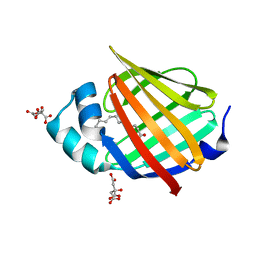
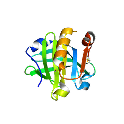
1A18
DownloadVisualize
BU of 1a18 by Molmil
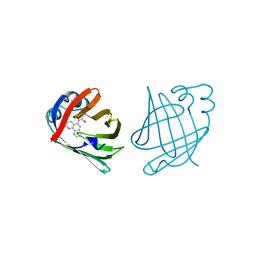
PHENANTHROLINE MODIFIED MURINE ADIPOCYTE LIPID BINDING PROTEIN
Descriptor: ADIPOCYTE LIPID BINDING PROTEIN
Authors:Ory, J, Mazhary, A, Kuang, H, Davies, R, Distefano, M, Banaszak, L.
Deposit date:1997-12-23
Release date:1998-07-01
Last modified:2023-08-02
Method:X-RAY DIFFRACTION (2.4 Å)
Cite:Structural characterization of two synthetic catalysts based on adipocyte lipid-binding protein.
Protein Eng., 11, 1998
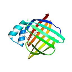
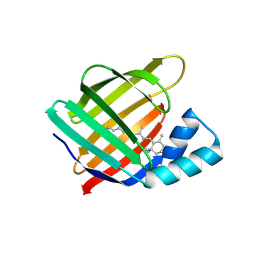
1BBP
DownloadVisualize
BU of 1bbp by Molmil
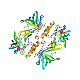
MOLECULAR STRUCTURE OF THE BILIN BINDING PROTEIN (BBP) FROM PIERIS BRASSICAE AFTER REFINEMENT AT 2.0 ANGSTROMS RESOLUTION.
Descriptor: BILIN BINDING PROTEIN, BILIVERDIN IX GAMMA CHROMOPHORE
Authors:Huber, R, Schneider, M, Mayr, I, Mueller, R, Deutzmann, R, Suter, F, Zuber, H, Falk, H, Kayser, H.
Deposit date:1990-09-19
Release date:1991-10-15
Last modified:2017-11-29
Method:X-RAY DIFFRACTION (2 Å)
Cite:Molecular structure of the bilin binding protein (BBP) from Pieris brassicae after refinement at 2.0 A resolution.
J.Mol.Biol., 198, 1987
1B8E
DownloadVisualize
BU of 1b8e by Molmil
HIGH RESOLUTION CRYSTAL STRUCTURE OF THE BOVINE BETA-LACTOGLOBULIN (ISOFORMS A AND B) IN ORTHOROMBIC SPACE GROUP
Descriptor: PROTEIN (BETA-LACTOGLOBULIN)
Authors:Oliveira, K.M.G, Sawyer, L, Polikarpov, I.
Deposit date:1999-01-30
Release date:2001-05-02
Last modified:2024-04-03
Method:X-RAY DIFFRACTION (1.95 Å)
Cite:Crystal structures of bovine beta-lactoglobulin in the orthorhombic space group C222(1). Structural differences between genetic variants A and B and features of the Tanford transition.
Eur.J.Biochem., 268, 2001
6VIT
DownloadVisualize
BU of 6vit by Molmil
The Crystal Structure of Apo Domain-Swapped dimer Q108K:T51D:I32C Variant of HCRBPII with an Engineered Disulfide Bond
Descriptor: Retinol-binding protein 2
Authors:Ghanbarpour, A, Geiger, J.
Deposit date:2020-01-13
Release date:2020-08-19
Last modified:2023-10-11
Method:X-RAY DIFFRACTION (3.2 Å)
Cite:Human Cellular Retinol Binding Protein II Forms a Domain-Swapped Trimer Representing a Novel Fold and a New Template for Protein Engineering.
Chembiochem, 21, 2020
6VID
DownloadVisualize
BU of 6vid by Molmil
The Crystal Structure of Aps Domain-Swapped Trimer Q108K:K40D:T53A:R58L:Q38F:Q4F Variant of HCRBPII
Descriptor: ACETATE ION, GLYCEROL, Retinol-binding protein 2
Authors:Ghanbarpour, A, Geiger, J.
Deposit date:2020-01-12
Release date:2020-08-19
Last modified:2023-10-11
Method:X-RAY DIFFRACTION (2.89 Å)
Cite:The Crystal Structure of Apo Domain Swapped Trimer Q108K:K40D:T53A:R58L:Q38F:Q4F variant of HCRBPII
To Be Published
6VIS
DownloadVisualize
BU of 6vis by Molmil
The Crystal Structure of Domain-Swapped Trimer Q108K:K40D:T53A:R58L:Q38F:Q4F:V62E Variant of HCRBPII
Descriptor: GLYCEROL, Retinol-binding protein 2
Authors:Ghanbarpour, A, Geiger, J.
Deposit date:2020-01-13
Release date:2020-09-09
Last modified:2023-10-11
Method:X-RAY DIFFRACTION (2.79 Å)
Cite:Human Cellular Retinol Binding Protein II Forms a Domain-Swapped Trimer Representing a Novel Fold and a New Template for Protein Engineering.
Chembiochem, 21, 2020
6WNF
DownloadVisualize
BU of 6wnf by Molmil
The Crystal Structure of Apo Domain-Swapped Dimer Q108K:K40D:T53A:R58L:Q38F:Q4F:F57H Variant of HCRBPII
Descriptor: ACETATE ION, GLYCEROL, Retinol-binding protein 2
Authors:Ghanbarpour, A, Geiger, J.
Deposit date:2020-04-22
Release date:2020-08-19
Last modified:2023-10-18
Method:X-RAY DIFFRACTION (1.67 Å)
Cite:Human Cellular Retinol Binding Protein II Forms a Domain-Swapped Trimer Representing a Novel Fold and a New Template for Protein Engineering.
Chembiochem, 21, 2020
6WNJ
DownloadVisualize
BU of 6wnj by Molmil
The Crystal Structure of Apo Domain-Swapped Trimer Q108K:T51D:A28C:I32C of HCRBPII
Descriptor: GLYCEROL, Retinol-binding protein 2, SULFATE ION, ...
Authors:Ghanbarpour, A, Geiger, J.
Deposit date:2020-04-22
Release date:2020-08-19
Last modified:2023-10-18
Method:X-RAY DIFFRACTION (2.1 Å)
Cite:Human Cellular Retinol Binding Protein II Forms a Domain-Swapped Trimer Representing a Novel Fold and a New Template for Protein Engineering.
Chembiochem, 21, 2020
6WP1
DownloadVisualize
BU of 6wp1 by Molmil
The Crystal Structure of Apo Domain-Swapped Trimer Q108K:K40L:T51K Variant of HCRBPII
Descriptor: ACETATE ION, GLYCEROL, Retinol-binding protein 2
Authors:Ghanbarpour, A, Geiger, J.
Deposit date:2020-04-26
Release date:2020-08-19
Last modified:2023-10-18
Method:X-RAY DIFFRACTION (2.99 Å)
Cite:Human Cellular Retinol Binding Protein II Forms a Domain-Swapped Trimer Representing a Novel Fold and a New Template for Protein Engineering.
Chembiochem, 21, 2020
6WP0
DownloadVisualize
BU of 6wp0 by Molmil
The Crystal Structure of Domain-Swapped Trimer Q108K:T51D variant of HCRBPII
Descriptor: ACETATE ION, GLYCEROL, Retinol-binding protein 2
Authors:Ghanbarpour, A, Geiger, J.
Deposit date:2020-04-26
Release date:2020-08-19
Last modified:2023-10-18
Method:X-RAY DIFFRACTION (2.78 Å)
Cite:Human Cellular Retinol Binding Protein II Forms a Domain-Swapped Trimer Representing a Novel Fold and a New Template for Protein Engineering.
Chembiochem, 21, 2020
6WP2
DownloadVisualize
BU of 6wp2 by Molmil
The Crystal Structure of Apo Zinc-Bound Domain Swapped-Trimer Q108K:K40D:T53A:R58L:Q38F:Q4F:F57H Variant of HCRBPII
Descriptor: ACETATE ION, GLYCEROL, Retinol-binding protein 2, ...
Authors:Ghanbarpour, A, Geiger, J.
Deposit date:2020-04-26
Release date:2020-08-19
Last modified:2023-10-18
Method:X-RAY DIFFRACTION (2.48 Å)
Cite:Human Cellular Retinol Binding Protein II Forms a Domain-Swapped Trimer Representing a Novel Fold and a New Template for Protein Engineering.
Chembiochem, 21, 2020
6XU5
DownloadVisualize
BU of 6xu5 by Molmil
Human myelin protein P2 mutant N2D
Descriptor: CITRIC ACID, Myelin P2 protein, PALMITIC ACID
Authors:Ruskamo, S, Lehtimaki, M, Kursula, P.
Deposit date:2020-01-17
Release date:2020-04-08
Last modified:2024-01-24
Method:X-RAY DIFFRACTION (1.65 Å)
Cite:Cryo-EM, X-ray diffraction, and atomistic simulations reveal determinants for the formation of a supramolecular myelin-like proteolipid lattice.
J.Biol.Chem., 295, 2020
6XUA
DownloadVisualize
BU of 6xua by Molmil
Human myelin protein P2 mutant K21Q
Descriptor: CITRIC ACID, Myelin P2 protein, PALMITIC ACID
Authors:Ruskamo, S, Lehtimaki, M, Kursula, P.
Deposit date:2020-01-17
Release date:2020-04-08
Last modified:2024-01-24
Method:X-RAY DIFFRACTION (2.3 Å)
Cite:Cryo-EM, X-ray diffraction, and atomistic simulations reveal determinants for the formation of a supramolecular myelin-like proteolipid lattice.
J.Biol.Chem., 295, 2020
6XW9
DownloadVisualize
BU of 6xw9 by Molmil
Human myelin protein P2 mutant K120S
Descriptor: CHLORIDE ION, Myelin P2 protein, PALMITIC ACID
Authors:Ruskamo, S, Lehtimaki, M, Kursula, P.
Deposit date:2020-01-23
Release date:2020-04-08
Last modified:2024-01-24
Method:X-RAY DIFFRACTION (2.9 Å)
Cite:Cryo-EM, X-ray diffraction, and atomistic simulations reveal determinants for the formation of a supramolecular myelin-like proteolipid lattice.
J.Biol.Chem., 295, 2020
6MQW
DownloadVisualize
BU of 6mqw by Molmil
Crystal Structure of All-trans Retinal-Bound R111K:Y134F:T54V:R132Q:P39Y:R59Y:L121E Human Cellular Retinoic Acid Binding Protein II Irradiated with 400 nm laser (30 seconds) and subsequently dark adapted (10 minutes) at 2.1 Angstrom Resolution
Descriptor: Cellular retinoic acid-binding protein 2, RETINAL
Authors:Ghanbarpour, A, Geiger, J.
Deposit date:2018-10-11
Release date:2019-01-09
Last modified:2023-10-11
Method:X-RAY DIFFRACTION (2.09 Å)
Cite:Mimicking Microbial Rhodopsin Isomerization in a Single Crystal.
J. Am. Chem. Soc., 141, 2019
6NOE
DownloadVisualize
BU of 6noe by Molmil
Crystal Structure of the All-Trans Retinal-Bound R111K:Y134F:T54V:R132Q:P39Y:R59Y:L121E:I63D Mutant of Human Cellular Retinoic Acid Binding Protein II in the Dark at 1.97 Angstrom Resolution
Descriptor: Cellular retinoic acid-binding protein 2, RETINAL
Authors:Ghanbarpour, A, Geiger, J.
Deposit date:2019-01-16
Release date:2019-01-30
Last modified:2023-10-11
Method:X-RAY DIFFRACTION (1.969 Å)
Cite:Crystal Structure of the All-Trans Retinal-Bound R111K:Y134F:T54V:R132Q:P39Y:R59Y:L121E:I63D Mutant of Human Cellular Retinoic Acid Binding Protein II in the Dark at 1.97 Angstrom Resolution
To Be Published
6NRE
DownloadVisualize
BU of 6nre by Molmil
Monomeric Lipocalin Can F 6
Descriptor: Lipocalin-Can f 6 allergen
Authors:Clayton G, M, Kappler J, W, Chan, S.
Deposit date:2019-01-23
Release date:2019-09-25
Method:X-RAY DIFFRACTION (2.06 Å)
Cite:Structural characteristics of lipocalin allergens: Crystal structure of the immunogenic dog allergen Can f 6.
Plos One, 14, 2019
6NKQ
DownloadVisualize
BU of 6nkq by Molmil
The structure of bovine beta-lactoglobulin in novel crystals grown at pH 3.8
Descriptor: Beta-lactoglobulin, CALCIUM ION
Authors:McPherson, A.
Deposit date:2019-01-07
Release date:2019-01-23
Last modified:2023-10-11
Method:X-RAY DIFFRACTION (2.3 Å)
Cite:The structure of bovine beta-lactoglobulin in crystals grown at pH 3.8 exhibiting novel threefold twinning.
Acta Crystallogr.,Sect.F, 75, 2019
6NNX
DownloadVisualize
BU of 6nnx by Molmil
Crystal Structure of the All-Trans Retinal-Bound R111K:Y134F:T54V:R132Q:P39Y:R59Y:L121M Mutant of Human Cellular Retinoic Acid Binding Protein II in the Dark at 1.87 Angstrom Resolution
Descriptor: Cellular retinoic acid-binding protein 2, RETINAL
Authors:Ghanbarpour, A, Geiger, J.
Deposit date:2019-01-15
Release date:2019-01-30
Last modified:2023-10-11
Method:X-RAY DIFFRACTION (1.87 Å)
Cite:Crystal Structure of the All-Trans Retinal-Bound R111K:Y134F:T54V:R132Q:P39Y:R59Y:L121M Mutant of Human Cellular Retionic Acid Binding Protein II in the Dark at 1.87 Angstrom Resolution
To Be Published
6NNY
DownloadVisualize
BU of 6nny by Molmil
Crystal Structure of the All-Trans Retinal-Bound R111K:Y134F:T54V:R132Q:P39E:R59Y:L121E Mutant of Human Cellular Retinoic Acid Binding Protein II in the Dark at 1.67 Angstrom Resolution
Descriptor: Cellular retinoic acid-binding protein 2, RETINAL
Authors:Ghanbarpour, A, Geiger, J.
Deposit date:2019-01-15
Release date:2019-01-30
Last modified:2023-10-11
Method:X-RAY DIFFRACTION (1.668 Å)
Cite:Crystal Structure of the All-Trans Retinal-Bound R111K:Y134F:T54V:R132Q:P39E:R59Y:L121E Mutant of Human Cellular Retinoic Acid Binding Protein II in the Dark at 1.68 Angstrom Resolution
To Be Published
6O5D
DownloadVisualize
BU of 6o5d by Molmil
PYOCHELIN
Descriptor: GLYCEROL, Neutrophil gelatinase-associated lipocalin, SULFATE ION
Authors:Rupert, P.B, Strong, R.K, Clifton, M.C, Edwards, T.E, Seattle Structural Genomics Center for Infectious Disease (SSGCID)
Deposit date:2019-03-01
Release date:2019-09-11
Last modified:2020-07-29
Method:X-RAY DIFFRACTION (2.4 Å)
Cite:Parsing the functional specificity of Siderocalin/Lipocalin 2/NGAL for siderophores and related small-molecule ligands.
J Struct Biol X, 2, 2019
6ON5
DownloadVisualize
BU of 6on5 by Molmil
Crystal Structure of the Zn-bound Domain-Swapped Dimer Q108K:T51D:A28C:L36C:F57H Mutant of Human Cellular Retinol Binding Protein II
Descriptor: Retinol-binding protein 2, ZINC ION
Authors:Ghanbarpour, A, Geiger, J.
Deposit date:2019-04-20
Release date:2019-10-16
Last modified:2023-10-11
Method:X-RAY DIFFRACTION (1.638 Å)
Cite:Engineering the hCRBPII Domain-Swapped Dimer into a New Class of Protein Switches.
J.Am.Chem.Soc., 141, 2019
6ON8
DownloadVisualize
BU of 6on8 by Molmil
Crystal Structure of the Reduced Form of Apo Domain-Swapped Dimer Q108K:T51D:A28C:L36C:F57H Mutant of Human Cellular Retinol Binding Protein II
Descriptor: Retinol-binding protein 2
Authors:Ghanbarpour, A, Geiger, J.
Deposit date:2019-04-20
Release date:2019-10-16
Last modified:2023-10-11
Method:X-RAY DIFFRACTION (2.395 Å)
Cite:Engineering the hCRBPII Domain-Swapped Dimer into a New Class of Protein Switches.
J.Am.Chem.Soc., 141, 2019

218853

PDB entries from 2024-04-24

PDB statisticsPDBj update infoContact PDBjnumon