5HSX
 
 | |
5CC8
 | |
5CM7
 | |
5DD7
 | |
6VH5
 | |
4YK2
 | |
6VS4
 | |
4H51
 | |
3HN6
 | |
8DOP
 | |
6WHP
 | |
6PBL
 | |
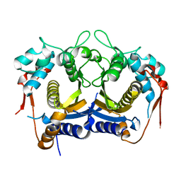
6WQM
 | | Crystal structure of 2,3,4,5-tetrahydropyridine-2-carboxylate N-succinyltransferase from Bartonella henselae | | Descriptor: | 2,3,4,5-tetrahydropyridine-2,6-dicarboxylate N-succinyltransferase, CHLORIDE ION | | Authors: | Seattle Structural Genomics Center for Infectious Disease (SSGCID) | | Deposit date: | 2020-04-29 | | Release date: | 2020-05-13 | | Last modified: | 2023-10-18 | | Method: | X-RAY DIFFRACTION (2.15 Å) | | Cite: | Crystal structure of 2,3,4,5-tetrahydropyridine-2-carboxylate N-succinyltransferase from Bartonella henselae
to be published
|
|
4ZR8
 | |
8EGM
 | |
6XDK
 | |
6XGS
 | |
3IX6
 | |
7S5O
 | |
3PGZ
 | |
8F87
 | |
3KM3
 | |
3LUZ
 | |
5CY4
 | |
3MEN
 | |


