5EL0
 
 | |
6VH5
 | |
3OA3
 | |
3OCA
 | |
4YWN
 | |
3OA1
 | |
5EKS
 | |
5EPF
 | |
5ER6
 | |
5EZ3
 | |
6VS4
 | |
4HJH
 | |
5F23
 | |
6PI4
 | |
4I1Y
 | |
6P81
 | |
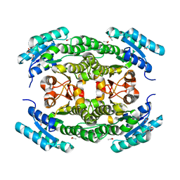
6WQM
 | | Crystal structure of 2,3,4,5-tetrahydropyridine-2-carboxylate N-succinyltransferase from Bartonella henselae | | Descriptor: | 2,3,4,5-tetrahydropyridine-2,6-dicarboxylate N-succinyltransferase, CHLORIDE ION | | Authors: | Seattle Structural Genomics Center for Infectious Disease (SSGCID) | | Deposit date: | 2020-04-29 | | Release date: | 2020-05-13 | | Last modified: | 2023-10-18 | | Method: | X-RAY DIFFRACTION (2.15 Å) | | Cite: | Crystal structure of 2,3,4,5-tetrahydropyridine-2-carboxylate N-succinyltransferase from Bartonella henselae
to be published
|
|
4DXL
 | |
6XGS
 | |
6XDK
 | |
6CXY
 | |
6XK2
 | |
4JE1
 | |
3KNU
 | |
4DZ4
 | |


