1NA7
 
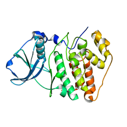 | |
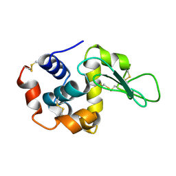
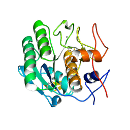
2AUB
 | | Lysozyme structure derived from thin-film-based crystals | | Descriptor: | Lysozyme C | | Authors: | Pechkova, E, Sivozhelezov, V, Tropiano, G, Fiordoro, S, Nicolini, C. | | Deposit date: | 2005-08-27 | | Release date: | 2005-12-06 | | Last modified: | 2011-07-13 | | Method: | X-RAY DIFFRACTION (1.7 Å) | | Cite: | Comparison of lysozyme structures derived from thin-film-based and classical crystals.
Acta Crystallogr.,Sect.D, 61, 2005
|
|
4DJ5
 | |
4DIY
 | |
4DJ0
 | |
4DJ1
 | |
4DIZ
 | |
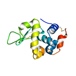
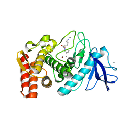
4HV1
 | | Laser-induced microfragmentation of lysozyme crystals allows X-ray nanodiffraction characterization of individual domains (lb4) | | Descriptor: | Lysozyme C | | Authors: | Pechkova, E, Belmonte, L, Riekel, C, Popov, D, Koenig, C, Nicolini, C. | | Deposit date: | 2012-11-05 | | Release date: | 2013-11-20 | | Last modified: | 2014-04-16 | | Method: | X-RAY DIFFRACTION (2.3 Å) | | Cite: | Laser-induced microfragmentation of lysozyme crystals allows X-ray nanodiffraction characterization of individual domains
To be Published
|
|
4HV2
 | | Laser-induced microfragmentation of lysozyme crystals allows X-ray nanodiffraction characterization of individual domains (lb5) | | Descriptor: | Lysozyme C | | Authors: | Pechkova, E, Belmonte, L, Riekel, C, Popov, D, Koenig, C, Nicolini, C. | | Deposit date: | 2012-11-05 | | Release date: | 2013-11-20 | | Last modified: | 2014-04-16 | | Method: | X-RAY DIFFRACTION (2.501 Å) | | Cite: | Laser-induced microfragmentation of lysozyme crystals allows X-ray nanodiffraction characterization of individual domains
To be Published
|
|
3DE1
 | |
3DDZ
 | |
3DE0
 | |
3D9Q
 | |
3DE2
 | |
3DE6
 | |
3DE3
 | |
3DO1
 | |
3DE5
 | |
3DO0
 | |
3DVR
 | |
3DWE
 | |
3DNZ
 | |
3DE4
 | |
3DE7
 | |
3DVS
 | |


