Loading
PDBj
MenuPDBj@FacebookPDBj@TwitterPDBj@YouTubewwPDB FoundationwwPDB
RCSB PDBPDBeBMRBAdv. SearchSearch help
PDB: 93 results

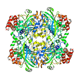
6PM7
DownloadVisualize
BU of 6pm7 by Molmil
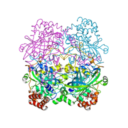
The structure of the triclinic crystal form of beef liver catalase at 1.85 A resolution
Descriptor: Catalase, NADPH DIHYDRO-NICOTINAMIDE-ADENINE-DINUCLEOTIDE PHOSPHATE, PROTOPORPHYRIN IX CONTAINING FE
Authors:McPherson, A.
Deposit date:2019-07-01
Release date:2020-05-20
Last modified:2024-03-13
Method:X-RAY DIFFRACTION (1.85 Å)
Cite:The structure of the triclinic crystal form of beef liver catalase at 1.85 A resolution
to be published
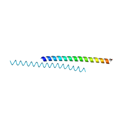
6DXR
DownloadVisualize
BU of 6dxr by Molmil
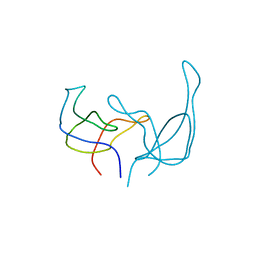
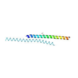
Structure of the Monoclinic-2 (Monocl-2) Crystal Form of Human Apolipoprotein C1
Descriptor: (4S)-2-METHYL-2,4-PENTANEDIOL, Apolipoprotein C-I
Authors:McPherson, A.
Deposit date:2018-06-29
Release date:2018-12-26
Last modified:2023-10-11
Method:X-RAY DIFFRACTION (2 Å)
Cite:The structure of human apolipoprotein C-1 in four different crystal forms.
J. Lipid Res., 60, 2019
6PO0
DownloadVisualize
BU of 6po0 by Molmil
The structure of the orthorhombic (P212121) crystal form of beef liver catalase at 1.85 A resolution
Descriptor: Catalase, NADPH DIHYDRO-NICOTINAMIDE-ADENINE-DINUCLEOTIDE PHOSPHATE, PROTOPORPHYRIN IX CONTAINING FE
Authors:McPherson, A.
Deposit date:2019-07-03
Release date:2020-05-20
Last modified:2024-03-13
Method:X-RAY DIFFRACTION (1.75 Å)
Cite:The structure of the triclinic crystal form of beef liver catalase at 1.85 A resolution
To be published
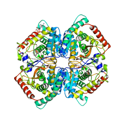
6P6U
DownloadVisualize
BU of 6p6u by Molmil
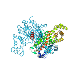
Crystal Structure of Monoclinic Rabbit Muscle Lactate Dehydrogenase with Four Tetramers as the Asymmetric Unit
Descriptor: ADENOSINE MONOPHOSPHATE, L-lactate dehydrogenase A chain
Authors:McPherson, A.
Deposit date:2019-06-04
Release date:2020-06-17
Last modified:2023-10-11
Method:X-RAY DIFFRACTION (2.42 Å)
Cite:Crystal Structure of Monoclinic Rabbit Muscle Lactate Dehydrogenase with Four Tetramers as the Asymmetric Unit
to be published
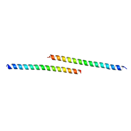
6DVU
DownloadVisualize
BU of 6dvu by Molmil
Structure of the Monoclinic-1 (Monocl-1) Crystal Form of Human Apolipoprotein C1
Descriptor: Apolipoprotein C-I
Authors:McPherson, A, Larson, S.B.
Deposit date:2018-06-25
Release date:2018-12-26
Last modified:2023-10-11
Method:X-RAY DIFFRACTION (1.8 Å)
Cite:The structure of human apolipoprotein C-1 in four different crystal forms.
J. Lipid Res., 60, 2019
6DZ6
DownloadVisualize
BU of 6dz6 by Molmil
Structure of the Orthorhombic (Orthrhmb) Crystal Form of Human Apolipoprotein C1
Descriptor: Apolipoprotein C-I
Authors:McPherson, A.
Deposit date:2018-07-03
Release date:2018-12-26
Last modified:2024-03-13
Method:X-RAY DIFFRACTION (3 Å)
Cite:The structure of human apolipoprotein C-1 in four different crystal forms.
J. Lipid Res., 60, 2019
6C6W
DownloadVisualize
BU of 6c6w by Molmil
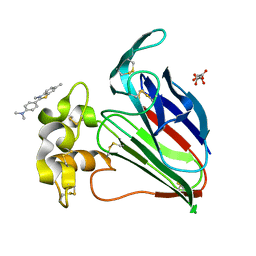
Structure of a Complex Between Thaumatin and Thioflavin T
Descriptor: 2-[4-(dimethylamino)phenyl]-3,6-dimethyl-1,3-benzothiazol-3-ium, L(+)-TARTARIC ACID, Thaumatin I
Authors:McPherson, A.
Deposit date:2018-01-19
Release date:2018-02-07
Last modified:2018-09-19
Method:X-RAY DIFFRACTION (2.23 Å)
Cite:Investigation into the binding of dyes within protein crystals.
Acta Crystallogr F Struct Biol Commun, 74, 2018
6E0D
DownloadVisualize
BU of 6e0d by Molmil
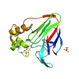
X-ray structure of a complex of thaumatin with xylene cyanol
Descriptor: (1R,2R,3S,4R,6S)-4,6-diamino-2,3-dihydroxycyclohexyl 2,6-diamino-2,6-dideoxy-alpha-D-glucopyranoside, L(+)-TARTARIC ACID, Thaumatin I
Authors:McPherson, A.
Deposit date:2018-07-06
Release date:2018-09-19
Last modified:2020-07-29
Method:X-RAY DIFFRACTION (2.24 Å)
Cite:Investigation into the binding of dyes within protein crystals.
Acta Crystallogr F Struct Biol Commun, 74, 2018
6OR3
DownloadVisualize
BU of 6or3 by Molmil
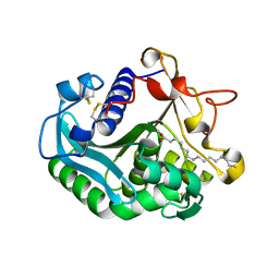
Structure of an Acyl Intermediate of Thermomyces Lanuginosa Lipase With Palmitic Acid in an Orthorhombic Crystal
Descriptor: 2-acetamido-2-deoxy-beta-D-glucopyranose, CALCIUM ION, Lipase, ...
Authors:McPherson, A.
Deposit date:2019-04-29
Release date:2020-05-20
Last modified:2023-10-11
Method:X-RAY DIFFRACTION (1.45 Å)
Cite:The crystal Structures of Thermomyces (Humicola) lanuginosa lipase in complex with enzymatic reactants
Current Enzyme Inhibition, 16, 2020
5BKN
DownloadVisualize
BU of 5bkn by Molmil
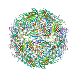
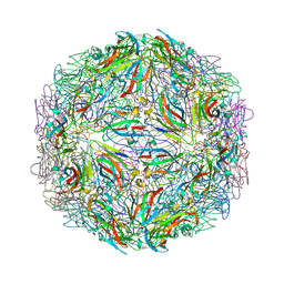
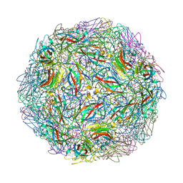
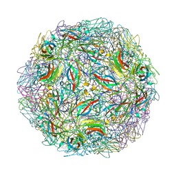
Crystallographic structure of a cubic crystal form of STMV (84.5 degree rotation) grown from chloride
Descriptor: CHLORIDE ION, Coat protein, MAGNESIUM ION, ...
Authors:McPherson, A.
Deposit date:2021-03-20
Release date:2021-12-15
Last modified:2023-09-27
Method:X-RAY DIFFRACTION (3 Å)
Cite:Structures of additional crystal forms of Satellite tobacco mosaic virus grown from a variety of salts.
Acta Crystallogr.,Sect.F, 77, 2021
5BKL
DownloadVisualize
BU of 5bkl by Molmil
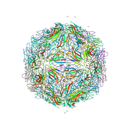
Crystallographic structure of the cubic crystal form of STMV (77.9 degree rotation) grown from NaCl
Descriptor: CHLORIDE ION, Coat protein, MAGNESIUM ION, ...
Authors:McPherson, A.
Deposit date:2021-03-20
Release date:2021-12-15
Last modified:2023-09-27
Method:X-RAY DIFFRACTION (2.94 Å)
Cite:Structures of additional crystal forms of Satellite tobacco mosaic virus grown from a variety of salts.
Acta Crystallogr.,Sect.F, 77, 2021
5BKQ
DownloadVisualize
BU of 5bkq by Molmil
Crystallographic structure of a cubic form of STMV grown from nitrate
Descriptor: Coat protein, MAGNESIUM ION, NITRATE ION
Authors:McPherson, A.
Deposit date:2021-03-21
Release date:2021-12-15
Last modified:2023-09-27
Method:X-RAY DIFFRACTION (3.19 Å)
Cite:Structures of additional crystal forms of Satellite tobacco mosaic virus grown from a variety of salts.
Acta Crystallogr.,Sect.F, 77, 2021
6CAF
DownloadVisualize
BU of 6caf by Molmil
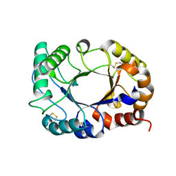
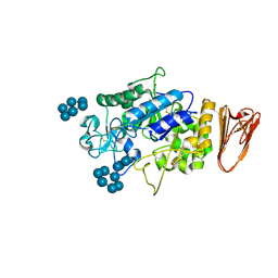
High Resolution Structure of Concanavalin B from Jack Bean (Canavalia ensiformis), A Chitinase-like Protein
Descriptor: Concanavalin B
Authors:McPherson, A.
Deposit date:2018-01-30
Release date:2018-03-28
Method:X-RAY DIFFRACTION (1.3 Å)
Cite:High Resolution Structure of Concanavalin B from Jack Bean (Canavalia ensiformis), a Chitinase-like Protein
To be published
6C4W
DownloadVisualize
BU of 6c4w by Molmil
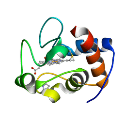
Structure of the yeast Pichia membranifaciens cytochrome c
Descriptor: HEME C, cytochrome c
Authors:McPherson, A.
Deposit date:2018-01-13
Release date:2018-02-07
Last modified:2023-10-04
Method:X-RAY DIFFRACTION (2.4 Å)
Cite:Characterization of two crystal forms of cytochrome c from Valida membranifaciens
to be published
4V99
DownloadVisualize
BU of 4v99 by Molmil
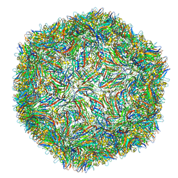
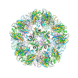
The Crystallographic Structure of Panicum Mosaic Virus
Descriptor: 5'-R(P*UP*UP*AP*AP*UP*AP*UP*UP*UP*UP*UP*AP*UP*UP*UP*UP*U)-3', CALCIUM ION, Capsid protein
Authors:Makino, D.L, Larson, S.B, McPherson, A.
Deposit date:2012-07-04
Release date:2014-07-09
Last modified:2023-09-20
Method:X-RAY DIFFRACTION (2.9 Å)
Cite:The crystallographic structure of Panicum Mosaic Virus (PMV).
J.Struct.Biol., 181, 2013
4OQ9
DownloadVisualize
BU of 4oq9 by Molmil
Satellite Tobacco Mosaic Virus Refined to 1.4 A Resolution using non-crystallographic symmetry restraints
Descriptor: Coat protein, MAGNESIUM ION, PHOSPHATE ION, ...
Authors:Larson, S.B, Day, J.S, McPherson, A.
Deposit date:2014-02-07
Release date:2014-09-10
Last modified:2023-09-20
Method:X-RAY DIFFRACTION (1.45 Å)
Cite:Satellite tobacco mosaic virus refined to 1.4 angstrom resolution.
Acta Crystallogr.,Sect.D, 70, 2014
4OQ8
DownloadVisualize
BU of 4oq8 by Molmil
Satellite Tobacco Mosaic Virus Refined to 1.4 A Resolution using icosahedral constraints
Descriptor: Coat protein, MAGNESIUM ION, PHOSPHATE ION, ...
Authors:Larson, S.B, Day, J.S, McPherson, A.
Deposit date:2014-02-07
Release date:2014-09-10
Last modified:2023-09-20
Method:X-RAY DIFFRACTION (1.45 Å)
Cite:Satellite tobacco mosaic virus refined to 1.4 angstrom resolution.
Acta Crystallogr.,Sect.D, 70, 2014
3JW1
DownloadVisualize
BU of 3jw1 by Molmil
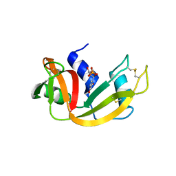
Crystal Structure of Bovine Pancreatic Ribonuclease Complexed with Uridine-5'-monophosphate at 1.60 A Resolution
Descriptor: Ribonuclease pancreatic, URIDINE-5'-MONOPHOSPHATE
Authors:Larson, S.B, Day, J.S, Nguyen, C, Cudney, R, Mcpherson, A, Center for High-Throughput Structural Biology (CHTSB)
Deposit date:2009-09-17
Release date:2009-10-06
Last modified:2023-09-06
Method:X-RAY DIFFRACTION (1.6 Å)
Cite:Structure of bovine pancreatic ribonuclease complexed with uridine 5'-monophosphate at 1.60 A resolution.
Acta Crystallogr.,Sect.F, 66, 2010
4NIA
DownloadVisualize
BU of 4nia by Molmil
Satellite Tobacco Mosaic Virus Refined at room temperature to 1.8 A Resolution using NCS Restraints
Descriptor: Coat protein, MAGNESIUM ION, PHOSPHATE ION, ...
Authors:Larson, S.B, Day, J.S, McPherson, A.
Deposit date:2013-11-05
Release date:2014-09-10
Last modified:2023-09-20
Method:X-RAY DIFFRACTION (1.82 Å)
Cite:Satellite tobacco mosaic virus refined to 1.4 angstrom resolution.
Acta Crystallogr.,Sect.D, 70, 2014
2GN5
DownloadVisualize
BU of 2gn5 by Molmil
REFINED STRUCTURE OF THE GENE 5 DNA BINDING PROTEIN FROM BACTERIOPHAGE FD
Descriptor: GENE V PROTEIN
Authors:Brayer, G.D, McPherson, A.
Deposit date:1986-01-13
Release date:1986-01-21
Last modified:2024-02-14
Method:X-RAY DIFFRACTION (2.3 Å)
Cite:Refined structure of the gene 5 DNA binding protein from bacteriophage fd.
J.Mol.Biol., 169, 1983
3L2L
DownloadVisualize
BU of 3l2l by Molmil
X-ray Crystallographic Analysis of Pig Pancreatic Alpha-Amylase with Limit Dextrin and Oligosaccharide
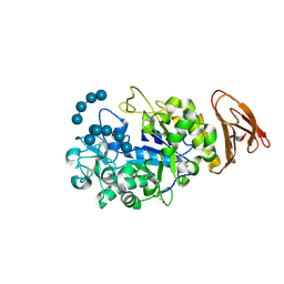
Descriptor: CALCIUM ION, CHLORIDE ION, Pancreatic alpha-amylase, ...
Authors:Larson, S.B, Day, J.S, McPherson, A.
Deposit date:2009-12-15
Release date:2010-04-14
Last modified:2020-07-29
Method:X-RAY DIFFRACTION (2.11 Å)
Cite:X-ray crystallographic analyses of pig pancreatic alpha-amylase with limit dextrin, oligosaccharide, and alpha-cyclodextrin.
Biochemistry, 49, 2010
1YC6
DownloadVisualize
BU of 1yc6 by Molmil
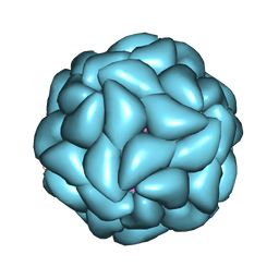
Crystallographic Structure of the T=1 Particle of Brome Mosaic Virus
Descriptor: Coat protein
Authors:Larson, S.B, Lucas, R.W, McPherson, A.
Deposit date:2004-12-22
Release date:2005-01-18
Last modified:2023-08-23
Method:X-RAY DIFFRACTION (2.9 Å)
Cite:Crystallographic structure of the T=1 particle of brome mosaic virus.
J.Mol.Biol., 346, 2005
3L2M
DownloadVisualize
BU of 3l2m by Molmil
X-ray Crystallographic Analysis of Pig Pancreatic Alpha-Amylase with Alpha-cyclodextrin
Descriptor: CALCIUM ION, CHLORIDE ION, Cyclohexakis-(1-4)-(alpha-D-glucopyranose), ...
Authors:Larson, S.B, Day, J.S, McPherson, A.
Deposit date:2009-12-15
Release date:2010-04-14
Last modified:2020-07-29
Method:X-RAY DIFFRACTION (1.97 Å)
Cite:X-ray crystallographic analyses of pig pancreatic alpha-amylase with limit dextrin, oligosaccharide, and alpha-cyclodextrin.
Biochemistry, 49, 2010
1ZA6
DownloadVisualize
BU of 1za6 by Molmil
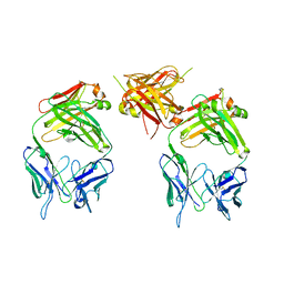
The structure of an antitumor CH2-domain-deleted humanized antibody
Descriptor: IGG Heavy chain, IGG Light chain
Authors:Larson, S.B, Day, J.S, Glaser, S, Braslawsky, G, McPherson, A.
Deposit date:2005-04-05
Release date:2005-05-10
Last modified:2023-08-23
Method:X-RAY DIFFRACTION (2.8 Å)
Cite:The Structure of an Antitumor C(H)2-domain-deleted Humanized Antibody.
J.Mol.Biol., 348, 2005
3ENJ
DownloadVisualize
BU of 3enj by Molmil
Structure of Pig Heart Citrate Synthase at 1.78 A resolution
Descriptor: CHLORIDE ION, CYSTEINE, Citrate synthase, ...
Authors:Larson, S.B, Day, J.S, Nguyen, C, Cudney, R, McPherson, A, Center for High-Throughput Structural Biology (CHTSB)
Deposit date:2008-09-25
Release date:2009-02-03
Last modified:2023-09-06
Method:X-RAY DIFFRACTION (1.78 Å)
Cite:Structure of pig heart citrate synthase at 1.78 A resolution.
Acta Crystallogr.,Sect.F, 65, 2009

220113

PDB entries from 2024-05-22

PDB statisticsPDBj update infoContact PDBjnumon