Loading
PDBj
MenuPDBj@FacebookPDBj@TwitterPDBj@YouTubewwPDB FoundationwwPDB
RCSB PDBPDBeBMRBAdv. SearchSearch help
Search by PDB author
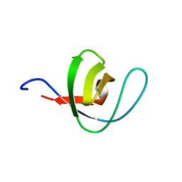
1BK2
DownloadVisualize
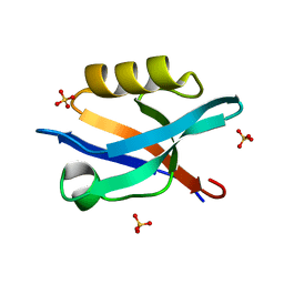
BU of 1bk2 by Molmil
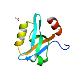
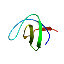
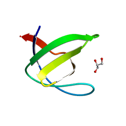
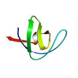
A-SPECTRIN SH3 DOMAIN D48G MUTANT
Descriptor: A-SPECTRIN
Authors:Martinez, J.C, Pisabarro, M.T, Serrano, L.
Deposit date:1998-07-14
Release date:1999-02-16
Last modified:2023-08-02
Method:X-RAY DIFFRACTION (2.01 Å)
Cite:Obligatory steps in protein folding and the conformational diversity of the transition state.
Nat.Struct.Biol., 5, 1998
1PWT
DownloadVisualize
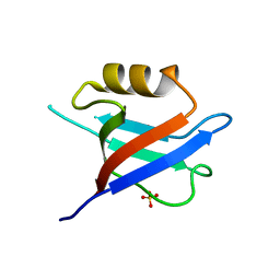
BU of 1pwt by Molmil
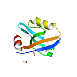
THERMODYNAMIC ANALYSIS OF ALPHA-SPECTRIN SH3 AND TWO OF ITS CIRCULAR PERMUTANTS WITH DIFFERENT LOOP LENGTHS: DISCERNING THE REASONS FOR RAPID FOLDING IN PROTEINS
Descriptor: ALPHA SPECTRIN
Authors:Martinez, J.C, Viguera, A.R, Berisio, R, Wilmanns, M, Mateo, P.L, Filmonov, V.V, Serrano, L.
Deposit date:1998-10-06
Release date:1999-05-11
Last modified:2023-08-09
Method:X-RAY DIFFRACTION (1.77 Å)
Cite:Thermodynamic analysis of alpha-spectrin SH3 and two of its circular permutants with different loop lengths: discerning the reasons for rapid folding in proteins.
Biochemistry, 38, 1999
2F0R
DownloadVisualize
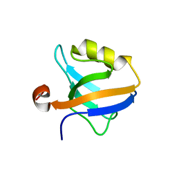
BU of 2f0r by Molmil
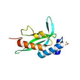
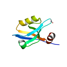
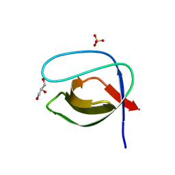
Crystallographic structure of human Tsg101 UEV domain
Descriptor: SULFATE ION, Tumor susceptibility gene 101 protein
Authors:Camara-Artigas, A, Luque, I, Palencia, A, Martinez, J.C, Mateo, P.L.
Deposit date:2005-11-13
Release date:2006-03-28
Last modified:2023-08-23
Method:X-RAY DIFFRACTION (2.26 Å)
Cite:Structure of human TSG101 UEV domain.
Acta Crystallogr.,Sect.D, 62, 2006
2JMC
DownloadVisualize
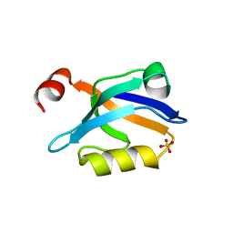
BU of 2jmc by Molmil
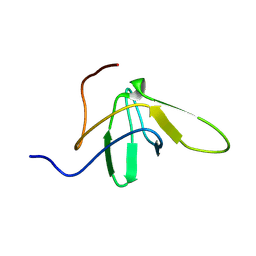
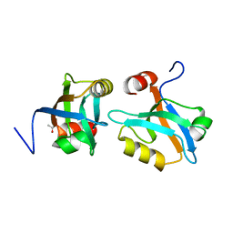
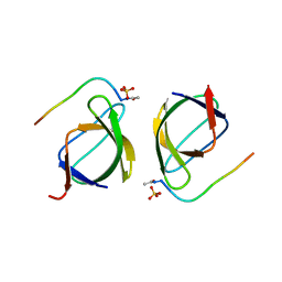
Chimer between Spc-SH3 and P41
Descriptor: Spectrin alpha chain, brain and P41 peptide chimera
Authors:van Nuland, N.A.J, Candel, A.M, Martinez, J.C, Conejero-Lara, F, Bruix, M.
Deposit date:2006-11-02
Release date:2007-04-24
Last modified:2023-12-20
Method:SOLUTION NMR
Cite:The high-resolution NMR structure of a single-chain chimeric protein mimicking a SH3-peptide complex
Febs Lett., 581, 2007
7ZX2
DownloadVisualize
BU of 7zx2 by Molmil
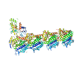
Tubulin-Pelophen B complex
Descriptor: (3R,4S,7S,9S,11S)-3,4,11-trihydroxy-7-((R,Z)-4-(hydroxymethyl)hex-2-en-2-yl)-9-methoxy-12,12-dimethyl-6-oxa-1(1,3)-benzenacyclododecaphan-5-one, 2-(N-MORPHOLINO)-ETHANESULFONIC ACID, CALCIUM ION, ...
Authors:Estevez-Gallego, J, Diaz, J.F, Van der Eycken, J, Oliva, M.A.
Deposit date:2022-05-20
Release date:2022-11-23
Last modified:2024-02-07
Method:X-RAY DIFFRACTION (2.5 Å)
Cite:Chemical modulation of microtubule structure through the laulimalide/peloruside site.
Structure, 31, 2023
8A0L
DownloadVisualize
BU of 8a0l by Molmil
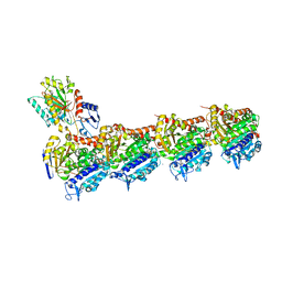
Tubulin-CW1-complex
Descriptor: (3~{S},4~{R},8~{S},10~{S},12~{S},14~{S})-14-[(~{Z},4~{R})-4-(hydroxymethyl)hex-2-en-2-yl]-4,12-dimethoxy-9,9-dimethyl-3,8,10-tris(oxidanyl)-1-oxacyclotetradecan-2-one, 2-(N-MORPHOLINO)-ETHANESULFONIC ACID, CALCIUM ION, ...
Authors:Prota, A.E, Diaz, J.F, Steinmetz, M.O, Oliva, M.A.
Deposit date:2022-05-28
Release date:2022-12-14
Last modified:2024-01-31
Method:X-RAY DIFFRACTION (1.9981 Å)
Cite:Chemical modulation of microtubule structure through the laulimalide/peloruside site.
Structure, 31, 2023
8AH5
DownloadVisualize
BU of 8ah5 by Molmil
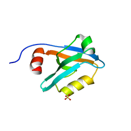
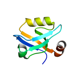
Crystal Structure of the third PDZ domain of PSD-95 protein in the space group P212121 at pH 4.6
Descriptor: ACETATE ION, cDNA FLJ50577, highly similar to Discs large homolog 4
Authors:Camara-Artigas, A, Salinas Garcia, M.C.
Deposit date:2022-07-20
Release date:2023-03-08
Last modified:2024-02-07
Method:X-RAY DIFFRACTION (1.25 Å)
Cite:pH-Driven Polymorphic Behaviour of the Third PDZ Domain of PSD95: The Role of Electrostatic Interactions
Crystals, 2023
8AH4
DownloadVisualize
BU of 8ah4 by Molmil
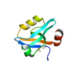
Crystal Structure of the third PDZ domain of PSD-95 protein in the space group P3112 at pH 4.0
Descriptor: ACETATE ION, cDNA FLJ50577, highly similar to Discs large homolog 4
Authors:Camara-Artigas, A, Salinas Garcia, M.C.
Deposit date:2022-07-20
Release date:2023-03-08
Last modified:2024-02-07
Method:X-RAY DIFFRACTION (1.48 Å)
Cite:pH-Driven Polymorphic Behaviour of the Third PDZ Domain of PSD95: The Role of Electrostatic Interactions
Crystals, 2023
8AH8
DownloadVisualize
BU of 8ah8 by Molmil
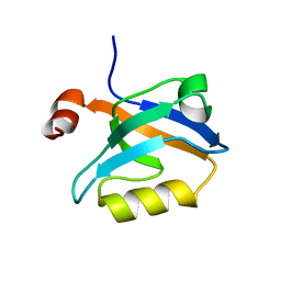
Crystal Structure of the third PDZ domain of PSD-95 protein in the space group P3121 at pH 3.7
Descriptor: cDNA FLJ50577, highly similar to Discs large homolog 4
Authors:Camara-Artigas, A, Salinas Garcia, M.C.
Deposit date:2022-07-20
Release date:2023-03-08
Last modified:2024-02-07
Method:X-RAY DIFFRACTION (1.5 Å)
Cite:pH-Driven Polymorphic Behaviour of the Third PDZ Domain of PSD95: The Role of Electrostatic Interactions
Crystals, 2023
8AH6
DownloadVisualize
BU of 8ah6 by Molmil
Crystal Structure of the third PDZ domain of PSD-95 protein in the space group P21 at pH 4.0
Descriptor: ACETATE ION, cDNA FLJ50577, highly similar to Discs large homolog 4
Authors:Camara-Artigas, A, Salinas Garcia, M.C.
Deposit date:2022-07-20
Release date:2023-03-08
Last modified:2024-02-07
Method:X-RAY DIFFRACTION (1.63 Å)
Cite:pH-Driven Polymorphic Behaviour of the Third PDZ Domain of PSD95: The Role of Electrostatic Interactions
Crystals, 2023
8AH7
DownloadVisualize
BU of 8ah7 by Molmil
Crystal Structure of the third PDZ domain of PSD-95 protein in the space group P212121 at pH 4.0
Descriptor: SULFATE ION, cDNA FLJ50577, highly similar to Discs large homolog 4
Authors:Camara-Artigas, A, Salinas-Garcia, M.C.
Deposit date:2022-07-20
Release date:2023-03-08
Last modified:2024-02-07
Method:X-RAY DIFFRACTION (1.25 Å)
Cite:pH-Driven Polymorphic Behaviour of the Third PDZ Domain of PSD95: The Role of Electrostatic Interactions
Crystals, 2023
1QKX
DownloadVisualize
BU of 1qkx by Molmil
Alpha-spectrin Src Homology 3 domain, N47A mutant in the distal loop.
Descriptor: SPECTRIN ALPHA CHAIN
Authors:Vega, M.C, Martinez, J, Serrano, L.
Deposit date:1999-08-16
Release date:2000-12-15
Last modified:2023-12-13
Method:X-RAY DIFFRACTION (1.8 Å)
Cite:Thermodynamic and structural characterization of Asn and Ala residues in the disallowed II' region of the Ramachandran plot.
Protein Sci., 9, 2000
1QKW
DownloadVisualize
BU of 1qkw by Molmil
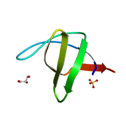
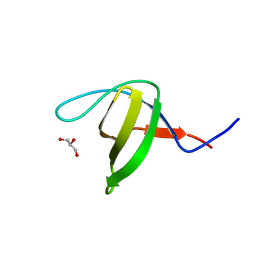
Alpha-spectrin Src Homology 3 domain, N47G mutant in the distal loop.
Descriptor: ALPHA II SPECTRIN, GLYCEROL, SULFATE ION
Authors:Vega, M.C, Martinez, J, Serrano, L.
Deposit date:1999-08-16
Release date:2000-08-18
Last modified:2023-12-13
Method:X-RAY DIFFRACTION (2 Å)
Cite:Thermodynamic and structural characterization of Asn and Ala residues in the disallowed II' region of the Ramachandran plot.
Protein Sci., 9, 2000
3EG1
DownloadVisualize
BU of 3eg1 by Molmil
Crystal structure of the N114Q mutant of ABL-SH3 domain complexed with a designed high-affinity peptide ligand: implications for SH3-ligand interactions
Descriptor: Proto-oncogene tyrosine-protein kinase ABL1, SULFATE ION, p41 peptide
Authors:Camara-Artigas, A.
Deposit date:2008-09-10
Release date:2009-09-15
Last modified:2023-08-30
Method:X-RAY DIFFRACTION (1.85 Å)
Cite:Role of interfacial water molecules in proline-rich ligand recognition by the Src homology 3 domain of Abl.
J.Biol.Chem., 285, 2010
3EGU
DownloadVisualize
BU of 3egu by Molmil
Crystal structure of the N114A mutant of ABL-SH3 domain
Descriptor: GLYCEROL, Proto-oncogene tyrosine-protein kinase ABL1, SULFATE ION
Authors:Camara-Artigas, A.
Deposit date:2008-09-11
Release date:2009-09-15
Last modified:2024-02-21
Method:X-RAY DIFFRACTION (2.25 Å)
Cite:Role of interfacial water molecules in proline-rich ligand recognition by the Src homology 3 domain of Abl.
J.Biol.Chem., 285, 2010
3EG2
DownloadVisualize
BU of 3eg2 by Molmil
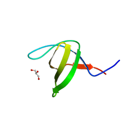
Crystal structure of the N114Q mutant of ABL-SH3 domain
Descriptor: GLYCEROL, Proto-oncogene tyrosine-protein kinase ABL1
Authors:Camara-Artigas, A.
Deposit date:2008-09-10
Release date:2009-09-15
Last modified:2023-08-30
Method:X-RAY DIFFRACTION (1.8 Å)
Cite:Role of interfacial water molecules in proline-rich ligand recognition by the Src homology 3 domain of Abl.
J.Biol.Chem., 285, 2010
3EG3
DownloadVisualize
BU of 3eg3 by Molmil
Crystal structure of the N114A mutant of ABL-SH3 domain
Descriptor: GLYCEROL, Proto-oncogene tyrosine-protein kinase ABL1
Authors:Camara-Artigas, A.
Deposit date:2008-09-10
Release date:2009-09-15
Last modified:2023-08-30
Method:X-RAY DIFFRACTION (1.4 Å)
Cite:Role of interfacial water molecules in proline-rich ligand recognition by the Src homology 3 domain of Abl.
J.Biol.Chem., 285, 2010
3EG0
DownloadVisualize
BU of 3eg0 by Molmil
Crystal structure of the N114T mutant of ABL-SH3 domain
Descriptor: GLYCEROL, Proto-oncogene tyrosine-protein kinase ABL1
Authors:Camara-Artigas, A.
Deposit date:2008-09-10
Release date:2009-09-15
Last modified:2023-08-30
Method:X-RAY DIFFRACTION (2.3 Å)
Cite:Role of interfacial water molecules in proline-rich ligand recognition by the Src homology 3 domain of Abl.
J.Biol.Chem., 285, 2010
6QJJ
DownloadVisualize
BU of 6qjj by Molmil
Crystal Structure of the third PDZ domain of PSD-95 protein: space group P3221
Descriptor: Disks large homolog 4, SULFATE ION
Authors:Camara-Artigas, A.
Deposit date:2019-01-24
Release date:2019-04-17
Last modified:2024-01-24
Method:X-RAY DIFFRACTION (1.7 Å)
Cite:Conformational changes in the third PDZ domain of the neuronal postsynaptic density protein 95.
Acta Crystallogr D Struct Biol, 75, 2019
6QJD
DownloadVisualize
BU of 6qjd by Molmil
Crystal Structure of the truncated form of the third PDZ domain of PSD-95: residues 302-392
Descriptor: Disks large homolog 4, SULFATE ION
Authors:Camara-Artigas, A.
Deposit date:2019-01-24
Release date:2019-04-17
Last modified:2024-01-24
Method:X-RAY DIFFRACTION (1.551 Å)
Cite:Conformational changes in the third PDZ domain of the neuronal postsynaptic density protein 95.
Acta Crystallogr D Struct Biol, 75, 2019
6QJN
DownloadVisualize
BU of 6qjn by Molmil
Crystal Structure of the third PDZ domain of PSD-95 protein D332G mutant: space group I4122
Descriptor: Disks large homolog 4
Authors:Camara-Artigas, A.
Deposit date:2019-01-24
Release date:2019-04-17
Last modified:2024-01-24
Method:X-RAY DIFFRACTION (1.8 Å)
Cite:Conformational changes in the third PDZ domain of the neuronal postsynaptic density protein 95.
Acta Crystallogr D Struct Biol, 75, 2019
6QJI
DownloadVisualize
BU of 6qji by Molmil
Crystal Structure of the third PDZ domain of PSD-95 protein: space group P3112
Descriptor: Disks large homolog 4, SULFATE ION
Authors:Camara-Artigas, A.
Deposit date:2019-01-24
Release date:2019-04-17
Last modified:2024-01-24
Method:X-RAY DIFFRACTION (1.5 Å)
Cite:Conformational changes in the third PDZ domain of the neuronal postsynaptic density protein 95.
Acta Crystallogr D Struct Biol, 75, 2019
6QJF
DownloadVisualize
BU of 6qjf by Molmil
Crystal Structure of the third PDZ domain of PSD-95 protein D332P mutant: space group C121, structure 1
Descriptor: Disks large homolog 4
Authors:Camara-Artigas, A.
Deposit date:2019-01-24
Release date:2019-04-17
Last modified:2024-01-24
Method:X-RAY DIFFRACTION (1.5 Å)
Cite:Conformational changes in the third PDZ domain of the neuronal postsynaptic density protein 95.
Acta Crystallogr D Struct Biol, 75, 2019
6QJL
DownloadVisualize
BU of 6qjl by Molmil
Crystal Structure of the third PDZ domain of PSD-95 protein D332G mutant: space group P21
Descriptor: Disks large homolog 4, GLYCEROL, SULFATE ION
Authors:Camara-Artigas, A.
Deposit date:2019-01-24
Release date:2019-04-17
Last modified:2024-01-24
Method:X-RAY DIFFRACTION (1.043 Å)
Cite:Conformational changes in the third PDZ domain of the neuronal postsynaptic density protein 95.
Acta Crystallogr D Struct Biol, 75, 2019
6QJG
DownloadVisualize
BU of 6qjg by Molmil
Crystal Structure of the third PDZ domain of PSD-95 protein D332P mutant: space group C121, structure 2
Descriptor: Disks large homolog 4
Authors:Camara-Artigas, A.
Deposit date:2019-01-24
Release date:2019-04-17
Last modified:2024-01-24
Method:X-RAY DIFFRACTION (2.003 Å)
Cite:Conformational changes in the third PDZ domain of the neuronal postsynaptic density protein 95.
Acta Crystallogr D Struct Biol, 75, 2019


123>

219140

PDB entries from 2024-05-01

PDB statisticsPDBj update infoContact PDBjnumon